Laser-based angle-resolved photoemission spectroscopy

Last updated

Laser-based angle-resolved photoemission spectroscopy is a form of angle-resolved photoemission spectroscopy that uses a laser as the light source. Photoemission spectroscopy is a powerful and sensitive experimental technique to study surface physics. [1] It is based on the photoelectric effect originally observed by Heinrich Hertz in 1887 and later explained by Albert Einstein in 1905 that when a material is shone by light, the electrons can absorb photons and escape from the material with the kinetic energy: , where is the incident photon energy, the work function of the material. Since the kinetic energy of ejected electrons are highly associated with the internal electronic structure, by analyzing the photoelectron spectroscopy one can realize the fundamental physical and chemical properties of the material, such as the type and arrangement of local bonding, electronic structure and chemical composition.

Contents

In addition, because electrons with different momentum will escape from the sample in different directions, angle-resolved photoemission spectroscopy is widely used to provide the dispersive energy-momentum spectrum. The photoemission experiment is conducted using synchrotron radiation light source with typical photon energy of 20 – 100 eV. Synchrotron light is ideal for investigating two-dimensional surface systems and offers unparalleled flexibility to continuously vary the incident photon energy. However, due to the high costs to construct and maintain this accelerator, high competition for beam time, as well as the universal minimum electron mean free path in the material around the operating photon energy (20–100 eV) which leads to the fundamental hindrance to the three-dimensional bulk materials sensitivity, an alternative photon source for angle-resolved photoemission spectroscopy is desirable.

If femtosecond lasers are used, the method can easily be extended to access excited electronic states and electron dynamics by introducing a pump-probe scheme, see also two-photon photoelectron spectroscopy.

Laser-based ARPES

Background

Table-top laser-based angle-resolved photoemission spectroscopy had been developed by some research groups. [2] [3] [4] Daniel Dessau of University of Colorado, Boulder, made the first demonstration and applied this technique to explore superconducting system. [2] The achievement not only greatly reduces the costs and size of facility, but also, most importantly, provides the unprecedented higher bulk sensitivity due to the low photon energy, typically 6 eV, and consequently the longer photoelectron mean free path (2–7 nm) in the sample. This advantage is extremely beneficial and powerful for the study of strongly correlated materials and high-Tc superconductors in which the physics of photoelectrons from the topmost layers might be different from the bulk. In addition to about one-order-of-magnitude improvement in the bulk sensitivity, the advance in the momentum resolution is also very significant: the photoelectrons will be more broadly dispersed in emission angle when the energy of incident photon decreases. In other words, for a given angular resolution of the electron spectrometer, the lower photon energy leads to higher momentum resolution.[ citation needed ] The typical momentum resolution of a 6 eV laser-based ARPES is approximately 8 times better than that of a 50 eV synchrotron radiation ARPES. Besides, the better momentum resolution due to low photon energy also results in less k-space accessible to ARPES which is helpful to the more precise spectrum analysis. For instance, in the 50 eV synchrotron ARPES, electrons from the first 4 Brillouin zones will be excited and scattered to contribute to the background of photoelectron analysis. However, the small momentum of 6 eV ARPES will only access some part of the first Brillouin zone and therefore only those electrons from small region of k-space can be ejected and detected as the background. The reduced inelastic scattering background is desirable while doing the measurement of weak physical quantities, in particular the high-Tc superconductors.

Experimental realization

The first 6 eV laser-based ARPES system used a Kerr mode-locked Ti: sapphire oscillator is used and pumped with another frequency doubled Nd:Vanadate laser of 5 W and then generates 70 fs and 6 nJ pulses which are tunable around 840 nm (1.5 eV) with the 1 MHz repetition rate.[ citation needed ] Two stages of non-linear second harmonic generation of light are carried out through type Ι phase matching in β-barium borate and then the quadruple light with 210 nm (~ 6 eV) is generated and finally focused and directed into the ultra-high vacuum chamber as the low-energy photon source to investigate the electronic structure of the sample.

In the first demonstration, Dessau’s group showed that the typical forth harmonic spectrum fits very well with the Gaussian profile with a full width at half maximum of 4.7 meV as well as presents a 200 μW power.[ citation needed ] The performance of high flux (~ 1014- 1015 photons/s) and narrow bandwidth makes the laser-based ARPES overwhelm the synchrotron radiation ARPES even though the best undulator beamlines are used.[ citation needed ] Another noticeable point is that one can make the quadruple light pass through either 1/4 wave plate or 1/2 wave plate which produces the circular polarization or any linear polarization light in the ARPES. Because the polarization of light can influence the signal to background ratio, the ability to control the polarization of light is a very significant improvement and advantage over the synchrotron ARPES. With the aforementioned favorable features, including lower costs for operating and maintenance, better energy and momentum resolution, and higher flux and ease of polarization control of photon source, the laser-based ARPES undoubtedly is an ideal candidate to be employed to conduct more sophisticated experiments in condensed matter physics.

Applications

High-Tc superconductor

One way to show the powerful ability of laser-based ARPES is to study high Tc superconductors. [3] The following figure references refer to this publication. Fig. 1 shows the experimental dispersion relation, binding energy vs. momentum, of the superconducting Bi2Sr2CaCu2O8+d along the nodal direction of the Brillouin zone. Fig. 1 (b) and Fig. 1 (c) are taken by the synchrotron light source of 28 eV and 52 eV, respectively, with the best undulator beamlines. The significantly sharper spectral peaks, the evidence of quasiparticles in the cuprate superconductor, by the powerful laser-based ARPES are shown in Fig. 1 (a). This is the first comparison of dispersive energy-momentum relation at low photon energy from table-top laser with higher energy from synchrotron ARPES. The much clearer dispersion in (a) indicates the improved energy-momentum resolution as well as many important physical features, such as overall band dispersion, Fermi surface, superconducting gaps, and a kink by electron-boson coupling, are successfully reproduced. It is foreseeable that in the near future the laser-based ARPES will be widely used to help condensed matter physicists get more detailed information about the nature of superconductivity in the exotic materials as well as other novel properties that cannot be observed by the state-of-the-art conventional experimental techniques.

Time-resolved electron dynamics

Femtosecond laser-based ARPES can be extended to give spectroscopic access to excited states in time-resolved photoemission and two-photon photoelectron spectroscopy. By pumping an electron to a higher level excited state with the first photon, the subsequent evolution and interactions of electronic states as a function of time can be studied by the second probing photon. The traditional pump-probe experiments usually measure the changes of some optical constants, which might be too complex to obtain the relevant physics. Since the ARPES can provide a lot of detailed information about the electronic structures and interactions, the pump-probe laser-based ARPES may study more complicated electronic systems with sub-picosecond resolution.

Summary and perspective

Even though the angle-resolved synchrotron radiation source is widely used to investigate the surface dispersive energy-momentum spectrum, the laser-based ARPES can even provide more detailed and bulk-sensitive electronic structures with much better energy and momentum resolution, which are critically necessary for studying the strongly correlated electronic system, high-Tc superconductor, and phase transition in exotic quantum system.[ citation needed ] In addition, the lower costs for operating and higher photon flux make laser-based ARPES easier to be handled and more versatile and powerful among other modern experimental techniques for surface science.

See also

Related Research Articles

Photoelectric effect Emission of electrons when light hits a material

The photoelectric effect is the emission of electrons when electromagnetic radiation, such as light, hits a material. Electrons emitted in this manner are called photoelectrons. The phenomenon is studied in condensed matter physics, and solid state and quantum chemistry to draw inferences about the properties of atoms, molecules and solids. The effect has found use in electronic devices specialized for light detection and precisely timed electron emission.

X-ray photoelectron spectroscopy Spectroscopic technique

X-ray photoelectron spectroscopy (XPS) is a surface-sensitive quantitative spectroscopic technique based on the photoelectric effect that can identify the elements that exist within a material or are covering its surface, as well as their chemical state, and the overall electronic structure and density of the electronic states in the material. XPS is a powerful measurement technique because it not only shows what elements are present, but also what other elements they are bonded to. The technique can be used in line profiling of the elemental composition across the surface, or in depth profiling when paired with ion-beam etching. It is often applied to study chemical processes in the materials in their as-received state or after cleavage, scraping, exposure to heat, reactive gasses or solutions, ultraviolet light, or during ion implantation.

Synchrotron light source

A synchrotron light source is a source of electromagnetic radiation (EM) usually produced by a storage ring, for scientific and technical purposes. First observed in synchrotrons, synchrotron light is now produced by storage rings and other specialized particle accelerators, typically accelerating electrons. Once the high-energy electron beam has been generated, it is directed into auxiliary components such as bending magnets and insertion devices in storage rings and free electron lasers. These supply the strong magnetic fields perpendicular to the beam which are needed to convert high energy electrons into photons.

Diamond Light Source UKs national synchrotron science facility located in Oxfordshire

Diamond Light Source is the UK's national synchrotron light source science facility located at the Harwell Science and Innovation Campus in Oxfordshire. Its purpose is to produce intense beams of light whose special characteristics are useful in many areas of scientific research. In particular it can be used to investigate the structure and properties of a wide range of materials from proteins, and engineering components to conservation of archeological artifacts.

In physics and physical chemistry, time-resolved spectroscopy is the study of dynamic processes in materials or chemical compounds by means of spectroscopic techniques. Most often, processes are studied after the illumination of a material occurs, but in principle, the technique can be applied to any process that leads to a change in properties of a material. With the help of pulsed lasers, it is possible to study processes that occur on time scales as short as 10−16 seconds.

Photoemission spectroscopy

Photoemission spectroscopy (PES), also known as photoelectron spectroscopy, refers to energy measurement of electrons emitted from solids, gases or liquids by the photoelectric effect, in order to determine the binding energies of electrons in the substance. The term refers to various techniques, depending on whether the ionization energy is provided by X-ray, XUV or UV photons. Regardless of the incident photon beam, however, all photoelectron spectroscopy revolves around the general theme of surface analysis by measuring the ejected electrons.

Photoemission electron microscopy is a type of electron microscopy that utilizes local variations in electron emission to generate image contrast. The excitation is usually produced by ultraviolet light, synchrotron radiation or X-ray sources. PEEM measures the coefficient indirectly by collecting the emitted secondary electrons generated in the electron cascade that follows the creation of the primary core hole in the absorption process. PEEM is a surface sensitive technique because the emitted electrons originate from a shallow layer. In physics, this technique is referred to as PEEM, which goes together naturally with low-energy electron diffraction (LEED), and low-energy electron microscopy (LEEM). In biology, it is called photoelectron microscopy (PEM), which fits with photoelectron spectroscopy (PES), transmission electron microscopy (TEM), and scanning electron microscopy (SEM).

In condensed matter physics, the Fermi surface is the surface in reciprocal space which separates occupied from unoccupied electron states at zero temperature. The shape of the Fermi surface is derived from the periodicity and symmetry of the crystalline lattice and from the occupation of electronic energy bands. The existence of a Fermi surface is a direct consequence of the Pauli exclusion principle, which allows a maximum of one electron per quantum state.

Extreme ultraviolet Ultraviolet light with a wavelength of 10–121nm

Extreme ultraviolet radiation or high-energy ultraviolet radiation is electromagnetic radiation in the part of the electromagnetic spectrum spanning wavelengths from 124 nm down to 10 nm, and therefore having photons with energies from 10 eV up to 124 eV. EUV is naturally generated by the solar corona and artificially by plasma, high harmonic generation sources and synchrotron light sources. Since UVC extends to 100 nm, there is some overlap in the terms.

High-energy X-rays or HEX-rays are very hard X-rays, with typical energies of 80–1000 keV (1 MeV), about one order of magnitude higher than conventional X-rays used for X-ray crystallography. They are produced at modern synchrotron radiation sources such as the beamline ID15 at the European Synchrotron Radiation Facility (ESRF). The main benefit is the deep penetration into matter which makes them a probe for thick samples in physics and materials science and permits an in-air sample environment and operation. Scattering angles are small and diffraction directed forward allows for simple detector setups.

Ultrafast laser spectroscopy is a spectroscopic technique that uses ultrashort pulse lasers for the study of dynamics on extremely short time scales. Different methods are used to examine the dynamics of charge carriers, atoms, and molecules. Many different procedures have been developed spanning different time scales and photon energy ranges; some common methods are listed below.

Electron spectroscopy refers to a group formed by techniques based on the analysis of the energies of emitted electrons such as photoelectrons and Auger electrons. This group includes X-ray photoelectron spectroscopy (XPS), which also known as Electron Spectroscopy for Chemical Analysis (ESCA), Electron energy loss spectroscopy (EELS), Ultraviolet photoelectron spectroscopy (UPS), and Auger electron spectroscopy (AES). These analytical techniques are used to identify and determine the elements and their electronic structures from the surface of a test sample. Samples can be solids, gases or liquids.

X-ray absorption near edge structure (XANES), also known as near edge X-ray absorption fine structure (NEXAFS), is a type of absorption spectroscopy that indicates the features in the X-ray absorption spectra (XAS) of condensed matter due to the photoabsorption cross section for electronic transitions from an atomic core level to final states in the energy region of 50–100 eV above the selected atomic core level ionization energy, where the wavelength of the photoelectron is larger than the interatomic distance between the absorbing atom and its first neighbour atoms.

Angle-resolved photoemission spectroscopy Experimental technique to determine the distribution of electrons in solids

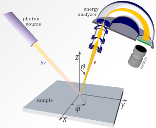
Angle-resolved photoemission spectroscopy (ARPES) is a powerful technique used in condensed matter physics to probe the structure of the electrons in a material, usually a crystalline solid. The technique is best suited for use in one- or two-dimensional materials. It is based on the photoelectric effect, in which an incoming photon of sufficient frequency dislodges an electron from the surface of a material. By directly measuring the kinetic energy and momentum distributions of the emitted photoelectrons, the technique can be used to map the electronic band structure, provide elemental information, and map Fermi surfaces. ARPES has been used by physicists to investigate high-temperature superconductors and materials exhibiting charge density waves.

Ultraviolet photoelectron spectroscopy (UPS) refers to the measurement of kinetic energy spectra of photoelectrons emitted by molecules which have absorbed ultraviolet photons, in order to determine molecular orbital energies in the valence region.

Resonant inelastic X-ray scattering

Resonant inelastic X-ray scattering (RIXS) is an X-ray spectroscopy technique used to investigate the electronic structure of molecules and materials.

National Synchrotron Light Source II

The National Synchrotron Light Source II (NSLS-II) at Brookhaven National Laboratory (BNL) in Upton, New York is a national user research facility funded primarily by the U.S. Department of Energy's (DOE) Office of Science. NSLS-II is one of the world's most advanced synchrotron light sources, designed to produce x-rays 10,000 times brighter than BNL's original light source, the National Synchrotron Light Source (NSLS). NSLS-II supports basic and applied research in energy security, advanced materials synthesis and manufacturing, environment, and human health.

Two-photon photoelectron spectroscopy

Time-resolved two-photon photoelectron (2PPE) spectroscopy is a time-resolved spectroscopy technique which is used to study electronic structure and electronic excitations at surfaces. The technique utilizes femtosecond to picosecond laser pulses in order to first photoexcite an electron. After a time delay, the excited electron is photoemitted into a free electron state by a second pulse. The kinetic energy and the emission angle of the photoelectron are measured in an electron energy analyzer. To facilitate investigations on the population and relaxation pathways of the excitation, this measurement is performed at different time delays.

Solaris (synchrotron)

Solaris is the first synchrotron built in Poland, under the auspices of the Jagiellonian University. It is located on the Campus of the 600th Anniversary of the Jagiellonian University Revival, in the southern part of Krakow. It is the central facility of the National Center of Synchrotron Radiation SOLARIS.

References

  1. K. Oura et al., Surface Science, An Introduction (Springer, Berlin, 2003).
  2. 1 2 J. Koralek; et al. (2007). "Experimental setup for low-energy laser-based angle resolved photoemission spectroscopy". Rev. Sci. Instrum. 78 (5): 053905. arXiv: 0706.1060 . Bibcode:2007RScI...78e3905K. doi:10.1063/1.2722413. PMID   17552839.
  3. 1 2 J. Koralek; et al. (2006). "Laser-based angle-resolved photoemission, the sudden approximation, and quasiparticle-like spectral peaks in Bi2Sr2CaCu2O8+d". Phys. Rev. Lett. 96 (1): 017005. arXiv: cond-mat/0508404 . Bibcode:2006PhRvL..96a7005K. doi:10.1103/PhysRevLett.96.017005. PMID   16486502.
  4. Guodong Liu; et al. (2008). "Development of a vacuum ultraviolet laser-based angle-resolved photoemission system with a superhigh energy resolution better than 1 meV". Rev. Sci. Instrum. 79 (2 Pt 1): 023105. arXiv: 0711.0282 . Bibcode:2008RScI...79b3105L. doi:10.1063/1.2835901. PMID   18315281.