Related Research Articles

In the human brain, the anterior cingulate cortex (ACC) is the frontal part of the cingulate cortex that resembles a "collar" surrounding the frontal part of the corpus callosum. It consists of Brodmann areas 24, 32, and 33.

The claustrum is a thin, bilateral collection of neurons and supporting glial cells, that connects to cortical and subcortical regions of the brain. It is located between the insula laterally and the putamen medially, separated by the extreme and external capsules respectively. The blood supply to the claustrum is fulfilled via the middle cerebral artery. It is considered to be the most densely connected structure in the brain, allowing for integration of various cortical inputs into one experience rather than singular events. The claustrum is difficult to study given the limited number of individuals with claustral lesions and the poor resolution of neuroimaging.
The pretectal area, or pretectum, is a midbrain structure composed of seven nuclei and comprises part of the subcortical visual system. Through reciprocal bilateral projections from the retina, it is involved primarily in mediating behavioral responses to acute changes in ambient light such as the pupillary light reflex, the optokinetic reflex, and temporary changes to the circadian rhythm. In addition to the pretectum's role in the visual system, the anterior pretectal nucleus has been found to mediate somatosensory and nociceptive information.

The insular cortex is a portion of the cerebral cortex folded deep within the lateral sulcus within each hemisphere of the mammalian brain.
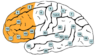
In mammalian brain anatomy, the prefrontal cortex (PFC) is the cerebral cortex which covers the front part of the frontal lobe. The PFC contains the Brodmann areas BA8, BA9, BA10, BA11, BA12, BA13, BA14, BA24, BA25, BA32, BA44, BA45, BA46, and BA47.

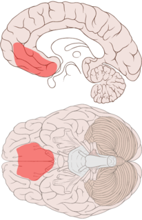
The posterior cingulate cortex (PCC) is the caudal part of the cingulate cortex, located posterior to the anterior cingulate cortex. This is the upper part of the "limbic lobe". The cingulate cortex is made up of an area around the midline of the brain. Surrounding areas include the retrosplenial cortex and the precuneus.

The reward system is a group of neural structures responsible for incentive salience, associative learning, and positively-valenced emotions, particularly ones involving pleasure as a core component. Reward is the attractive and motivational property of a stimulus that induces appetitive behavior, also known as approach behavior, and consummatory behavior. A rewarding stimulus has been described as "any stimulus, object, event, activity, or situation that has the potential to make us approach and consume it is by definition a reward". In operant conditioning, rewarding stimuli function as positive reinforcers; however, the converse statement also holds true: positive reinforcers are rewarding.
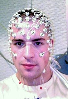
The psychological and physiological effects of meditation have been studied. In recent years, studies of meditation have increasingly involved the use of modern instruments, such as fMRI and EEG, which are able to observe brain physiology and neural activity in living subjects, either during the act of meditation itself or before and after meditation. Correlations can thus be established between meditative practices and brain structure or function.
Simulation theory of empathy is a theory that holds that humans anticipate and make sense of the behavior of others by activating mental processes that, if carried into action, would produce similar behavior. This includes intentional behavior as well as the expression of emotions. The theory states that children use their own emotions to predict what others will do. Therefore, we project our own mental states onto others.
Simulation theory is not primarily a theory of empathy, but rather a theory of how people understand others—that they do so by way of a kind of empathetic response. This theory uses more biological evidence than other theories of mind, such as the theory-theory.
The ventromedial prefrontal cortex (vmPFC) is a part of the prefrontal cortex in the mammalian brain. The ventral medial prefrontal is located in the frontal lobe at the bottom of the cerebral hemispheres and is implicated in the processing of risk and fear, as it is critical in the regulation of amygdala activity in humans. It also plays a role in the inhibition of emotional responses, and in the process of decision-making and self-control. It is also involved in the cognitive evaluation of morality.

In neuroscience, the default mode network (DMN), also known as the default network, default state network, or anatomically the medial frontoparietal network (M-FPN), is a large-scale brain network primarily composed of the medial prefrontal cortex, posterior cingulate cortex/precuneus and angular gyrus. It is best known for being active when a person is not focused on the outside world and the brain is at wakeful rest, such as during daydreaming and mind-wandering. It can also be active during detailed thoughts related to external task performance. Other times that the DMN is active include when the individual is thinking about others, thinking about themselves, remembering the past, and planning for the future.

The somatosensory system is the network of neural structures in the brain and body that produce the perception of touch, as well as temperature, body position (proprioception), and pain. It is a subset of the sensory nervous system, which also represents visual, auditory, olfactory, and gustatory stimuli. Somatosensation begins when mechano- and thermosensitive structures in the skin or internal organs sense physical stimuli such as pressure on the skin. Activation of these structures, or receptors, leads to activation of peripheral sensory neurons that convey signals to the spinal cord as patterns of action potentials. Sensory information is then processed locally in the spinal cord to drive reflexes, and is also conveyed to the brain for conscious perception of touch and proprioception. Note, somatosensory information from the face and head enters the brain through peripheral sensory neurons in the cranial nerves, such as the trigeminal nerve.
The biology of obsessive–compulsive disorder (OCD) refers biologically based theories about the mechanism of OCD. Cognitive models generally fall into the category of executive dysfunction or modulatory control. Neuroanatomically, functional and structural neuroimaging studies implicate the prefrontal cortex (PFC), basal ganglia (BG), insula, and posterior cingulate cortex (PCC). Genetic and neurochemical studies implicate glutamate and monoamine neurotransmitters, especially serotonin and dopamine.

Meditation and its effect on brain activity and the central nervous system became a focus of collaborative research in neuroscience, psychology and neurobiology during the latter half of the 20th century. Research on meditation sought to define and characterize various practices. Meditation's effect on the brain can be broken up into two categories: state changes and trait changes, respectively alterations in brain activities during the act of meditating and changes that are the outcome of long-term practice.
Pain empathy is a specific subgroup of empathy that involves recognizing and understanding another person's pain. Empathy is the mental ability that allows one person to understand another person's mental and emotional state and how to effectively respond to that person. When a person receives cues that another person is in pain, neural pain circuits within the brain are activated. There are several cues that can communicate pain to another person: visualization of the injury causing event, the injury itself, behavioral efforts of the injured to avoid further harm, and displays of pain and distress such as facial expressions, crying, and screaming. From an evolutionary perspective, pain empathy is beneficial for human group survival since it provides motivation for non-injured people to offer aid to the injured and to avoid injury themselves.

Mindfulness has been defined in modern psychological terms as "paying attention to relevant aspects of experience in a nonjudgmental manner", and maintaining attention on present moment experience with an attitude of openness and acceptance. Meditation is a platform used to achieve mindfulness. Both practices, mindfulness and meditation, have been "directly inspired from the Buddhist tradition" and have been widely promoted by Jon Kabat-Zinn. Mindfulness meditation has been shown to have a positive impact on several psychiatric problems such as depression and therefore has formed the basis of mindfulness programs such as mindfulness-based cognitive therapy, mindfulness-based stress reduction and mindfulness-based pain management. The applications of mindfulness meditation are well established, however the mechanisms that underlie this practice are yet to be fully understood. Many tests and studies on soldiers with PTSD have shown tremendous positive results in decreasing stress levels and being able to cope with problems of the past, paving the way for more tests and studies to normalize and accept mindful based meditation and research, not only for soldiers with PTSD, but numerous mental inabilities or disabilities.

Interoception is contemporarily defined as the sense of the internal state of the body. This can be both conscious and non-conscious. It encompasses the brain's process of integrating signals relayed from the body into specific subregions—like the brainstem, thalamus, insula, somatosensory, and anterior cingulate cortex—allowing for a nuanced representation of the physiological state of the body. This is important for maintaining homeostatic conditions in the body and, potentially, facilitating self-awareness.

Bipolar disorder is an affective disorder characterized by periods of elevated and depressed mood. The cause and mechanism of bipolar disorder is not yet known, and the study of its biological origins is ongoing. Although no single gene causes the disorder, a number of genes are linked to increase risk of the disorder, and various gene environment interactions may play a role in predisposing individuals to developing bipolar disorder. Neuroimaging and postmortem studies have found abnormalities in a variety of brain regions, and most commonly implicated regions include the ventral prefrontal cortex and amygdala. Dysfunction in emotional circuits located in these regions have been hypothesized as a mechanism for bipolar disorder. A number of lines of evidence suggests abnormalities in neurotransmission, intracellular signalling, and cellular functioning as possibly playing a role in bipolar disorder.
Social cognitive neuroscience is the scientific study of the biological processes underpinning social cognition. Specifically, it uses the tools of neuroscience to study "the mental mechanisms that create, frame, regulate, and respond to our experience of the social world". Social cognitive neuroscience uses the epistemological foundations of cognitive neuroscience, and is closely related to social neuroscience. Social cognitive neuroscience employs human neuroimaging, typically using functional magnetic resonance imaging (fMRI). Human brain stimulation techniques such as transcranial magnetic stimulation and transcranial direct-current stimulation are also used. In nonhuman animals, direct electrophysiological recordings and electrical stimulation of single cells and neuronal populations are utilized for investigating lower-level social cognitive processes.

Consoling touch is a pro-social behavior involving physical contact between a distressed individual and a caregiver. The physical contact, most commonly recognized in the form of a hand hold or embrace, is intended to comfort one or more of the participating individuals. Consoling touch is intended to alleviate or lessen emotional or physical pain. This type of social support has been observed across species and cultures. Studies have found little difference in the applications of consoling touch, with minor differences in frequency occurrence across cultures. These findings suggest a degree of universality. It remains unclear whether the relationship between social touch and interpersonal emotional bonds reflect biologically driven or culturally normative behavior. Evidence of consoling touch in non-human primates, who embrace one another following distressing events, suggest a biological basis. Numerous studies of consoling touch in humans and animals unveil a consistent physiological response. An embrace from a friend, relative, or even stranger can trigger the release of oxytocin, dopamine, and serotonin into the bloodstream. These neurotransmitters are associated with positive mood, numerous health benefits, and longevity. Cortisol, a stress hormone, also decreases. Studies have found that the degree of intimacy and quality of relationship between consoler and the consoled mediates physiological effects. In other words, while subjects experience reduced cortisol levels while holding the hand of a stranger, they exhibit a larger effect when receiving comfort from a trusted friend, and greater still, when holding the hand of a high quality romantic partner.
References
- 1 2 3 4 5 6 7 8 9 10 11 Nakata, Hiroki; Sakamoto, Kiwako; Kakigi, Ryusuke (2014-12-16). "Meditation reduces pain-related neural activity in the anterior cingulate cortex, insula, secondary somatosensory cortex, and thalamus". Frontiers in Psychology. 5: 1489. doi: 10.3389/fpsyg.2014.01489 . ISSN 1664-1078. PMC 4267182 . PMID 25566158.
- 1 2 3 4 5 6 Fox, Kieran C.R.; Dixon, Matthew L.; Nijeboer, Savannah; Girn, Manesh; Floman, James L.; Lifshitz, Michael; Ellamil, Melissa; Sedlmeier, Peter; Christoff, Kalina (2016). "Functional neuroanatomy of meditation: A review and meta-analysis of 78 functional neuroimaging investigations". Neuroscience & Biobehavioral Reviews. 65: 208–228. arXiv: 1603.06342 . doi:10.1016/j.neubiorev.2016.03.021. PMID 27032724. S2CID 9451371.
- 1 2 3 4 5 6 Mitsi, Vasiliki; Zachariou, Venetia (2016). "Modulation of pain, nociception, and analgesia by the brain reward center". Neuroscience. 338: 81–92. doi:10.1016/j.neuroscience.2016.05.017. PMC 5083150 . PMID 27189881.
- 1 2 3 4 Tang, Yi-Yuan; Hölzel, Britta K.; Posner, Michael I. (April 2015). "The neuroscience of mindfulness meditation". Nature Reviews. Neuroscience. 16 (4): 213–225. doi:10.1038/nrn3916. ISSN 1471-0048. PMID 25783612. S2CID 54521922.