Pulmonary artery stenosis

Last updated
Stenosis of pulmonary artery
Stenosis of right pulmonary artery.png
Stenosis of the right pulmonary artery in a patient which was due to a case of congenital rubella.
Specialty Medical genetics   OOjs UI icon edit-ltr-progressive.svg

Pulmonary artery stenosis (PAS) is a narrowing of the pulmonary artery. The pulmonary artery is a blood vessel moving blood from the right side of the heart to the lungs. This narrowing can be due to many causes, including infection during pregnancy, a congenital heart defect, a problem with blood clotting in childhood or early adulthood, or a genetic change. [1]

Contents

The narrowing can occur at many points along the pulmonary artery. The specific location indicates severity and affects treatment options. Most people with high-risk PAS are neonates, newborns, and young children. The more severe the disease the more likely it is to present with symptoms. With high-risk patients, it is necessary and acceptable to actively treat, to avoid worsening blood pressure, poor heart function, and worsening vessel disease across the body. [2]

Blood flows in a methodical way through the body. Blood that has already delivered oxygen returns to the heart. It arrives at the right upper chamber, gets pumped though the tricuspid valve and into the lower right chamber. It travels through the pulmonary valve to the pulmonary artery and toward the lungs. Oxygenated blood returns to the left side of the heart before it is pumped throughout the body. [3] When the pulmonary artery narrows, it increases blood pressure on the right side of the heart and causes the heart to work harder. [1]

PAS is not the same as pulmonary valve stenosis. The pulmonary valve is the opening between the right heart and the pulmonary artery. Valve narrowing has similar effects. However, treatment is different. The long term consequences of each condition also vary and present with other comorbidities. [4]

Symptoms

Pulmonary artery stenosis symptoms depend on the location and cause of the narrowing. Common symptoms include: [5]

Other symptoms are caused by lack of oxygenated blood flow. This causes individuals to present with: [5]

When blood cannot reach the lungs because of the narrowing, blood can back up. This can lead to swelling in body parts such as the hands and feet and can present similarly to right sided heart failure. [5]

Causes

Although 40% of individuals have no other underlying heart problems, PAS can still occur in 2-3% of individuals with congenital heart disease:

Genetic and infectious conditions:

Diagnosis

There is no immediate way to know if a child has PAS, but abnormal heart sounds, also known as murmurs, heard on physical exam, dictate further testing.

Treatment

The three treatments are balloon angioplasty, cutting balloon angioplasty, and stenting.

Balloon angioplasty

Balloon angioplasty expands the diameter of a vessel to counter narrowing. In this treatment a catheter with a balloon on the end is inserted into a larger, peripheral vessel and moved to the narrowing site. The goal is to tear two of the three layers of the artery, which increases the vessel's diameter and blood flow. A relatively stiff balloon achieves 72% efficacy with no risk increase. [4]

After the procedure is performed, several factors help determine success. These are:

The major reasons for simple balloon angioplasty failure are inability to tear the vessel wall, restenosis, and areas of narrowing due to compression by another body part rather than issues with the vessel itself. Even given evidence of vessel wall tearing, the rate of restenosis is between 15 and 44%. Studies reported that the rate of restenosis increases as time from the procedure increases. Rates of complications overall are estimated around 22%, including 10% higher risk complications. Other complications from this procedure are likely from too much vessel damage and include full vessel tears, deep vein thrombosis (clot in the vessel), complete artery rupture, and pulmonary edema. [4]

Younger patients are typically treated with balloon angioplasty until they are older and the risk for metal stents is significantly reduced. [2] [4]

Cutting balloon angioplasty

Cutting balloon angioplasty was invented to help reduce vessel damage. This method utilizes balloons with a blade that can cut through vessel walls rather than simply causing vessel wall tearing via crushing/expansion of the balloon. When the balloon is not inflated the blades are protected inside the balloon folds, ensuring they will not accidentally damage other vessels. [4] After the use of the cutting balloon, a larger, higher-pressure balloon can be used to improve efficacy. Multicenter studies reported the same safety profiles for simple versus cutting, with adverse effect rates of 2% and 3%, respectively. When a high-pressure balloon was used after cutting balloon angioplasty the rate of effectiveness increased from 52% to 85%. [2] Cutting balloon angioplasty provides more areas of vessel damage especially in vessels further from the heart. Overall cutting balloon angioplasty has similar complications and restenosis rates as simple, but offers a more effective treatment, and is a better option for smaller areas. [4]

Metal stents

To solve restenosis and external compression, metal stents were introduced. These stents are deployed via catheter. They are expanded at the site. The stents form a rigid structure that remain at the site. Newer studies reported successful dilation in 90% of patients on average, with newer studies indicating a 100% success rate. The benefits of stents is long term efficacy and the prevention of long term heart disease. Stents improved right side heart pressures, blood flow to the lungs, and pressure difference between the right and left sides. Stents can last up to 15 years, much longer than balloon angioplasty. Stents have the lowest rate of restenosis, at 2-3%. [4]

Stents allow for normal vessel growth during childhood and adolescent years, and do not damage the heart or vasculature. 30-50% of young children required replacement with a larger stent in the first 2 years of placement. Complication rates are around 12% and include drift due to blood flow, which can damage vessels, clot formation on the stent. Because of these concerns and necessary removal of the stent if these were to occur, the stent must be monitored frequently after placement. [4]

Another concern is fracturing, or breaking, due to repetitive pressure. Patients experience a 13% adverse event rate with major events such as a fracture at 1.2%. Studies reported that the adverse event rate decreased with patient age, likely due to less growth in the vessel. Replacement rates are as high as 43%, and increases over time. [2] [4]

History

Prior to the development of balloon angioplasty, surgical angioplasty was the main treatment method. [4] Because of lack of efficacy, limited accessibility to certain areas of stenosis, increased risk for scarring, and a high rate of repeat stenosis, surgical angioplasty is only used if other methods fail or if surgeons observe the narrowing while repairing another defect. Some studies reported a 62% surgical success rate for surgical angioplasty. Alternative treatments would be required for 4/10 patients while exposing them to risks of surgery. [2]

Initially a soft balloon was used, but only about 60% of patients experienced even a 50% increase in blood flow. This efficacy was similar to surgical angioplasty with a large risk reduction. Later, a stiffer balloon achieved better results,

Related Research Articles

<span class="mw-page-title-main">Angioplasty</span> Procedure to widen narrow arteries or veins

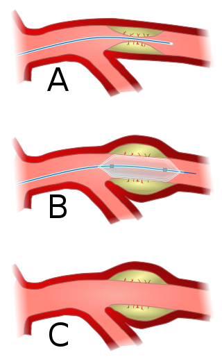
Angioplasty, also known as balloon angioplasty and percutaneous transluminal angioplasty (PTA), is a minimally invasive endovascular procedure used to widen narrowed or obstructed arteries or veins, typically to treat arterial atherosclerosis.

<span class="mw-page-title-main">Coronary catheterization</span> Radiography of heart and blood vessels

A coronary catheterization is a minimally invasive procedure to access the coronary circulation and blood filled chambers of the heart using a catheter. It is performed for both diagnostic and interventional (treatment) purposes.

dextro-Transposition of the great arteries Medical condition

dextro-Transposition of the great arteries is a potentially life-threatening birth defect in the large arteries of the heart. The primary arteries are transposed.

<span class="mw-page-title-main">Mitral stenosis</span> Heart disease with narrowing of valve

Mitral stenosis is a valvular heart disease characterized by the narrowing of the opening of the mitral valve of the heart. It is almost always caused by rheumatic valvular heart disease. Normally, the mitral valve is about 5 cm2 during diastole. Any decrease in area below 2 cm2 causes mitral stenosis. Early diagnosis of mitral stenosis in pregnancy is very important as the heart cannot tolerate increased cardiac output demand as in the case of exercise and pregnancy. Atrial fibrillation is a common complication of resulting left atrial enlargement, which can lead to systemic thromboembolic complications such as stroke.

<span class="mw-page-title-main">Restenosis</span> Recurrence of stenosis, a narrowing of a blood vessel

Restenosis is the recurrence of stenosis, a narrowing of a blood vessel, leading to restricted blood flow. Restenosis usually pertains to an artery or other large blood vessel that has become narrowed, received treatment to clear the blockage, and subsequently become re-narrowed. This is usually restenosis of an artery, or other blood vessel, or possibly a vessel within an organ.

<span class="mw-page-title-main">Stenosis</span> Abnormal narrowing of a blood vessel or other tubular organ or structure

Stenosis is the abnormal narrowing of a blood vessel or other tubular organ or structure such as foramina and canals. It is also sometimes called a stricture.

<span class="mw-page-title-main">Interventional cardiology</span> Catheter-based treatment of structural heart diseases

Interventional cardiology is a branch of cardiology that deals specifically with the catheter based treatment of structural heart diseases. Andreas Gruentzig is considered the father of interventional cardiology after the development of angioplasty by interventional radiologist Charles Dotter.

<span class="mw-page-title-main">Cardiac catheterization</span> Insertion of a catheter into a chamber or vessel of the heart

Cardiac catheterization is the insertion of a catheter into a chamber or vessel of the heart. This is done both for diagnostic and interventional purposes.

<span class="mw-page-title-main">Renovascular hypertension</span> Medical condition

Renovascular hypertension is a condition in which high blood pressure is caused by the kidneys' hormonal response to narrowing of the arteries supplying the kidneys. When functioning properly this hormonal axis regulates blood pressure. Due to low local blood flow, the kidneys mistakenly increase blood pressure of the entire circulatory system. It is a form of secondary hypertension - a form of hypertension whose cause is identifiable.

<span class="mw-page-title-main">Pulmonary valve stenosis</span> Heart valve disorder

Pulmonary valve stenosis (PVS) is a heart valve disorder. Blood going from the heart to the lungs goes through the pulmonary valve, whose purpose is to prevent blood from flowing back to the heart. In pulmonary valve stenosis this opening is too narrow, leading to a reduction of flow of blood to the lungs.

<span class="mw-page-title-main">Aortic valvuloplasty</span>

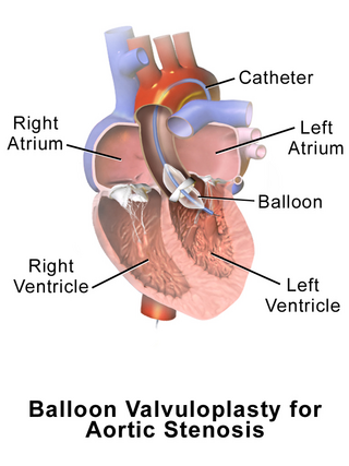
Aortic valvuloplasty, also known as balloon aortic valvuloplasty (BAV), is a procedure used to improve blood flow through the aortic valve in conditions that cause aortic stenosis, or narrowing of the aortic valve. It can be performed in various patient populations including fetuses, newborns, children, adults, and pregnant women. The procedure involves using a balloon catheter to dilate the narrowed aortic valve by inflating the balloon.

<span class="mw-page-title-main">Carotid stenting</span> Medical intervention

Carotid artery stenting is an endovascular procedure where a stent is deployed within the lumen of the carotid artery to treat narrowing of the carotid artery and decrease the risk of stroke. It is used to treat narrowing of the carotid artery in high-risk patients, when carotid endarterectomy is considered too risky.

<span class="mw-page-title-main">Lutembacher's syndrome</span> Medical condition

Lutembacher's syndrome is a very rare form of congenital heart disease that affects one of the chambers of the heart as well as a valve. It is commonly known as both congenital atrial septal defect (ASD) and acquired mitral stenosis (MS). Congenital atrial septal defect refers to a hole being in the septum or wall that separates the two atria; this condition is usually seen in fetuses and infants. Mitral stenosis refers to mitral valve leaflets sticking to each other making the opening for blood to pass from the atrium to the ventricles very small. With the valve being so small, blood has difficulty passing from the left atrium into the left ventricle. Septal defects that may occur with Lutembacher's syndrome include: Ostium primum atrial septal defect or ostium secundum which is more prevalent.

<span class="mw-page-title-main">Coronary stent</span> Medical stent implanted into coronary arteries

A coronary stent is a tube-shaped device placed in the coronary arteries that supply blood to the heart, to keep the arteries open in patients suffering from coronary heart disease. The vast majority of stents used in modern interventional cardiology are drug-eluting stents (DES). They are used in a medical procedure called percutaneous coronary intervention (PCI). Coronary stents are divided into two broad types: drug-eluting and bare metal stents. As of 2023, drug-eluting stents were used in more than 90% of all PCI procedures. Stents reduce angina and have been shown to improve survival and decrease adverse events after a patient has suffered a heart attack—medically termed an acute myocardial infarction.

The following outline is provided as an overview of and topical guide to cardiology, the branch of medicine dealing with disorders of the human heart. The field includes medical diagnosis and treatment of congenital heart defects, coronary artery disease, heart failure, valvular heart disease and electrophysiology. Physicians who specialize in cardiology are called cardiologists.

<span class="mw-page-title-main">Congenital stenosis of vena cava</span> Medical condition

Congenital stenosis of vena cava is a congenital anomaly in which the superior vena cava or inferior vena cava has an aberrant interruption or coarctation.

<span class="mw-page-title-main">Arterial embolism</span> Interruption of blood flow to an organ

Arterial embolism is a sudden interruption of blood flow to an organ or body part due to an embolus adhering to the wall of an artery blocking the flow of blood, the major type of embolus being a blood clot (thromboembolism). Sometimes, pulmonary embolism is classified as arterial embolism as well, in the sense that the clot follows the pulmonary artery carrying deoxygenated blood away from the heart. However, pulmonary embolism is generally classified as a form of venous embolism, because the embolus forms in veins. Arterial embolism is the major cause of infarction.

Pulmonary Artery Banding (PAB) was introduced by Muller and Dammann in 1951 as a surgical technique to reduce excessive pulmonary blood flow in infants suffering from congenital heart defects. PAB is a palliative operation as it does not correct the problems, but attempts to improve abnormal heart function, relieve symptoms and reduce high pressure in the lungs. The use of PAB has decreased over the years due to advancements in definitive surgical repairs, however PAB still has widespread clinical use. PAB is commonly used in patients when definitive surgical repair is not feasible.

<span class="mw-page-title-main">Hybrid cardiac surgery</span>

A hybrid cardiac surgical procedure in a narrow sense is defined as a procedure that combines a conventional, more invasive surgical part with an interventional part, using some sort of catheter-based procedure guided by fluoroscopy imaging in a hybrid operating room (OR) without interruption. The hybrid technique has a reduced risk of surgical complications and has shown decreased recovery time. It can be used to treat numerous heart diseases and conditions and with the increasing complexity of each case, the hybrid surgical technique is becoming more common.

<span class="mw-page-title-main">Balloon pulmonary angioplasty</span> Medical procedure

Balloon pulmonary angioplasty (BPA) is an emerging minimally invasive procedure to treat chronic thromboembolic pulmonary hypertension (CTEPH) in people who are not suitable for pulmonary thromboendarterectomy (PTE) or still have residual pulmonary hypertension and areas of narrowing in the pulmonary arterial tree following previous PTE.

References

  1. 1 2 "Pulmonary Artery Stenosis". ucsfbenioffchildrens.org. University of California San Francisco. Retrieved 2023-11-02.
  2. 1 2 3 4 5 Patel, Anuj B.; Ratnayaka, Kanishka; Bergersen, Lisa (2019). "A review: Percutaneous pulmonary artery stenosis therapy: State-of-the-art and look to the future". Cardiology in the Young. 29 (2): 93–99. doi:10.1017/S1047951118001087. PMID   30587259. S2CID   58650748.
  3. "Pulmonary Artery Stenosis | Interventional Cardiology Program | UPMC Children's". Children's Hospital of Pittsburgh. Retrieved 2023-11-02.
  4. 1 2 3 4 5 6 7 8 9 10 11 12 13 14 Kim, Chan W.; Aronow, Wilbert S.; Dutta, Tanya; Spevack, Daniel M.; Frishman, William H. (2021). "Treatment of Peripheral Pulmonary Artery Stenosis". Cardiology in Review. 29 (3): 115–119. doi:10.1097/crd.0000000000000300. PMID   32053544. S2CID   211113778.
  5. 1 2 3 4 5 6 7 "Pulmonary Artery Stenosis: Causes, Symptoms and Treatment". Cleveland Clinic. Retrieved 2023-11-02.
  6. 1 2 Leitman, Ellen M.; McDermott, Shaunagh (2019). "Pulmonary arteries: Imaging of pulmonary embolism and beyond". Cardiovascular Diagnosis and Therapy. 9 (Suppl 1): S37–S58. doi: 10.21037/cdt.2018.08.05 . PMC   6732114 . PMID   31559153.