Leptin | |||||||||||
---|---|---|---|---|---|---|---|---|---|---|---|
Identifiers | |||||||||||
Symbol | Leptin | ||||||||||
Pfam | PF02024 | ||||||||||
Pfam clan | CL0053 | ||||||||||
InterPro | IPR000065 | ||||||||||
SCOP2 | 1ax8 / SCOPe / SUPFAM | ||||||||||
|
Teleost leptins are a family of peptide hormones found in fish (teleostei) that are orthologs of the mammalian hormone leptin. The teleost and mammalian leptins appear to have similar functions, namely, regulation of energy intake and expenditure.
The leptin (LEP) hormone was long thought to be specific to mammals, but in recent years the gene (lep) has been found in amphibia such as the tiger salamander (Ambystoma tigrinum), [2] [3] and the African clawed frog (Xenopus laevi). [4] The discovery of lep in puffer fish (Takifugu rubripes) [5] demonstrates the ancient ancestry of this hormone.
There are two closely related lep paralogues in Atlantic salmon (Salmo salar). [6] A single lep gene has been documented for green-spotted pufferfish (Tetraodon nigroviridis), [5] rainbow trout (Oncorhynchus mykiss), [7] Arctic charr (Salvelinus alpinus), [8] silver carp (Hypophthalmichthys molitrix), and grass carp (Ctenopharyngodon idellus). [9] In other species there are reports of two closely related lep paralogues, including common carp (Cyprinus carpio) [10] and Atlantic salmon. [6] More distantly related lep genes have been found in medaka (Oryzias latipes) [11] and zebrafish (Danio rerio). [12] At least 2 leptin genes (lepa and lepb) exist in the crown-clade (Fig. 1). [5] [10] [11] [12] Early findings have shown that lepa and lepb share low interspecies aa identity, and are argued to have arisen through whole genome duplication, [5] which occurred early in the teleost lineage. [13] The duplicity of genes has been described for Atlantic salmon, Japanese medaka, common carp and zebrafish. [6] [10] [11] [12] Both lep paralogues [6] cluster with lepa, and therefore suggest that at least one or more form (lepb) may exist in this species, since it is tetraploid. [14] However, previous attempts using genomic synteny have only found the putative genomic duplicates in medaka and zebrafish paralogue. [11] [12] Currently it remains unclear, whether lepb exists in other teleosts due to the degenerative nature of this paralogue.
The large differences among endothermic (warm-blooded) mammalian and ectothermic (cold-blooded) teleost leptins raised the question of whether the energy homeostatic functions of the teleost leptins are conserved. Initial phylogenetic analysis has revealed that amino acid conservation with other vertebrate Lep orthologues is low, with only 13.2% sequence identity between torafugu and human LEP. [5] Subsequent investigations have confirmed the low amino acid identity of teleost leps compared to mammalian LEP. [5] [10] [15] [16]
The three-dimensional homology modeling predicts strong conservation of the tertiary structure between Atlantic salmon and other teleost Leps compared to their mammalian orthologues (Fig. 2). [5] [6] [7] [11] [12]
Both lepa1 and lepa2 have two characteristic cysteine residues which predict the formation of a disulfide bond in Lep, which is a pre-equisite for this 3D configuration and bioactivity of human LEP. [19] [20] The models suggest that the bonding of lepa2 might be different from lepa1. There are several differences between the 3D structures of lepa1 and lepa2; e.g. α-helix 5 is considerable shorter in lepa1 than lepa2. Furthermore, α-helix 1 for lepa2 appears to be split by a short-disordered region, and may therefore have a poorer affinity. However, considering that it is a predicted model based upon the structure mask of human LEP, the significance of these putative conformational adjustments remains to be tested.
The importance of the conserved tertiary structure of Lep is most likely explained by requirements for specific LepR-binding affinity and is constrained by the structure of the receptor-binding pocket. [4] This might also explain some of the results from studies on teleost using heterologous mammalian Lep. E.g. treatment with the mammalian hormone caused an anorexic effect in goldfish (Carassius auratus) [21] [22] and green sunfish (Lepomis cyanellus), [23] but not in Coho salmon (Oncorhynchus kisutch), [24] channel catfish (Ictalurus punctatus) [25] and green sunfish. [26] These contradicting results have been explained by the relatively large differences in amino acid sequences observed between mammals and fish. [4] [5] [10]
Rønnestad and colleagues [6] recently detected five isoforms of the leptin receptor (lepr) that have differences in 3'-end of the mRNA sequence. Of these, only the longest form conserved all functionally important domains (such as three fibronectin type III domains, the Ig C2-like domain, a pair of WSXWS motifs, two JAK2-binding motif boxes, and a STAT-binding domain), [6] while the other four forms have only the intra-cellular region. The long form of mammalian LepR has a function for full signal transduction through the JAK/STAT pathways, whereas the shorter forms exhibit partial or no signaling capabilities. [27] [28] The biological importance of long form LepR via the JAK/STAT pathway in maintaining body weight and energy homeostasis has been demonstrated. [29] Previous studies in teleosts have only identified a single lepr. [11] [15] [30] Rønnestad et al., [6] is the first to report that plural LepR transcripts in any ectotherm species. When looking at the available motif for lepr, the model suggests that it would bind easily to lepa1 and not lepa2 (Fig. 2). Furthermore, the relatively ubiquitous expression of lepr in salmon tissues supports diverse roles of lep in teleosts. [6]
The study on torafugu [5] indicated that lep is mainly expressed in the liver in contrasts to the adipose secretion in mammals. [31] [32] However recent studies have shown that lep is expressed in several peripheral tissues, including intestine, kidney, ovary, muscle and adipose tissue. [6] [7] [11] The multiplicity of lep genes and their low conservation in Teleostei. [10] [11] [12] suggest that their physiological roles may be more divergent than reported for mammals.
The tissue expression pattern for the Atlantic salmon lep paralogues differs substantially (Fig.3) [6] and hence indicates a possible difference in function. With the exception of the results presented here, and those for zebrafish and Japanese medaka. [6] [11] [12] Few studies have investigated the broad tissue distribution of lep in teleost fishes. The more distantly related lep genes (lepa and lepb) showed distinct differences in tissue distribution, as shown in e.g. medaka, where lepa is being expressed in liver and muscle, while lepb is more highly expressed in the brain and eye. However, these differences are also observed for more closely related lep paralogues, such as lepa1 in Atlantic salmon, being more highly expressed in brain, liver and white muscle, while lepa2 is mainly expressed in the stomach and midgut. (Fig. 3).
The observations that long-term feed restriction does not significantly affect lep expression in Atlantic salmon [6] has also been noted in other teleosts. However it is likely that prolonged feed restriction can influence several endocrine parameters to adapt to the nutritional condition. For example, in common carp, a rapid response in ob gene expression in hepatic tissue of common carp shortly after feeding, but no changes in expression in response to different long-term feeding regime was observed. [10] These authors suggested that this effect could be explained by the fact that starved fish do not lose weight as rapidly as mammals, a consequence of being ectothermic and possessing a much lower standard metabolic rate, and therefore can withstand longer periods of starvation. A similar study on grass carp showed that chronic injection of species-specific Lep did not affect long-term food intake and body weight, while acute injection decreased food intake. [9] Conversely, Murashita et al., (unpublished results) observed increased proopiomelanocortin a1(pomca1) levels following chronic injection of Lep in Atlantic salmon, which suggests that chronic exposure to elevated Lep levels can decrease food intake through the Pomc pathway in this species. Recent studies in Atlantic salmon did not observe any difference of feed restriction in circulating plasma levels, [18] which contrasts recent results in rainbow trout [33] and suggest that the relation between circulating lep levels and energy status differs from that in mammals. However, salmon RIA appears to allow interspecies assessment of plasma lep levels. [33] This only confirms that more comprehensive studies are needed for conclusive data interpretation. Studies on rainbow trout also implicated Lep as an anorectic hormone as in mammals. Injection of rainbow trout with recombinant trout leptin (rt-leptin) resulted in a significantly reduced appetite over two days that coincided with a decrease in hypothalamic mRNA expression of neuropeptide Y (npy) and increase of pomc mRNAs, respectively. [7] Whether these observations are due to species-specific differences in long-term leptin regulation of appetite or growth is not known, however, consensus data indicate that the effects of Lep on appetite regulation may be short-term in teleosts.
Recent studies on short-term effects of a meal or the absence of a meal [18] has revealed that lepa1 expression specifically peaks in the peripheral tissues after 6 – 9 hr in the unfed fish. This suggests that the transcript specific response could be associated with the absence of food. Conversely, since the unfed fish had not received food for 33 hr (24 + 9 hr), the peaks could represent an unrelated effect. Each lepa1 peak occurred during a phase of falling plasma Lep, and since this occurred in both fed and unfed fish, the temporal upregulation of lepa1 does not in fact appear to be specifically related to the absence of food.
The earliest peak of lepa1 occurred in the white muscle, which represents an important lipid reservoir in Atlantic salmon. [34] Unlike pufferfish, which utilizes the liver as a major lipid repository, [5] Atlantic salmon shows that despite a high visceral lipid content, hepatocytes contain few lipid droplets compared to other fish species, [35] yet are an important site for leptin expression. [5] [6] [7] [10] [12] [18] Moen and colleagues [18] reported that both lepa1 and lepa2 peaked at 9 hr in the liver of unfed fish. By contrast, however, studies in common carp demonstrated a peak in leptin-I(lepa1) and leptin–II (lepa2) in liver at 3 and 6 hr post feeding respectively. [10] The earlier expression response of leptins in common carp likely reflects the higher temperature under which the experiments were conducted, but contrasts the findings of upregulation of lepa1 due to the absence of food. [18] Similarly, in mice, a postprandial increase in hepatic leptin expression has also been reported. [36] However, in grass carp, intraperitoneal injection of recombinant leptin only alters the appetite on the first day, and does not influence food intake during the ensuing 12 days. [16] At present, the data for Atlantic salmon are therefore quite different and suggest that leptin expression in this species may have a complex lipostatic function.
Pro-opiomelanocortin (POMC) is a precursor polypeptide with 241 amino acid residues. POMC is synthesized in corticotrophs of the anterior pituitary from the 267-amino-acid-long polypeptide precursor pre-pro-opiomelanocortin (pre-POMC), by the removal of a 26-amino-acid-long signal peptide sequence during translation. POMC is part of the central melanocortin system.
Leptin is a protein hormone predominantly made by adipocytes. Its primary role is likely to regulate long-term energy balance.
Neuropeptide Y (NPY) is a 36 amino-acid neuropeptide that is involved in various physiological and homeostatic processes in both the central and peripheral nervous systems. It is secreted alongside other neurotransmitters such as GABA and glutamate.
The immunoglobulin heavy chain (IgH) is the large polypeptide subunit of an antibody (immunoglobulin). In human genome, the IgH gene loci are on chromosome 14.
Melanin-concentrating hormone (MCH), also known as pro-melanin stimulating hormone (PMCH), is a cyclic 19-amino acid orexigenic hypothalamic peptide originally isolated from the pituitary gland of teleost fish, where it controls skin pigmentation. In mammals it is involved in the regulation of feeding behavior, mood, sleep-wake cycle and energy balance.
GnRH2, also known as gonadotropin-releasing hormone II or LHRH-II. Its gene is located on human chromosome 20.
Urocortin is a protein that in humans is encoded by the UCN gene. Urocortin belongs to the corticotropin-releasing factor (CRF) family of proteins which includes CRF, urotensin I, sauvagine, urocortin II and urocortin III. Urocortin is involved in the mammalian stress response, and regulates aspects of appetite and stress response.
Peptide YY (PYY), also known as peptide tyrosine tyrosine, is a peptide that in humans is encoded by the PYY gene. Peptide YY is a short peptide released from cells in the ileum and colon in response to feeding. In the blood, gut, and other elements of periphery, PYY acts to reduce appetite; similarly, when injected directly into the central nervous system, PYY is also anorexigenic, i.e., it reduces appetite.
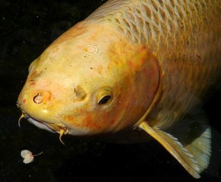
In fish anatomy and turtle anatomy, a barbel is a slender, whiskerlike sensory organ near the mouth. Fish that have barbels include the catfish, the carp, the goatfish, the hagfish, the sturgeon, the zebrafish, the black dragonfish and some species of shark such as the sawshark. Barbels house the taste buds of such fish and are used to search for food in murky water.
The Japanese rice fish, also known as the medaka, is a member of genus Oryzias (ricefish), the only genus in the subfamily Oryziinae. This small native of Japan is a denizen of rice paddies, marshes, ponds, slow-moving streams and tide pools. It is euryhaline, occurring in both brackish and freshwater. It became popular as an aquarium fish because of its hardiness and pleasant coloration: its coloration varies from creamy-white to yellowish in the wild to white, creamy-yellow, or orange in aquarium-bred individuals. Bright yellow, red or green transgenic populations, similar to GloFish, have also been developed, but are banned from sale in the EU. The medaka has been a popular pet since the 17th century in Japan. After fertilization, the female carries her eggs attached anterior to the anal fin for a period before depositing them on plants or similar things.
The type-I interferons (IFN) are cytokines which play essential roles in inflammation, immunoregulation, tumor cells recognition, and T-cell responses. In the human genome, a cluster of thirteen functional IFN genes is located at the 9p21.3 cytoband over approximately 400 kb including coding genes for IFNα, IFNω (IFNW1), IFNɛ (IFNE), IFNк (IFNK) and IFNβ (IFNB1), plus 11 IFN pseudogenes.
Leptin receptor, also known as LEP-R or OB-R, is a type I cytokine receptor, a protein that in humans is encoded by the LEPR gene. LEP-R functions as a receptor for the fat cell-specific hormone leptin. LEP-R has also been designated as CD295. Its location is the cell membrane, and it has extracellular, trans-membrane and intracellular sections.
The central melanocortin system is defined anatomically as a collection of central nervous system circuits which include:
Genetically modified fish are organisms from the taxonomic clade which includes the classes Agnatha, Chondrichthyes and Osteichthyes whose genetic material (DNA) has been altered using genetic engineering techniques. In most cases, the aim is to introduce a new trait to the fish which does not occur naturally in the species, i.e. transgenesis.
Fish are exposed to large oxygen fluctuations in their aquatic environment since the inherent properties of water can result in marked spatial and temporal differences in the concentration of oxygen. Fish respond to hypoxia with varied behavioral, physiological, and cellular responses to maintain homeostasis and organism function in an oxygen-depleted environment. The biggest challenge fish face when exposed to low oxygen conditions is maintaining metabolic energy balance, as 95% of the oxygen consumed by fish is used for ATP production releasing the chemical energy of nutrients through the mitochondrial electron transport chain. Therefore, hypoxia survival requires a coordinated response to secure more oxygen from the depleted environment and counteract the metabolic consequences of decreased ATP production at the mitochondria.
Relaxin family peptide hormones in humans are represented by seven members: three relaxin-like (RLN) and four insulin-like (INSL) peptides: RLN1, RLN2, RNL3, INSL3, INSL4, INSL5, INSL6. This subdivision into two classes is based primarily on early findings, and does not reflect the evolutionary origins or physiological differences between peptides. For example, it is known that the genes coding for RLN3 and INSL5 arose from one ancestral gene, and INSL3 shares origin with RLN2 and its multiple duplicates: RLN1, INSL4, INSL6.
Retinal homeobox protein Rx also known as retina and anterior neural fold homeobox is a protein that in humans is encoded by the RAX gene. The RAX gene is located on chromosome 18 in humans, mice, and rats.
Stanniocalcin is a family of hormones which regulate calcium and phosphate balance in the body. The first stanniocalcin discovered was from fish and was identified as the principal calcium-reducing (hypocalcaemic) factor. It was isolated from special organs in fish called corpuscles of Stannius, hence the name stanniocalcin. Chemically, stanniocalcins are glycosylated proteins having a molecular mass of 50 kDa. They exist in molecular pairs (homodimers) and are joined together by disulfide linkage. Stanniocalcins are made up of approximately 250 amino acids.
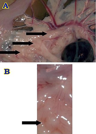
Brockmann body is an endocrine organ in some teleost fish, and is composed of a collection of islet tissues. The islet tissues are in turn composed of endocrine cells which are the principal sites of insulin synthesis. They are distributed around the spleen and the large intestine. They also secrete other hormones such as glucagon and somatostatin. Hence, Brochmann body is the centre of control of blood glucose level in these fishes. Glucagon is also produced from the intestine, but Brockmann body is the major source. Increased level of glucose stimulate the Brockmann body to release insulin, while inhibiting glucagon. Somatostatin released from Brockmann body inhibits cells to produce insulin and glucagon. In addition it inhibits release of growth hormone from the pituitary. It is named after a German physician Heinrich Brochmann who discovered it in 1848.
The food-entrainable oscillator (FEO) is a circadian clock that can be entrained by varying the time of food presentation. It was discovered when a rhythm was found in rat activity. This was called food anticipatory activity (FAA), and this is when the wheel-running activity of mice decreases after feeding, and then rapidly increases in the hours leading up to feeding. FAA appears to be present in non-mammals (pigeons/fish), but research heavily focuses on its presence in mammals. This rhythmic activity does not require the suprachiasmatic nucleus (SCN), the central circadian oscillator in mammals, implying the existence of an oscillator, the FEO, outside of the SCN, but the mechanism and location of the FEO is not yet known. There is ongoing research to investigate if the FEO is the only non-light entrainable oscillator in the body.