Platelet-derived growth factor receptor

Last updated
Platelet-derived growth factor receptor
PDGFR-beta 3MJG.png
Ribbon image of two molecules of human PDGF receptor beta (yellow and magenta) in complex with dimeric PDGF-B (cyan and green). [1]
Identifiers
SymbolPDGFR
Pfam PF04692
InterPro IPR006782
Membranome 1204
Available protein structures:
Pfam   structures / ECOD  
PDB RCSB PDB; PDBe; PDBj
PDBsum structure summary
platelet-derived growth factor receptor, alpha polypeptide
Identifiers
Symbol PDGFRA
NCBI gene 5156
HGNC 8803
OMIM 173490
RefSeq NM_006206
UniProt P16234
Other data
Locus Chr. 4 q12
Search for
Structures Swiss-model
Domains InterPro
platelet-derived growth factor receptor, beta polypeptide
Identifiers
Symbol PDGFRB
Alt. symbolsPDGFR
NCBI gene 5159
HGNC 8804
OMIM 173410
RefSeq NM_002609
UniProt P09619
Other data
Locus Chr. 5 q31-q32
Search for
Structures Swiss-model
Domains InterPro

Platelet-derived growth factor receptors (PDGF-R) are cell surface tyrosine kinase receptors for members of the platelet-derived growth factor (PDGF) family. PDGF subunits -A and -B are important factors regulating cell proliferation, cellular differentiation, cell growth, development and many diseases including cancer. [2] There are two forms of the PDGF-R, alpha and beta each encoded by a different gene. [3] Depending on which growth factor is bound, PDGF-R homo- or heterodimerizes. [4]

Contents

Mechanism of action

The PDGF family consists of PDGF-A, -B, -C and -D, which form either homo- or heterodimers (PDGF-AA, -AB, -BB, -CC, -DD). The four PDGFs are inactive in their monomeric forms. The PDGFs bind to the protein tyrosine kinase receptors PDGF receptor-α and -β. These two receptor isoforms dimerize upon binding the PDGF dimer, leading to three possible receptor combinations, namely -αα, -ββ and -αβ. The extracellular region of the receptor consists of five immunoglobulin-like domains while the intracellular part is a tyrosine kinase domain. The ligand-binding sites of the receptors are located to the three first immunoglobulin-like domains. PDGF-CC specifically interacts with PDGFR-αα and -αβ, but not with -ββ, and thereby resembles PDGF-AB. PDGF-DD binds to PDGFR-ββ with high affinity, and to PDGFR-αβ to a markedly lower extent and is therefore regarded as PDGFR-ββ specific. PDGF-AA binds only to PDGFR-αα, while PDGF-BB is the only PDGF that can bind all three receptor combinations with high affinity. [5]

Dimerization is a prerequisite for the activation of the kinase. Kinase activation is visualized as tyrosine phosphorylation of the receptor molecules, which occurs between the dimerized receptor molecules (transphosphorylation). In conjunction with dimerization and kinase activation, the receptor molecules undergo conformational changes, which allow a basal kinase activity to phosphorylate a critical tyrosine residue, thereby "unlocking" the kinase, leading to full enzymatic activity directed toward other tyrosine residues in the receptor molecules as well as other substrates for the kinase. Expression of both receptors and each of the four PDGFs is under independent control, giving the PDGF/PDGFR system a high flexibility. Different cell types vary greatly in the ratio of PDGF isoforms and PDGFRs expressed. Different external stimuli such as inflammation, embryonic development or differentiation modulate cellular receptor expression allowing binding of some PDGFs but not others. Additionally, some cells display only one of the PDGFR isoforms while other cells express both isoforms, simultaneously or separately.

Interaction with signal transduction molecules

Tyrosine phosphorylation sites in growth factor receptors serve two major purposes—to control the state of activity of the kinase and to create binding sites for downstream signal transduction molecules, which in many cases also are substrates for the kinase. The second part of the tyrosine kinase domain in the PDGFβ receptor is phosphorylated at Tyr-857, and mutant receptors carrying phenylalanine at this position have reduced kinase activity. Tyr-857 has therefore been assigned a role in positive regulation of kinase activity. [6] Sites of tyrosine phosphorylation involved in binding signal transduction molecules have been identified in the juxtamembrane domain, the kinase insert, and in the C-terminal tail in the PDGFβ receptor. The phosphorylated tyrosine residue and in general three adjacent C-terminal amino acid residues form specific binding sites for signal transduction molecules. Binding to these sites involves a common conserved stretches, denoted the Src homology (SH) 2 domain and/or Phosphotyrosine Binding Domains (PTB). The specificity of these interactions appears to be very high, since mutant receptors carrying phenylalanine residues in one or several of the different phosphorylation sites generally lack the capacity to bind the targeted signal transduction molecule. The signal transduction molecules are either equipped with different enzymatic activities, or they are adaptor molecules, which in some but not all cases are found in complexes with subunits that carry a catalytic activity. Upon interaction with the activated receptor, the catalytic activities become up-regulated, through tyrosine phosphorylation or other mechanisms, generating a signal that may be unique for each type of signal transduction molecule.

Examination of the different signaling cascades induced by RTKs established Ras/mitogen-activated protein kinase (MAPK), PI-3 kinase, and phospholipase-γ (PLCγ) pathways as key downstream mediators of PDGFR signaling. [7] [8] In addition, reactive oxygen species (ROS)-dependent STAT3 activation has been established to be a key downstream mediator of PDGFR signaling in vascular smooth muscle cells. [9]

MAPK pathway

The adaptor protein Grb2 forms a complex with Sos by the Grb2 SH3 domain. Grb2 (or the Grb2/Sos complex) is recruited to the membrane by the Grb2 SH2 domain binding to activated PDGFR-bound SHP2 (also known as PTPN11, a cytosolic PTP), thereby allowing interaction with Ras and the exchange of GDP for GTP on Ras. Whereas the interaction between Grb2 and PDGFR occurs through interaction with the SHP2 protein, Grb2 instead binds to activated EGFR through Shc, another adaptor protein that forms a complex with many receptors via its PTB domain. [10] Once activated, Ras interacts with several proteins, namely Raf. Activated Raf stimulates MAPK-kinase (MAPKK or MEK) by phosphorylating a serine residue in its activation loop. MAPKK then phosphorylates MAPK (ERK1/2) on T and Y residues at the activation-loop leading to its activation. Activated MAPK phosphorylates a variety of cytoplasmic substrates, as well as transcription factors, when translocated into the nucleus. MAPK family members have been found to regulate various biological functions by phosphorylation of particular target molecules (such as transcription factors, other kinases etc.) located in cell membrane, cytoplasm and nucleus, and thus contribute to the regulation of different cellular processes such as cell proliferation, differentiation, apoptosis and immunoresponses.

PI3K pathway

The class IA phospholipid kinase, PI-3 kinase, is activated by the majority of RTKs. Similarly to other SH2 domain-containing proteins, PI-3 kinase forms a complex with PY sites on activated receptors. The main function of PI3K activation is the generation of PIP3, which functions as a second messenger to activate downstream tyrosine kinases Btk and Itk, the Ser/Thr kinases PDK1 and Akt (PKB). The major biological functions of Akt activation can be classified into three categories – survival, proliferation and cell growth. Akt is also known to be implicated in several cancers, particularly breast. PLCγ is immediately recruited by an activated RTK through the binding of its SH2 domains to phosphotyrosine sites of the receptor. After activation, PLCγ hydrolyses its substrate PtdIns(4,5)P2 and forms two second messengers, diacylglycerol and Ins(1,4,5)P3. Ins(1,4,5)P3 stimulates the release of Ca 2+ from intracellular supplies. Ca 2+ then binds to calmodulin, which subsequently activates a family of calmodulindependent protein kinases (CamKs). In addition, both diacylglycerol and Ca 2+ activate members of the PKC family. The second messengers generated by PtdIns(4,5)P2 hydrolysis stimulate a variety of intracellular processes such as proliferation, angiogenesis, cell motility.

See also

Related Research Articles

<span class="mw-page-title-main">Protein kinase</span> Enzyme that adds phosphate groups to other proteins

A protein kinase is a kinase which selectively modifies other proteins by covalently adding phosphates to them (phosphorylation) as opposed to kinases which modify lipids, carbohydrates, or other molecules. Phosphorylation usually results in a functional change of the target protein (substrate) by changing enzyme activity, cellular location, or association with other proteins. The human genome contains about 500 protein kinase genes and they constitute about 2% of all human genes. There are two main types of protein kinase. The great majority are serine/threonine kinases, which phosphorylate the hydroxyl groups of serines and threonines in their targets. Most of the others are tyrosine kinases, although additional types exist. Protein kinases are also found in bacteria and plants. Up to 30% of all human proteins may be modified by kinase activity, and kinases are known to regulate the majority of cellular pathways, especially those involved in signal transduction.

<span class="mw-page-title-main">Tyrosine kinase</span> Class hi residues

A tyrosine kinase is an enzyme that can transfer a phosphate group from ATP to the tyrosine residues of specific proteins inside a cell. It functions as an "on" or "off" switch in many cellular functions.

<span class="mw-page-title-main">Insulin receptor</span> Mammalian protein found in Homo sapiens

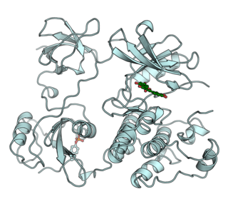
The insulin receptor (IR) is a transmembrane receptor that is activated by insulin, IGF-I, IGF-II and belongs to the large class of receptor tyrosine kinase. Metabolically, the insulin receptor plays a key role in the regulation of glucose homeostasis; a functional process that under degenerate conditions may result in a range of clinical manifestations including diabetes and cancer. Insulin signalling controls access to blood glucose in body cells. When insulin falls, especially in those with high insulin sensitivity, body cells begin only to have access to lipids that do not require transport across the membrane. So, in this way, insulin is the key regulator of fat metabolism as well. Biochemically, the insulin receptor is encoded by a single gene INSR, from which alternate splicing during transcription results in either IR-A or IR-B isoforms. Downstream post-translational events of either isoform result in the formation of a proteolytically cleaved α and β subunit, which upon combination are ultimately capable of homo or hetero-dimerisation to produce the ≈320 kDa disulfide-linked transmembrane insulin receptor.

The JAK-STAT signaling pathway is a chain of interactions between proteins in a cell, and is involved in processes such as immunity, cell division, cell death, and tumour formation. The pathway communicates information from chemical signals outside of a cell to the cell nucleus, resulting in the activation of genes through the process of transcription. There are three key parts of JAK-STAT signalling: Janus kinases (JAKs), signal transducer and activator of transcription proteins (STATs), and receptors. Disrupted JAK-STAT signalling may lead to a variety of diseases, such as skin conditions, cancers, and disorders affecting the immune system.

<span class="mw-page-title-main">Platelet-derived growth factor</span> Signaling glycoprotein regulating cell proliferation

Platelet-derived growth factor (PDGF) is one among numerous growth factors that regulate cell growth and division. In particular, PDGF plays a significant role in blood vessel formation, the growth of blood vessels from already-existing blood vessel tissue, mitogenesis, i.e. proliferation, of mesenchymal cells such as fibroblasts, osteoblasts, tenocytes, vascular smooth muscle cells and mesenchymal stem cells as well as chemotaxis, the directed migration, of mesenchymal cells. Platelet-derived growth factor is a dimeric glycoprotein that can be composed of two A subunits (PDGF-AA), two B subunits (PDGF-BB), or one of each (PDGF-AB).

<span class="mw-page-title-main">T-cell receptor</span> Protein complex on the surface of T cells that recognises antigens

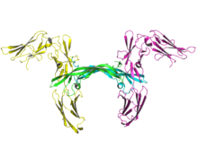
The T-cell receptor (TCR) is a protein complex found on the surface of T cells, or T lymphocytes, that is responsible for recognizing fragments of antigen as peptides bound to major histocompatibility complex (MHC) molecules. The binding between TCR and antigen peptides is of relatively low affinity and is degenerate: that is, many TCRs recognize the same antigen peptide and many antigen peptides are recognized by the same TCR.

<span class="mw-page-title-main">Nerve growth factor</span> Mammalian protein found in Homo sapiens

Nerve growth factor (NGF) is a neurotrophic factor and neuropeptide primarily involved in the regulation of growth, maintenance, proliferation, and survival of certain target neurons. It is perhaps the prototypical growth factor, in that it was one of the first to be described. Since it was first isolated by Nobel Laureates Rita Levi-Montalcini and Stanley Cohen in 1956, numerous biological processes involving NGF have been identified, two of them being the survival of pancreatic beta cells and the regulation of the immune system.

Biological crosstalk refers to instances in which one or more components of one signal transduction pathway affects another. This can be achieved through a number of ways with the most common form being crosstalk between proteins of signaling cascades. In these signal transduction pathways, there are often shared components that can interact with either pathway. A more complex instance of crosstalk can be observed with transmembrane crosstalk between the extracellular matrix (ECM) and the cytoskeleton.

The MAPK/ERK pathway is a chain of proteins in the cell that communicates a signal from a receptor on the surface of the cell to the DNA in the nucleus of the cell.

<span class="mw-page-title-main">Receptor tyrosine kinase</span> Class of enzymes

Receptor tyrosine kinases (RTKs) are the high-affinity cell surface receptors for many polypeptide growth factors, cytokines, and hormones. Of the 90 unique tyrosine kinase genes identified in the human genome, 58 encode receptor tyrosine kinase proteins. Receptor tyrosine kinases have been shown not only to be key regulators of normal cellular processes but also to have a critical role in the development and progression of many types of cancer. Mutations in receptor tyrosine kinases lead to activation of a series of signalling cascades which have numerous effects on protein expression. Receptor tyrosine kinases are part of the larger family of protein tyrosine kinases, encompassing the receptor tyrosine kinase proteins which contain a transmembrane domain, as well as the non-receptor tyrosine kinases which do not possess transmembrane domains.

<span class="mw-page-title-main">GRB2</span> Protein-coding gene in the species Homo sapiens

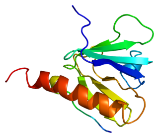
Growth factor receptor-bound protein 2 also known as Grb2 is an adaptor protein involved in signal transduction/cell communication. In humans, the GRB2 protein is encoded by the GRB2 gene.

<span class="mw-page-title-main">Linker for activation of T cells</span> Protein-coding gene in the species Homo sapiens

The Linker for activation of T cells, also known as linker of activated T cells or LAT, is a protein involved in the T-cell antigen receptor signal transduction pathway which in humans is encoded by the LAT gene. Alternative splicing results in multiple transcript variants encoding different isoforms.

<span class="mw-page-title-main">Fibroblast growth factor receptor 1</span> Protein-coding gene in the species Homo sapiens

Fibroblast growth factor receptor 1 (FGFR1), also known as basic fibroblast growth factor receptor 1, fms-related tyrosine kinase-2 / Pfeiffer syndrome, and CD331, is a receptor tyrosine kinase whose ligands are specific members of the fibroblast growth factor family. FGFR1 has been shown to be associated with Pfeiffer syndrome, and clonal eosinophilias.

<span class="mw-page-title-main">GAB2</span> Protein-coding gene in the species Homo sapiens

GRB2-associated-binding protein 2 also known as GAB2 is a protein that in humans is encoded by the GAB2 gene.

<span class="mw-page-title-main">IRS1</span> Protein-coding gene in the species Homo sapiens

Insulin receptor substrate 1 (IRS-1) is a signaling adapter protein that in humans is encoded by the IRS-1 gene. It is a 131 kDa protein with amino acid sequence of 1242 residues. It contains a single pleckstrin homology (PH) domain at the N-terminus and a PTB domain ca. 40 residues downstream of this, followed by a poorly conserved C-terminus tail. Together with IRS2, IRS3 (pseudogene) and IRS4, it is homologous to the Drosophila protein chico, whose disruption extends the median lifespan of flies up to 48%. Similarly, Irs1 mutant mice experience moderate life extension and delayed age-related pathologies.

<span class="mw-page-title-main">PDGFRB</span> Protein-coding gene in the species Homo sapiens

Platelet-derived growth factor receptor beta is a protein that in humans is encoded by the PDGFRB gene. Mutations in PDGFRB are mainly associated with the clonal eosinophilia class of malignancies.

<span class="mw-page-title-main">FRS2</span> Protein-coding gene in the species Homo sapiens

Fibroblast growth factor receptor substrate 2 is a protein that in humans is encoded by the FRS2 gene.

A non-receptor tyrosine kinase (nRTK) is a cytosolic enzyme that is responsible for catalysing the transfer of a phosphate group from a nucleoside triphosphate donor, such as ATP, to tyrosine residues in proteins. Non-receptor tyrosine kinases are a subgroup of protein family tyrosine kinases, enzymes that can transfer the phosphate group from ATP to a tyrosine residue of a protein (phosphorylation). These enzymes regulate many cellular functions by switching on or switching off other enzymes in a cell.

Src kinase family is a family of non-receptor tyrosine kinases that includes nine members: Src, Yes, Fyn, and Fgr, forming the SrcA subfamily, Lck, Hck, Blk, and Lyn in the SrcB subfamily, and Frk in its own subfamily. Frk has homologs in invertebrates such as flies and worms, and Src homologs exist in organisms as diverse as unicellular choanoflagellates, but the SrcA and SrcB subfamilies are specific to vertebrates. Src family kinases contain six conserved domains: a N-terminal myristoylated segment, a SH2 domain, a SH3 domain, a linker region, a tyrosine kinase domain, and C-terminal tail.

<span class="mw-page-title-main">Tyrosine phosphorylation</span> Phosphorylation of peptidyl-tyrosine

Tyrosine phosphorylation is the addition of a phosphate (PO43−) group to the amino acid tyrosine on a protein. It is one of the main types of protein phosphorylation. This transfer is made possible through enzymes called tyrosine kinases. Tyrosine phosphorylation is a key step in signal transduction and the regulation of enzymatic activity.

References

  1. PDB: 3MJG ; Shim AH, Liu H, Focia PJ, Chen X, Lin PC, He X (June 2010). "Structures of a platelet-derived growth factor/propeptide complex and a platelet-derived growth factor/receptor complex". Proceedings of the National Academy of Sciences of the United States of America. 107 (25): 11307–12. Bibcode:2010PNAS..10711307H. doi: 10.1073/pnas.1000806107 . PMC   2895058 . PMID   20534510.; rendered using PyMOL.
  2. Williams LT (March 1989). "Signal transduction by the platelet-derived growth factor receptor". Science. 243 (4898): 1564–70. Bibcode:1989Sci...243.1564W. doi:10.1126/science.2538922. PMID   2538922.
  3. Heldin CH, Westermark B (April 1989). "Platelet-derived growth factor: three isoforms and two receptor types". Trends in Genetics. 5 (4): 108–11. doi:10.1016/0168-9525(89)90040-1. PMID   2543106.
  4. Heldin CH, Ostman A, Eriksson A, Siegbahn A, Claesson-Welsh L, Westermark B (March 1992). "Platelet-derived growth factor: isoform-specific signalling via heterodimeric or homodimeric receptor complexes". Kidney International. 41 (3): 571–4. doi: 10.1038/ki.1992.84 . PMID   1315403.
  5. Cao Y, Cao R, Hedlund EM (July 2008). "R Regulation of tumor angiogenesis and metastasis by FGF and PDGF signaling pathways". Journal of Molecular Medicine. 86 (7): 785–9. doi:10.1007/s00109-008-0337-z. PMID   18392794. S2CID   21872247.
  6. Kazlauskas A, Cooper JA (September 1989). "Autophosphorylation of the PDGF receptor in the kinase insert region regulates interactions with cell proteins". Cell. 58 (6): 1121–33. doi:10.1016/0092-8674(89)90510-2. PMID   2550144. S2CID   25586248.
  7. Valius M, Kazlauskas A (April 1993). "Phospholipase C-gamma 1 and phosphatidylinositol 3 kinase are the downstream mediators of the PDGF receptor's mitogenic signal". Cell. 73 (2): 321–34. doi:10.1016/0092-8674(93)90232-F. PMID   7682895. S2CID   36305139.
  8. Montmayeur JP, Valius M, Vandenheede J, Kazlauskas A (December 1997). "The platelet-derived growth factor beta receptor triggers multiple cytoplasmic signaling cascades that arrive at the nucleus as distinguishable inputs". The Journal of Biological Chemistry. 272 (51): 32670–8. doi: 10.1074/jbc.272.51.32670 . PMID   9405485.
  9. Blazevic T, Schwaiberger AV, Schreiner CE, Schachner D, Schaible AM, Grojer CS, Atanasov AG, Werz O, Dirsch VM, Heiss EH (December 2013). "12/15-lipoxygenase contributes to platelet-derived growth factor-induced activation of signal transducer and activator of transcription 3". The Journal of Biological Chemistry. 288 (49): 35592–603. doi: 10.1074/jbc.M113.489013 . PMC   3853304 . PMID   24165129.
  10. Schlessinger, J. SH2/SH3 Signaling Proteins. Curr. Op. Gen. Dev. 1994, 4(1):25-30.