Placental growth factor

Last updated
PGF
PDB 1rv6 EBI.jpg
Available structures
PDB Ortholog search: PDBe RCSB
Identifiers
Aliases PGF , D12S1900, PGFL, PLGF, PlGF-2, SHGC-10760, Placental growth factor, PIGF
External IDs OMIM: 601121 MGI: 105095 HomoloGene: 1978 GeneCards: PGF
Orthologs
SpeciesHumanMouse
Entrez
Ensembl
UniProt
RefSeq (mRNA)

NM_002632
NM_001207012
NM_001293643

NM_001271705
NM_008827

RefSeq (protein)

NP_001193941
NP_001280572
NP_002623

NP_001258634
NP_032853

Location (UCSC) Chr 14: 74.94 – 74.96 Mb Chr 12: 85.21 – 85.22 Mb
PubMed search [3] [4]
Wikidata
View/Edit Human View/Edit Mouse

Placental growth factor(PlGF) is a protein that in humans is encoded by the PGF gene. [5] [6]

Contents

Placental growth factor (PGF) is a member of the VEGF (vascular endothelial growth factor) sub-family - a key molecule in angiogenesis and vasculogenesis, in particular during embryogenesis. The main source of PGF during pregnancy is the placental trophoblast. PGF is also expressed in many other tissues, including the villous trophoblast. [7]

The placental growth factor (PGF) gene is a protein-coding gene and a member of the vascular endothelial growth factor (VEGF) family. The PGF gene is expressed only in human umbilical vein endothelial cells (HUVE) and the placenta. PGF is ultimately associated with angiogenesis. Specifically, PGF plays a role in trophoblast growth and differentiation. Trophoblast cells, specifically extravillous trophoblast cells, are responsible for invading the uterine wall and the maternal spiral arteries. The extravillous trophoblast cells produce a blood vessel of larger diameter for the developing fetus that is independent of maternal vasoconstriction. This is essential for increased blood flow and reduced resistance. [8] Proper development of blood vessels in the placenta is crucial for the higher blood requirement of the fetus later in pregnancy. Under normal physiologic conditions, PGF is also expressed at a low level in other organs including the heart, lung, thyroid, and skeletal muscle.

Isoform tissue specificity

There are three isoforms of this protein: PGF-1, PGF-2, PGF-3. PGF-1 is specifically found in the colon as well as mammary carcinomas, while PGF-2 is only found in early placenta up until the 8th week of development. PGF-2 is the only isoform able to bind to heparin. PGF-3 is found mainly in placental tissues. [9] [10]

Clinical significance

Placental growth factor-expression within human atherosclerotic lesions is associated with plaque inflammation and neovascular growth. [11] [12]

Serum levels of PGF and sFlt-1 (soluble fms-like tyrosine kinase-1, also known as soluble VEGF receptor-1) are altered in women with preeclampsia. Studies show that in both early and late onset preeclampsia, maternal serum levels of sFlt-1 are higher and PGF lower in women presenting with preeclampsia. In addition, placental sFlt-1 levels were significantly increased and PGF decreased in women with preeclampsia as compared to those with uncomplicated pregnancies. This suggests that placental concentrations of sFlt-1 and PGF mirror the maternal serum changes. This is consistent with the view that the placenta is the main source of sFlt-1 and PGF during pregnancy.1

PGF is a potential biomarker for preeclampsia, a condition in which blood vessels in the placenta are too narrow, resulting in high blood pressure. As mentioned before, extravillous trophoblast cells invade maternal arteries. Improper differentiation may result in hypo-invasion of these arteries and thus failure to widen enough. Studies have found low levels of PGF in women who were diagnosed with preeclampsia later in their pregnancy.

Associated diseases

Placental insufficiency, otherwise known as uteroplacental vascular insufficiency, results from insufficient blood supply to the placenta. This disease is characterized by an alteration in the PGF gene and its GPCR and ERK signaling pathways. [13] Alterations in the PGF and the PGF receptor mRNA expression prevent the normal development of placental vasculature. [14]

Twin-to-twin transfusion syndrome is another disease associated with the PGF gene. This is a rare disease occurring primarily in identical twins where blood from one twin is transferred to the other. Typically, the twin whose blood is being transferred is born smaller and with anemia while the other twin is born larger with too much blood and at increased risk for heart failure. The PGF gene pathways primarily affected are the TGF-Beta pathway and AKT signaling pathway. [15]

Related Research Articles

<span class="mw-page-title-main">Angiogenesis</span> Blood vessel formation, when new vessels emerge from existing vessels

Angiogenesis is the physiological process through which new blood vessels form from pre-existing vessels, formed in the earlier stage of vasculogenesis. Angiogenesis continues the growth of the vasculature mainly by processes of sprouting and splitting, but processes such as coalescent angiogenesis, vessel elongation and vessel cooption also play a role. Vasculogenesis is the embryonic formation of endothelial cells from mesoderm cell precursors, and from neovascularization, although discussions are not always precise. The first vessels in the developing embryo form through vasculogenesis, after which angiogenesis is responsible for most, if not all, blood vessel growth during development and in disease.

<span class="mw-page-title-main">Pre-eclampsia</span> Hypertension occurring during pregnancy

Pre-eclampsia is a disorder of pregnancy characterized by the onset of high blood pressure and often a significant amount of protein in the urine. When it arises, the condition begins after 20 weeks of pregnancy. In severe cases of the disease there may be red blood cell breakdown, a low blood platelet count, impaired liver function, kidney dysfunction, swelling, shortness of breath due to fluid in the lungs, or visual disturbances. Pre-eclampsia increases the risk of undesirable as well as lethal outcomes for both the mother and the fetus including preterm labor. If left untreated, it may result in seizures at which point it is known as eclampsia.

<span class="mw-page-title-main">Platelet-derived growth factor</span> Signaling glycoprotein regulating cell proliferation

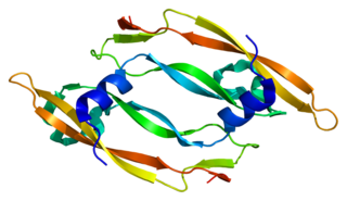
Platelet-derived growth factor (PDGF) is one among numerous growth factors that regulate cell growth and division. In particular, PDGF plays a significant role in blood vessel formation, the growth of blood vessels from already-existing blood vessel tissue, mitogenesis, i.e. proliferation, of mesenchymal cells such as fibroblasts, osteoblasts, tenocytes, vascular smooth muscle cells and mesenchymal stem cells as well as chemotaxis, the directed migration, of mesenchymal cells. Platelet-derived growth factor is a dimeric glycoprotein that can be composed of two A subunits (PDGF-AA), two B subunits (PDGF-BB), or one of each (PDGF-AB).

Vascular endothelial growth factor, originally known as vascular permeability factor (VPF), is a signal protein produced by many cells that stimulates the formation of blood vessels. To be specific, VEGF is a sub-family of growth factors, the platelet-derived growth factor family of cystine-knot growth factors. They are important signaling proteins involved in both vasculogenesis and angiogenesis.

An angiogenesis inhibitor is a substance that inhibits the growth of new blood vessels (angiogenesis). Some angiogenesis inhibitors are endogenous and a normal part of the body's control and others are obtained exogenously through pharmaceutical drugs or diet.

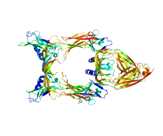
Soluble fms-like tyrosine kinase-1 is a tyrosine kinase protein with antiangiogenic properties. A non-membrane associated splice variant of VEGF receptor 1 (Flt-1), sFlt-1 binds the angiogenic factors VEGF and PlGF, reducing blood vessel growth through reduction of free VEGF and PlGF concentrations. In humans, sFlt-1 is important in the regulation of blood vessel formation in diverse tissues, including the kidneys, cornea, and uterus. Abnormally high levels of sFlt-1 have been implicated in the pathogenesis of preeclampsia.

<span class="mw-page-title-main">Decidua</span> Part of uterus modified in pregnancy

The decidua is the modified mucosal lining of the uterus that forms every month, in preparation for pregnancy. It is shed off each month when there is no fertilised egg to support. The decidua is under the influence of progesterone. Endometrial cells become highly characteristic. The decidua forms the maternal part of the placenta and remains for the duration of the pregnancy. After birth the decidua is shed together with the placenta.

<span class="mw-page-title-main">Angiopoietin</span> Protein family

Angiopoietin is part of a family of vascular growth factors that play a role in embryonic and postnatal angiogenesis. Angiopoietin signaling most directly corresponds with angiogenesis, the process by which new arteries and veins form from preexisting blood vessels. Angiogenesis proceeds through sprouting, endothelial cell migration, proliferation, and vessel destabilization and stabilization. They are responsible for assembling and disassembling the endothelial lining of blood vessels. Angiopoietin cytokines are involved with controlling microvascular permeability, vasodilation, and vasoconstriction by signaling smooth muscle cells surrounding vessels. There are now four identified angiopoietins: ANGPT1, ANGPT2, ANGPTL3, ANGPT4.

<span class="mw-page-title-main">VEGF receptor</span> Protein family

VEGF receptors (VEGFRs) are receptors for vascular endothelial growth factor (VEGF). There are three main subtypes of VEGFR, numbered 1, 2 and 3. Depending on alternative splicing, they may be membrane-bound (mbVEGFR) or soluble (sVEGFR).

<span class="mw-page-title-main">Angiopoietin 1</span> Protein-coding gene in the species Homo sapiens

Angiopoietin 1 is a type of angiopoietin and is encoded by the gene ANGPT1.

<span class="mw-page-title-main">VEGFR1</span> Protein-coding gene in the species Homo sapiens

Vascular endothelial growth factor receptor 1 is a protein that in humans is encoded by the FLT1 gene.

<span class="mw-page-title-main">Kinase insert domain receptor</span> Protein-coding gene in the species Homo sapiens

Kinase insert domain receptor also known as vascular endothelial growth factor receptor 2 (VEGFR-2) is a VEGF receptor. KDR is the human gene encoding it. KDR has also been designated as CD309. KDR is also known as Flk1.

<span class="mw-page-title-main">Vascular endothelial growth factor C</span> Growth factor protein found in humans

Vascular endothelial growth factor C (VEGF-C) is a protein that is a member of the platelet-derived growth factor / vascular endothelial growth factor (PDGF/VEGF) family. It is encoded in humans by the VEGFC gene, which is located on chromosome 4q34.

<span class="mw-page-title-main">Neuropilin 1</span> Protein-coding gene in the species Homo sapiens

Neuropilin-1 is a protein that in humans is encoded by the NRP1 gene. In humans, the neuropilin 1 gene is located at 10p11.22. This is one of two human neuropilins.

<span class="mw-page-title-main">C-fos-induced growth factor</span> Mammalian protein found in humans

C-fos-induced growth factor (FIGF) is a vascular endothelial growth factor that in humans is encoded by the FIGF gene.

<span class="mw-page-title-main">Vascular endothelial growth factor B</span> Protein-coding gene in the species Homo sapiens

Vascular endothelial growth factor B also known as VEGF-B is a protein that, in humans, is encoded by the VEGF-B gene. VEGF-B is a growth factor that belongs to the vascular endothelial growth factor family, of which VEGF-A is the best-known member.

<span class="mw-page-title-main">Vascular endothelial growth factor A</span> Protein involved in blood vessel growth

Vascular endothelial growth factor A (VEGF-A) is a protein that in humans is encoded by the VEGFA gene.

<span class="mw-page-title-main">Placental disease</span> Medical condition

A placental disease is any disease, disorder, or pathology of the placenta.

<span class="mw-page-title-main">AEE788</span> Chemical compound

AEE788 is a multitargeted human epidermal receptor (HER) 1/2 and vascular endothelial growth factor receptor (VEGFR) 1/2 receptor family tyrosine kinases inhibitor with IC50 of 2, 6, 77, 59 nM for EGFR, ErbB2, KDR, and Flt-1. In cells, growth factor-induced EGFR and ErbB2 phosphorylation was also efficiently inhibited with IC50s of 11 and 220 nM, respectively. It efficiently inhibited growth factor-induced EGFR and ErbB2 phosphorylation in tumors for >72 h, a phenomenon correlating with the antitumor efficacy of intermittent treatment schedules. It also inhibits VEGF-induced angiogenesis in a murine implant model. It has potential as an anticancer agent targeting deregulated tumor cell proliferation as well as angiogenic parameters.

<span class="mw-page-title-main">Asif Ahmed (scientist)</span>

Asif Ahmed FRSB is a British-Indian vascular scientist, whose research focuses on reducing the risk of mortality and morbidity in pregnancy. He is the founder and former Pro-Vice-Chancellor and Executive Dean of Aston Medical School, Birmingham, and established the Aston Medical Research Institute, a university-wide multidisciplinary translational research entity at Aston University.

References

  1. 1 2 3 GRCh38: Ensembl release 89: ENSG00000119630 - Ensembl, May 2017
  2. 1 2 3 GRCm38: Ensembl release 89: ENSMUSG00000004791 - Ensembl, May 2017
  3. "Human PubMed Reference:". National Center for Biotechnology Information, U.S. National Library of Medicine.
  4. "Mouse PubMed Reference:". National Center for Biotechnology Information, U.S. National Library of Medicine.
  5. "Entrez Gene: PGF placental growth factor, vascular endothelial growth factor-related protein".
  6. Maglione D, Guerriero V, Viglietto G, Ferraro MG, Aprelikova O, Alitalo K, Del Vecchio S, Lei KJ, Chou JY, Persico MG (April 1993). "Two alternative mRNAs coding for the angiogenic factor, placenta growth factor (PlGF), are transcribed from a single gene of chromosome 14". Oncogene. 8 (4): 925–31. PMID   7681160.
  7. Khalil A, Muttukrishna S, Harrington K, Jauniaux E (July 2008). Lumbiganon P (ed.). "Effect of antihypertensive therapy with alpha methyldopa on levels of angiogenic factors in pregnancies with hypertensive disorders". PLOS ONE. 3 (7): e2766. Bibcode:2008PLoSO...3.2766K. doi: 10.1371/journal.pone.0002766 . PMC   2447877 . PMID   18648513.
  8. Carlson, Bruce (2009). Human Embryology and Developmental Biology. Elsevier. ISBN   978-0-323-05385-3.
  9. "UniProtKB - P49763 (PLGF_HUMAN)". UniProt.
  10. "Placental Growth Factor". Online Mendelian Inheritance in Man.
  11. Khurana R, Moons L, Shafi S, Luttun A, Collen D, Martin JF, Carmeliet P, Zachary IC (May 2005). "Placental growth factor promotes atherosclerotic intimal thickening and macrophage accumulation" (PDF). Circulation. 111 (21): 2828–36. doi: 10.1161/CIRCULATIONAHA.104.495887 . PMID   15911697.
  12. Shibuya M (April 2008). "Vascular endothelial growth factor-dependent and -independent regulation of angiogenesis". BMB Reports. 41 (4): 278–86. doi: 10.5483/BMBRep.2008.41.4.278 . PMID   18452647.
  13. "Placental Insufficiency". MalaCards.
  14. Regnault, T. R.; Orbus, R. J.; De Vrijer, B.; Davidsen, M. L.; Galan, H. L.; Wilkening, R. B.; Anthony, R. V. (2002). "Placental expression of VEGF, PlGF and their receptors in a model of placental insufficiency-intrauterine growth restriction (PI-IUGR)". Placenta. 23 (2–3): 132–144. doi:10.1053/plac.2001.0757. PMID   11945079.
  15. "Twin-to-Twin Transfusion Syndrome". MalaCards.

Further reading