Blastulation

Last updated • 6 min readFrom Wikipedia, The Free Encyclopedia

Blastulation
Blastulation.png
Blastulation: from 1. morula to 2. blastula
Details
Days4
Precursor Morula
Gives rise to Gastrula
Anatomical terminology
A. Morula and B. cross section of a blastula displaying the blastocoel and blastoderm of early animal embryonic development Blastula (PSF).jpg
A. Morula and B. cross section of a blastula displaying the blastocoel and blastoderm of early animal embryonic development

Blastulation is the stage in early animal embryonic development that produces the blastula. In mammalian development, the blastula develops into the blastocyst with a differentiated inner cell mass and an outer trophectoderm. The blastula (from Greek βλαστός (blastos meaning sprout)) is a hollow sphere of cells known as blastomeres surrounding an inner fluid-filled cavity called the blastocoel. [1] [2] Embryonic development begins with a sperm fertilizing an egg cell to become a zygote, which undergoes many cleavages to develop into a ball of cells called a morula. Only when the blastocoel is formed does the early embryo become a blastula. The blastula precedes the formation of the gastrula in which the germ layers of the embryo form. [3]

Contents

A common feature of a vertebrate blastula is that it consists of a layer of blastomeres, known as the blastoderm, which surrounds the blastocoel. [4] [5] In mammals, the blastocyst contains an embryoblast (or inner cell mass) that will eventually give rise to the definitive structures of the fetus, and a trophoblast which goes on to form the extra-embryonic tissues. [3] [6]

During blastulation, a significant amount of activity occurs within the early embryo to establish cell polarity, cell specification, axis formation, and to regulate gene expression. [7] In many animals, such as Drosophila and Xenopus , the mid blastula transition (MBT) is a crucial step in development during which the maternal mRNA is degraded and control over development is passed to the embryo. [8] Many of the interactions between blastomeres are dependent on cadherin expression, particularly E-cadherin in mammals and EP-cadherin in amphibians. [7]

The study of the blastula, and of cell specification has many implications in stem cell research, and assisted reproductive technology. [6] In Xenopus, blastomeres behave as pluripotent stem cells which can migrate down several pathways, depending on cell signaling. [9] By manipulating the cell signals during the blastula stage of development, various tissues can be formed. This potential can be instrumental in regenerative medicine for disease and injury cases. In vitro fertilisation involves the transfer of an embryo into a uterus for implantation. [10]

Development

The blastula stage of early embryo development begins with the appearance of the blastocoel. The origin of the blastocoel in Xenopus has been shown to be from the first cleavage furrow, which is widened and sealed with tight junctions to create a cavity. [11]

In many organisms the development of the embryo up to this point and for the early part of the blastula stage is controlled by maternal mRNA, so called because it was produced in the egg prior to fertilization and is therefore exclusively from the mother. [12] [13]

Midblastula transition

In many organisms including Xenopus and Drosophila, the midblastula transition usually occurs after a particular number of cell divisions for a given species, and is defined by the ending of the synchronous cell division cycles of the early blastula development, and the lengthening of the cell cycles by the addition of the G1 and G2 phases. Prior to this transition, cleavage occurs with only the synthesis and mitosis phases of the cell cycle. [13] The addition of the two growth phases into the cell cycle allows for the cells to increase in size, as up to this point the blastomeres undergo reductive divisions in which the overall size of the embryo does not increase, but more cells are created. This transition begins the growth in size of the organism. [3]

The mid-blastula transition is also characterized by a marked increase in transcription of new, non-maternal mRNA transcribed from the genome of the organism. Large amounts of the maternal mRNA are destroyed at this point, either by proteins such as SMAUG in Drosophila [14] or by microRNA. [15] These two processes shift the control of the embryo from the maternal mRNA to the nuclei.

Structure

A blastula (blastocyst in mammals), is a sphere of cells surrounding a fluid-filled cavity called the blastocoel. The blastocoel contains amino acids, proteins, growth factors, sugars, ions and other components which are necessary for cellular differentiation. The blastocoel also allows blastomeres to move during the process of gastrulation. [16]

In Xenopus embryos, the blastula is composed of three different regions. The animal cap forms the roof of the blastocoel and goes on primarily to form ectodermal derivatives. The equatorial or marginal zone, which compose the walls of the blastocoel differentiate primarily into mesodermal tissue. The vegetal mass is composed of the blastocoel floor and primarily develops into endodermal tissue. [7]

In the mammalian blastocyst there are three lineages that give rise to later tissue development. The epiblast gives rise to the fetus itself while the trophoblast develops into part of the placenta and the primitive endoderm becomes the yolk sac. [6] In the mouse embryo, blastocoel formation begins at the 32-cell stage. During this process, water enters the embryo, aided by an osmotic gradient which is the result of sodium–potassium pumps that produce a high sodium gradient on the basolateral side of the trophectoderm. This movement of water is facilitated by aquaporins. A seal is created by tight junctions of the epithelial cells that line the blastocoel. [6]

Cellular adhesion

Tight junctions are very important in embryo development. In the blastula, these cadherin mediated cell interactions are essential to development of epithelium which are most important to paracellular transport, maintenance of cell polarity and the creation of a permeability seal to regulate blastocoel formation. These tight junctions arise after the polarity of epithelial cells is established which sets the foundation for further development and specification. Within the blastula, inner blastomeres are generally non-polar while epithelial cells demonstrate polarity. [16]

Mammalian embryos undergo compaction around the 8-cell stage where E-cadherins as well as alpha and beta catenins are expressed. This process makes a ball of embryonic cells which are capable of interacting, rather than a group of diffuse and undifferentiated cells. E-cadherin adhesion defines the apico-basal axis in the developing embryo and turns the embryo from an indistinct ball of cells to a more polarized phenotype which sets the stage for further development into a fully formed blastocyst. [16]

Xenopus membrane polarity is established with the first cell cleavage. Amphibian EP-cadherin and XB/U cadherin perform a similar role as E-cadherin in mammals establishing blastomere polarity and solidifying cell-cell interactions which are crucial for further development. [16]

Clinical implications

Fertilization technologies

Experiments with implantation in mice show that hormonal induction, superovulation and artificial insemination successfully produce preimplantation mouse embryos. In the mice, ninety percent of the females were induced by mechanical stimulation to undergo pregnancy and implant at least one embryo. [17] These results prove to be encouraging because they provide a basis for potential implantation in other mammalian species, such as humans.

Stem cells

Blastula-stage cells can behave as pluripotent stem cells in many species. Pluripotent stem cells are the starting point to produce organ specific cells that can potentially aid in repair and prevention of injury and degeneration. Combining the expression of transcription factors and locational positioning of the blastula cells can lead to the development of induced functional organs and tissues. Pluripotent Xenopus cells, when used in an in vivo strategy, were able to form into functional retinas. By transplanting them to the eye field on the neural plate, and by inducing several mis-expressions of transcription factors, the cells were committed to the retinal lineage and could guide vision based behavior in the Xenopus. [18]

See also

Related Research Articles

<span class="mw-page-title-main">Ontogeny</span> Origination and development of an organism

Ontogeny is the origination and development of an organism, usually from the time of fertilization of the egg to adult. The term can also be used to refer to the study of the entirety of an organism's lifespan.

<span class="mw-page-title-main">Embryo</span> Multicellular diploid eukaryote in its earliest stage of development

An embryo is the initial stage of development for a multicellular organism. In organisms that reproduce sexually, embryonic development is the part of the life cycle that begins just after fertilization of the female egg cell by the male sperm cell. The resulting fusion of these two cells produces a single-celled zygote that undergoes many cell divisions that produce cells known as blastomeres. The blastomeres are arranged as a solid ball that when reaching a certain size, called a morula, takes in fluid to create a cavity called a blastocoel. The structure is then termed a blastula, or a blastocyst in mammals.

<span class="mw-page-title-main">Gastrulation</span> Stage in embryonic development in which germ layers form

Gastrulation is the stage in the early embryonic development of most animals, during which the blastula, or in mammals the blastocyst, is reorganized into a two-layered or three-layered embryo known as the gastrula. Before gastrulation, the embryo is a continuous epithelial sheet of cells; by the end of gastrulation, the embryo has begun differentiation to establish distinct cell lineages, set up the basic axes of the body, and internalized one or more cell types including the prospective gut.

Mammalian embryogenesis is the process of cell division and cellular differentiation during early prenatal development which leads to the development of a mammalian embryo.

<span class="mw-page-title-main">Blastocyst</span> Structure formed around day 5 of mammalian embryonic development

The blastocyst is a structure formed in the early embryonic development of mammals. It possesses an inner cell mass (ICM) also known as the embryoblast which subsequently forms the embryo, and an outer layer of trophoblast cells called the trophectoderm. This layer surrounds the inner cell mass and a fluid-filled cavity or lumen known as the blastocoel. In the late blastocyst, the trophectoderm is known as the trophoblast. The trophoblast gives rise to the chorion and amnion, the two fetal membranes that surround the embryo. The placenta derives from the embryonic chorion and the underlying uterine tissue of the mother. The corresponding structure in non-mammalian animals is an undifferentiated ball of cells called the blastula.

<i>Drosophila</i> embryogenesis Embryogenesis of the fruit fly Drosophila, a popular model system

Drosophila embryogenesis, the process by which Drosophila embryos form, is a favorite model system for genetics and developmental biology. The study of its embryogenesis unlocked the century-long puzzle of how development was controlled, creating the field of evolutionary developmental biology. The small size, short generation time, and large brood size make it ideal for genetic studies. Transparent embryos facilitate developmental studies. Drosophila melanogaster was introduced into the field of genetic experiments by Thomas Hunt Morgan in 1909.

In biology, a blastomere is a type of cell produced by cell division (cleavage) of the zygote after fertilization; blastomeres are an essential part of blastula formation, and blastocyst formation in mammals.

<span class="mw-page-title-main">Blastocoel</span> Fluid-filled or yolk-filled cavity that forms in the blastula

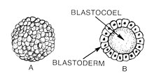
The blastocoel, also spelled blastocoele and blastocele, and also called cleavage cavity, or segmentation cavity is a fluid-filled or yolk-filled cavity that forms in the blastula during very early embryonic development. At this stage in mammals the blastula is called the blastocyst, which consists of an outer epithelium, the trophectoderm, enveloping the inner cell mass and the blastocoel.

<span class="mw-page-title-main">Animal embryonic development</span> Process by which the embryo forms and develops

In developmental biology, animal embryonic development, also known as animal embryogenesis, is the developmental stage of an animal embryo. Embryonic development starts with the fertilization of an egg cell (ovum) by a sperm cell (spermatozoon). Once fertilized, the ovum becomes a single diploid cell known as a zygote. The zygote undergoes mitotic divisions with no significant growth and cellular differentiation, leading to development of a multicellular embryo after passing through an organizational checkpoint during mid-embryogenesis. In mammals, the term refers chiefly to the early stages of prenatal development, whereas the terms fetus and fetal development describe later stages.

In embryology, cleavage is the division of cells in the early development of the embryo, following fertilization. The zygotes of many species undergo rapid cell cycles with no significant overall growth, producing a cluster of cells the same size as the original zygote. The different cells derived from cleavage are called blastomeres and form a compact mass called the morula. Cleavage ends with the formation of the blastula, or of the blastocyst in mammals.

<span class="mw-page-title-main">Inner cell mass</span> Early embryonic mass that gives rise to the fetus

The inner cell mass (ICM) or embryoblast is a structure in the early development of an embryo. It is the mass of cells inside the blastocyst that will eventually give rise to the definitive structures of the fetus. The inner cell mass forms in the earliest stages of embryonic development, before implantation into the endometrium of the uterus. The ICM is entirely surrounded by the single layer of trophoblast cells of the trophectoderm.

In the field of developmental biology, regional differentiation is the process by which different areas are identified in the development of the early embryo. The process by which the cells become specified differs between organisms.

In developmental biology, midblastula or midblastula transition (MBT) occurs during the blastula stage of embryonic development in non-mammals. During this stage, the embryo is referred to as a blastula. The series of changes to the blastula that characterize the midblastula transition include activation of zygotic gene transcription, slowing of the cell cycle, increased asynchrony in cell division, and an increase in cell motility.

<span class="mw-page-title-main">Human embryonic development</span> Development and formation of the human embryo

Human embryonic development or human embryogenesis is the development and formation of the human embryo. It is characterised by the processes of cell division and cellular differentiation of the embryo that occurs during the early stages of development. In biological terms, the development of the human body entails growth from a one-celled zygote to an adult human being. Fertilization occurs when the sperm cell successfully enters and fuses with an egg cell (ovum). The genetic material of the sperm and egg then combine to form the single cell zygote and the germinal stage of development commences. Human embryonic development covers the first eight weeks of development, which have 23 stages, called Carnegie stages. At the beginning of the ninth week, the embryo is termed a fetus. In comparison to the embryo, the fetus has more recognizable external features and a more complete set of developing organs.

<span class="mw-page-title-main">Cavitation (embryology)</span> Process in early embryonic development

Cavitation is a process in early embryonic development that follows cleavage. Cavitation is the formation of the blastocoel, a fluid-filled cavity that defines the blastula, or in mammals the blastocyst. After fertilization, cell division of the zygote occurs which results in the formation of a solid ball of cells (blastomeres) called the morula. Further division of cells increases their number in the morula, and the morula differentiates them into two groups. The internal cells become the inner cell mass, and the outer cells become the trophoblast. Before cell differentiation takes place there are two transcription factors, Oct-4 and nanog that are uniformly expressed on all of the cells, but both of these transcription factors are turned off in the trophoblast once it has formed.

Maternal to zygotic transition (MZT), also known as embryonic genome activation, is the stage in embryonic development during which development comes under the exclusive control of the zygotic genome rather than the maternal (egg) genome. The egg contains stored maternal genetic material mRNA which controls embryo development until the onset of MZT. After MZT the diploid embryo takes over genetic control. This requires both zygotic genome activation (ZGA), and degradation of maternal products. This process is important because it is the first time that the new embryonic genome is utilized and the paternal and maternal genomes are used in combination. The zygotic genome now drives embryo development.

Sir Richard Lavenham Gardner, FRSB, FRS is a British embryologist and geneticist. He is currently an Emeritus Professor at the University of York, and was previously a Royal Society Research Professor.

Magdalena Żernicka-Goetz is a Polish-British developmental biologist. She is Professor of Mammalian Development and Stem Cell Biology in the Department of Physiology, Development and Neuroscience and Fellow of Sidney Sussex College, Cambridge. She also serves as Bren Professor of Biology and Biological Engineering at California Institute of Technology (Caltech).

<span class="mw-page-title-main">Smaug (protein)</span> RNA-binding protein in Drosophila

Smaug is a RNA-binding protein in Drosophila that helps in maternal to zygotic transition (MZT). The protein is named after the fictional character Smaug, the dragon in J.R.R. Tolkien's 1937 novel The Hobbit. The MZT ends with the midblastula transition (MBT), which is defined as the first developmental event in Drosophila that depends on zygotic mRNA. In Drosophila, the initial developmental events are controlled by maternal mRNAs like Hsp83, nanos, string, Pgc, and cyclin B mRNA. Degradation of these mRNAs, which is expected to terminate maternal control and enable zygotic control of embryogenesis, happens at interphase of nuclear division cycle 14. During this transition smaug protein targets the maternal mRNA for destruction using miRs. Thus activating the zygotic genes. Smaug is expected to play a role in expression of three miRNAs – miR-3, miR-6, miR-309 and miR-286 during MZT in Drosophila. Among them smaug dependent expression of miR-309 is needed for destabilization of 410 maternal mRNAs. In smaug mutants almost 85% of maternal mRNA is found to be stable. Smaug also binds to 3′ untranslated region (UTR) elements known as SMG response elements (SREs) on nanos mRNA and represses its expression by recruiting a protein called Cup(an eIF4E-binding protein that blocks the binding of eIF4G to eIF4E). There after it recruits deadenylation complex CCR4-Not on to the nanos mRNA which leads to deadenylation and subsequent decay of the mRNA. It is also found to be involved in degradation and repression of maternal Hsp83 mRNA by recruiting CCR4/POP2/NOT deadenylase to the mRNA. The human Smaug protein homologs are SAMD4A and SAMD4B.

This glossary of developmental biology is a list of definitions of terms and concepts commonly used in the study of developmental biology and related disciplines in biology, including embryology and reproductive biology, primarily as they pertain to vertebrate animals and particularly to humans and other mammals. The developmental biology of invertebrates, plants, fungi, and other organisms is treated in other articles; e.g terms relating to the reproduction and development of insects are listed in Glossary of entomology, and those relating to plants are listed in Glossary of botany.

References

  1. "Blastulation". web-books.com.
  2. "Blastula". Encyclopædia Britannica. 2013.
  3. 1 2 3 Gilbert, Scott (2010). Developmental Biology 9th Ed + Devbio Labortatory Vade Mecum3. Sinauer Associates Inc. pp. 243–247, 161. ISBN   978-0-87893-558-1.[ permanent dead link ]
  4. Lombardi, Julian (1998). "Embryogenesis". Comparative vertebrate reproduction. Springer. p. 226. ISBN   978-0-7923-8336-9.
  5. Forgács & Newman, 2005: p. 27
  6. 1 2 3 4 Cockburn, Katie; Rossant, Janet (1 April 2010). "Making the blastocyst: lessons from the mouse". Journal of Clinical Investigation. 120 (4): 995–1003. doi:10.1172/JCI41229. PMC   2846056 . PMID   20364097.
  7. 1 2 3 Heasman, J (November 1997). "Patterning the Xenopus blastula". Development. 124 (21): 4179–91. doi:10.1242/dev.124.21.4179. PMID   9334267.
  8. Tadros, Wael; Lipshitz, Howard D. (1 March 2004). "Setting the stage for development: mRNA translation and stability during oocyte maturation and egg activation in Drosophila". Developmental Dynamics. 232 (3): 593–608. doi: 10.1002/dvdy.20297 . PMID   15704150.
  9. Gourdon, John B.; Standley, Henrietta J. (December 2002). "Uncommitted Xenopus blastula cells can be directed to uniform muscle gene expression by gradient interpretation and a community effect". The International Journal of Developmental Biology (Cambridge, UK). 46 (8): 993–8. PMID   12533022.
  10. Toth, Attila. "Treatment: Addressing the Causes of Infertility in Men and Women". Macleod Laboratory. Retrieved 22 March 2013.
  11. Kalt, MR (August 1971). "The relationship between cleavage and blastocoel formation in Xenopus laevis . I. Light microscopic observations". Journal of Embryology and Experimental Morphology. 26 (1): 37–49. PMID   5565077.
  12. Tadros, W; Lipshitz, HD (March 2005). "Setting the stage for development: mRNA translation and stability during oocyte maturation and egg activation in Drosophila". Developmental Dynamics. 232 (3): 593–608. doi: 10.1002/dvdy.20297 . PMID   15704150.
  13. 1 2 Etkin LD (1988) [1985]. "Regulation of the Mid-Blastula Transition in Amphibians". In Browder LW (ed.). The Molecular Biology of Cell Determination and Cell Differentiation. Developmental Biology. Vol. 5. New York. pp. 209–25. doi:10.1007/978-1-4615-6817-9_7. ISBN   978-1-4615-6819-3. PMID   3077975.{{cite book}}: CS1 maint: location missing publisher (link)
  14. Tadros, W; Westwood, JT; Lipshitz, HD (June 2007). "The mother-to-child transition". Developmental Cell. 12 (6): 847–9. doi: 10.1016/j.devcel.2007.05.009 . PMID   17543857.
  15. Weigel, D; Izaurralde, E (24 March 2006). "A tiny helper lightens the maternal load". Cell. 124 (6): 1117–8. doi: 10.1016/j.cell.2006.03.005 . PMID   16564001.
  16. 1 2 3 4 Fleming, Tom P.; Papenbrock, Tom; Fesenko, Irina; Hausen, Peter; Sheth, Bhavwanti (1 August 2000). "Assembly of tight junctions during early vertebrate development". Seminars in Cell & Developmental Biology. 11 (4): 291–299. doi:10.1006/scdb.2000.0179. PMID   10966863.
  17. Watson, J.G. (October 1977). "Collection and Transfer of Preimplantation Mouse Embryos". Biology of Reproduction. 17 (3): 453–8. doi: 10.1095/biolreprod17.3.453 . PMID   901897.
  18. Viczian, Andrea S.; Solessio, Eduardo C.; Lyou, Yung; Zuber, Michael E. (August 2009). "Generation of Functional Eyes from Pluripotent Cells". PLOS Biology. 7 (8): e1000174. doi: 10.1371/journal.pbio.1000174 . PMC   2716519 . PMID   19688031.

Bibliography