Bowman's capsule (or the Bowman capsule, capsula glomeruli, or glomerular capsule) is a cup-like sac at the beginning of the tubular component of a nephron in the mammalian kidney that performs the first step in the filtration of blood to form urine. A glomerulus is enclosed in the sac. Fluids from blood in the glomerulus are collected in the Bowman's capsule.
Bowman's space (or "urinary space", or "capsular space")—Between the visceral and parietal layers, into which the filtrate enters after passing through the filtration slits.[1]
Filtration barrier—The filtration barrier is composed of the fenestrated endothelium of the glomerular capillaries, the fused basal lamina of the endothelial cells and podocytes, and the filtration slits of the podocytes. The barrier permits the passage of water, ions, and small molecules from the bloodstream into the Bowman's space. The barrier prevents the passage of large and/or negatively charged proteins (such as albumin). The basal lamina of the filtration barrier is composed of three layers. The first layer is the lamina rara externa, adjacent to the podocyte processes. The second layer is the lamina rara interna, adjacent to the endothelial cells. The final layer is the lamina densa which is a darker central zone of the basal lamina. It consists of the meshwork of type IV collagen and laminin which act as a selective macromolecular filter.[citation needed]
Function
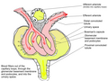
Diagram showing Bowman's capsule as part of the renal corpuscle
The process of filtration of the blood in the Bowman's capsule is ultrafiltration, and the normal rate of filtration is 125 ml/min, equivalent to 80 times the daily blood volume.[citation needed] It is a major site for blood filtration (including glomerulus)
Any proteins under roughly 30 kilodaltons can pass freely through the membrane, although there is some extra hindrance for negatively charged molecules due to the negative charge of the basement membrane and the podocytes.[citation needed]
As a result, the filtrate leaving the Bowman's capsule is very similar to blood plasma (filtrate or glomerular filtrate is composed of blood plasma minus plasma protein i.e. it contains all the components of blood plasma except the proteins) in composition as it passes into the proximal convoluted tubule.[citation needed]
Clinical significance
Micrograph of proteinaceous material in Bowman's space, which is unspecific. It is present in about 5% of people aged over 60 years, but also in for example hypertensive kidney disease.
A number of diseases can result in various problems within the glomerulus. Examples include acute proliferative (endocapillary) glomerulonephritis, mesangioproliferative glomerulonephritis, mesangiocapillary (membranoproliferative) glomerulonephritis, acute crescentic glomerulonephritis, focal segmental glomerulonephritis, and diabetic glomerulosclerosis.[citation needed]
History
Bowman's capsule is named after Sir William Bowman (1816–1892), a British surgeon and anatomist.[4] However, thorough microscopical anatomy of kidney including the nephronic capsule was first described by a Ukrainian surgeon and anatomist from the Russian Empire, Prof. Alexander Schumlansky (1748–1795), in his 1782 doctoral thesis "De structura renum" ("About Kidney Structure", in Latin); thus, much prior to Bowman.[5]
↑ Romagnani, Paola; Anders, Hans-Joachim (2019). "Excretory System". In Brüne, Martin; Schiefenhövel, Wulf (eds.). Oxford Handbook of Evolutionary Medicine. Oxford University Press. ISBN978-0198789666.
↑ Bowman, William; Royal Society of London. Philosophical transactions, v. 32, p. 57-80, 1842 (1842). On the structure and use of the Malpighian bodies of the kidney: with observations on the circulation through that gland. London: Taylor. OCLC7714131.{{cite book}}: CS1 maint: multiple names: authors list (link) CS1 maint: numeric names: authors list (link)
This page is based on this Wikipedia article Text is available under the CC BY-SA 4.0 license; additional terms may apply. Images, videos and audio are available under their respective licenses.


