Colic flexures

Last updated
Colic flexures
2420 Large Intestine.jpg
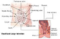
The hepatic and splenic flexures labelled at either side of transverse colon
Gray1098.png
Front of abdomen, showing surface markings for liver, stomach, duodenum, pancreas, colon
Details
Precursor Hindgut
Identifiers
Latin flexura coli
FMA 14555
Anatomical terminology

In the anatomy of the human digestive tract, there are two colic flexures, or curvatures in the transverse colon. The right colic flexure is also known as the hepatic flexure, and the left colic flexure is also known as the splenic flexure. [1] Note that "right" refers to the patient's anatomical right, which may be depicted on the left of a diagram.

Contents

Structure

Right colic flexure

The right colic flexure or hepatic flexure (as it is next to the liver) is the sharp bend between the ascending colon and the transverse colon. The hepatic flexure lies in the right upper quadrant of the human abdomen. It receives blood supply from the superior mesenteric artery.

Left colic flexure

The left colic flexure or splenic flexure (as it is close to the spleen) is the sharp bend between the transverse colon and the descending colon. The splenic flexure receives dual blood supply from the terminal branches of the superior mesenteric artery and the inferior mesenteric artery. [2]

Clinical significance

The splenic flexure is the last and highest positioned flexure in the colon. Gas can build up at this flexure and give abdominal pain giving rise to a condition known as splenic flexure syndrome. Splenic flexure syndrome is often found in those with irritable bowel syndrome (IBS), and is considered by some practitioners to be a type of IBS since it can also result from stress. [3]

The splenic flexure is a watershed region as it receives dual blood supply from the terminal branches of the superior mesenteric artery and the inferior mesenteric artery, thus making it prone to ischemic damage in cases of low blood pressure because it does not have its own primary source of blood. In the context of bowel ischemia in particular ischemic colitis, the splenic flexure is sometimes referred to as Griffith's point, along with the upper rectum (Sudeck's point). [4] [2]

Additional images

Related Research Articles

<span class="mw-page-title-main">Large intestine</span> Last part of the digestive system in vertebrates

The large intestine, also known as the large bowel, is the last part of the gastrointestinal tract and of the digestive system in tetrapods. Water is absorbed here and the remaining waste material is stored in the rectum as feces before being removed by defecation. The colon is the longest portion of the large intestine, and the terms are often used interchangeably but most sources define the large intestine as the combination of the cecum, colon, rectum, and anal canal. Some other sources exclude the anal canal.

<span class="mw-page-title-main">Portal vein</span> Short thick vein formed by the union of the superior mesenteric vein and the splenic vein

The portal vein or hepatic portal vein (HPV) is a blood vessel that carries blood from the gastrointestinal tract, gallbladder, pancreas and spleen to the liver. This blood contains nutrients and toxins extracted from digested contents. Approximately 75% of total liver blood flow is through the portal vein, with the remainder coming from the hepatic artery proper. The blood leaves the liver to the heart in the hepatic veins.

<span class="mw-page-title-main">Mesentery</span> Contiguous fold of tissues that supports the intestines

The mesentery is an organ that attaches the intestines to the posterior abdominal wall and is formed by the double fold of peritoneum. It helps in storing fat and allowing blood vessels, lymphatics, and nerves to supply the intestines, among other functions.

<span class="mw-page-title-main">Abdominal aorta</span> Largest artery in the abdomen

In human anatomy, the abdominal aorta is the largest artery in the abdominal cavity. As part of the aorta, it is a direct continuation of the descending aorta.

<span class="mw-page-title-main">Superior mesenteric artery</span> Artery which supplies blood to the intestines and pancreas

In human anatomy, the superior mesenteric artery (SMA) is an artery which arises from the anterior surface of the abdominal aorta, just inferior to the origin of the celiac trunk, and supplies blood to the intestine from the lower part of the duodenum through two-thirds of the transverse colon, as well as the pancreas.

<span class="mw-page-title-main">Inferior mesenteric artery</span>

In human anatomy, the inferior mesenteric artery (IMA) is the third main branch of the abdominal aorta and arises at the level of L3, supplying the large intestine from the distal transverse colon to the upper part of the anal canal. The regions supplied by the IMA are the descending colon, the sigmoid colon, and part of the rectum.

<span class="mw-page-title-main">Superior mesenteric vein</span> Vein which drains blood from the small intestine

In human anatomy, the superior mesenteric vein (SMV) is a blood vessel that drains blood from the small intestine. Behind the neck of the pancreas, the superior mesenteric vein combines with the splenic vein to form the portal vein that carries blood to the liver. The superior mesenteric vein lies to the right of the similarly named artery, the superior mesenteric artery, which originates from the abdominal aorta.

<span class="mw-page-title-main">Marginal artery of the colon</span> Human artery

In human anatomy, the marginal artery of the colon, also known as the marginal artery of Drummond, the artery of Drummond, and simply as the marginal artery, is an artery that connects the inferior mesenteric artery with the superior mesenteric artery. It is sometimes absent, as an anatomical variant.

<span class="mw-page-title-main">Inferior mesenteric vein</span> Vein which drains blood from the large intestine

In human anatomy, the inferior mesenteric vein (IMV) is a blood vessel that drains blood from the large intestine. It usually terminates when reaching the splenic vein, which goes on to form the portal vein with the superior mesenteric vein (SMV).

<span class="mw-page-title-main">Hepatic artery proper</span>

The hepatic artery proper is the artery that supplies the liver and gallbladder. It raises from the common hepatic artery, a branch of the celiac artery.

<span class="mw-page-title-main">Cystic artery</span>

The cystic artery is (usually) a branch of the right hepatic artery that provides arterial supply to the gallbladder and contributes arterial supply to the extrahepatic bile ducts.

<span class="mw-page-title-main">Ischemic colitis</span> Medical condition

Ischemic colitis is a medical condition in which inflammation and injury of the large intestine result from inadequate blood supply. Although uncommon in the general population, ischemic colitis occurs with greater frequency in the elderly, and is the most common form of bowel ischemia. Causes of the reduced blood flow can include changes in the systemic circulation or local factors such as constriction of blood vessels or a blood clot. In most cases, no specific cause can be identified.

<span class="mw-page-title-main">Right colic artery</span> Artery supplying the colon

The right colic artery is an artery of the abdomen, a branch of the superior mesenteric artery supplying the ascending colon. It divides into two terminal branches - an ascending branch and a descending branch - which form anastomoses with the middle colic artery, and ileocolic artery (respectively).

<span class="mw-page-title-main">Middle colic artery</span> Artery supplying the colon

The middle colic artery is an artery of the abdomen; a branch of the superior mesenteric artery distributed to parts of the ascending and transverse colon. It usually divides into two terminal branches - a left one and a right one - which go on to form anastomoses with the left colic artery, and right colic artery (respectively), thus participating in the formation of the marginal artery of the colon.

<span class="mw-page-title-main">Left colic artery</span> Artery

The left colic artery is a branch of the inferior mesenteric artery distributed to the descending colon, and left part of the transverse colon. It ends by dividing into an ascending branch and a descending branch; the terminal branches of the two branches go on to form anastomoses with the middle colic artery, and a sigmoid artery (respectively).

<span class="mw-page-title-main">Ileocolic artery</span> Artery in the abdomen

The ileocolic artery is the lowest branch arising from the concavity of the superior mesenteric artery. It supplies the cecum, ileum, and appendix.

<span class="mw-page-title-main">Transverse colon</span> Longest section of the large intestine

In human anatomy, the transverse colon is the longest and most movable part of the colon.

An acute abdomen refers to a sudden, severe abdominal pain. It is in many cases a medical emergency, requiring urgent and specific diagnosis. Several causes need immediate surgical treatment.

<span class="mw-page-title-main">Superior rectal artery</span>

The superior rectal artery is an artery that descends into the pelvis to supply blood to the rectum.

Watershed area is the medical term referring to regions of the body, that receive dual blood supply from the most distal branches of two large arteries, such as the splenic flexure of the large intestine. The term refers metaphorically to a geological watershed, or drainage divide, which separates adjacent drainage basins.For example, the watershed area of colon includes the griffith point and sudeck’s point.

References

  1. Jones, Jeremy. "Transverse colon | Radiology Reference Article | Radiopaedia.org". Radiopaedia. Retrieved 2 January 2022.
  2. 1 2 Dixon, Andrew. "Griffiths point | Radiology Reference Article | Radiopaedia.org". Radiopaedia. Retrieved 2 January 2022.
  3. "What Is the Splenic Flexure?". WebMD. Retrieved 2 January 2022.
  4. Amini, Afshin; Nagalli, Shivaraj (2021). "Bowel Ischemia". StatPearls. StatPearls Publishing. Retrieved 2 January 2022.