Cyathus striatus

Last updated

Cyathus striatus
2012-10-22 Cyathus striatus (Huds.) Willd 274333.jpg
Scientific classification
Kingdom:
Division:
Class:
Order:
Family:
Genus:
Species:
C. striatus
Binomial name
Cyathus striatus
(Huds.) Willd. (1787)
Synonyms [1]
  • Peziza striataHuds. (1778)
  • Nidularia striata(Huds.) With. (1792) [2]
  • Cyathella striata(Huds.) Brot. (1804) [3]
  • Nidularia striata var. pusilla Berk. (1839) [4]
Cyathus striatus
Information icon.svg
Gleba icon.png glebal hymenium
Infundibuliform cap icon.svg cap is infundibuliform
NA cap icon.svg hymenium attachment is not applicable
NA cap icon.svglacks a stipe
Saprotrophic fungus.svgecology is saprotrophic
Mycomorphbox Inedible.pngedibility: inedible

Cyathus striatus, commonly known as the fluted bird's nest, [5] [6] is a common saprobic bird's nest fungus with a widespread distribution throughout temperate regions of the world. This fungus resembles a miniature bird's nest with numerous tiny "eggs"; the eggs, or peridioles, are actually lens-shaped bodies that contain spores. C. striatus can be distinguished from most other bird's nest fungi by its hairy exterior and grooved ( striated ) inner walls. Although most frequently found growing on dead wood in open forests, it also grows on wood chip mulch in urban areas. The fruiting bodies are encountered from summer until early winter. The color and size of this species can vary somewhat, but they are typically less than a centimeter wide and tall, and grey or brown in color. Another common name given to C. striatus, splash cups, alludes to the method of spore dispersal: the sides of the cup are angled such that falling drops of water can dislodge the peridioles and eject them from the cup. [7] [8] The specific epithet is derived from the Latin stria, meaning "with fine ridges or grooves". [9]

Contents

Taxonomy

Cyathus striatus was first described by William Hudson in his 1778 work Flora Anglica as Peziza striata. [10] Carl Ludwig Willdenow transferred it to Cyathus in 1787. [11]

Description

a) young and mature fruiting bodies in longitudinal section; (b), (c) single peridioles-entire, and in section Cyathus striatus Buller.jpg
a) young and mature fruiting bodies in longitudinal section; (b), (c) single peridioles–entire, and in section

The "nest", or peridium, is usually about 7 to 10 mm in height and 6 to 8 mm in width, [8] but the size is somewhat variable and specimens have been found with heights and widths of up to 1.5 cm (0.59 in). [7] The shape typically resembles a vase or inverted cone. The outer surface (exoperidium) ranges in color from slightly brownish to grayish buff to deep brown; the exoperidium has a shaggy or hairy texture (a tomentum), with the hairs mostly pointing downward. The inner surface of the peridium (the endoperidium) is striated or grooved, and shiny. Young specimens have a lid, technically called an epiphragm, a thin membrane that covers the cup opening. The epiphragm is hairy like the rest of the exoperidial surface, but the hairs often wear off leaving behind a thin white layer stretched across the lid of the cup. As the peridium matures and expands, this membrane breaks and falls off, exposing the peridioles within. [13] The peridium is attached to its growing surface by a mass of closely packed hyphae called an emplacement; in C. striatus the maximum diameter of the emplacement is typically 8–12 mm, and often incorporating small fragments of the growing surface into its structure. [14] The species is inedible. [15]

Peridiole structure

The peridioles are about 1 to 1.5 mm wide and rarely up to 2 mm wide. They are disc-shaped, but may appear angular due to pressure from neighbouring peridioles. Peridioles may be dark, or a drab gray if still covered with a thin membrane called a tunica. [16]

A peridiole and attached funiculus in cross-section Peridiole cross section.jpg
A peridiole and attached funiculus in cross-section

Peridioles in C. striatus are sheathed and attached to the endoperidium by complex cords of mycelia known as a funiculus in the singular. The funiculus is differentiated into three regions: the basal piece, which attaches it to the inner wall of the peridium, the middle piece, and an upper sheath, called the purse, connected to the lower surface of the peridiole. Inside the purse and middle piece is a coiled thread of interwoven hyphae called the funicular cord, attached at one end to the peridiole and at the other end to an entangled mass of hyphae called the hapteron. When dry the funiculus is brittle, but when wet it is capable of long extension. [13]

Microscopic characteristics

The basidia, the spore-bearing cells, are club-shaped with long stalks. They typically hold 4 spores that are sessile, that is, attached directly to the surface of the basidium, rather than by a short stalk (a sterigmata). [17] Spores measure about 15 to 20  μm long by 8 to 12 μm wide. They are elliptical, smooth, hyaline, and notched at one end. [7] [8] During development, the spores are separated from the basidia when the latter collapse and gelatinize along with other cells lining the inner walls of the peridiole. The spores expand in size somewhat after being detached from the basidia. [17]

Habitat and distribution

Cyathus striatus is a saprobic fungus, deriving its nutrition from decaying organic material, and is typically found growing in clusters on small twigs or other woody debris. It is also common on mulch under shrubs. [18] The features of the microenvironment largely influence the appearance of C. striatus; all else being equal, it is more likely to be found in moist, shallow depressions than elevated areas. [19] It is very widespread in temperate areas throughout the world, [16] growing in summer and fall. [20] The fungus has been recorded from Asia, Europe, North America, Central America, South America, and New Zealand. [21]

Life cycle

The outer surface of C. striatus is covered with a shaggy or woolly tomentum. Cyathus striatus Uurrepesasieni H7618 C.jpg
The outer surface of C. striatus is covered with a shaggy or woolly tomentum.

Cyathus striatus can reproduce both asexually (via vegetative spores), or sexually (with meiosis), typical of taxa in the basidiomycetes that contain both haploid and diploid stages. Basidiospores produced in the peridioles each contain a single haploid nucleus. After the spores have been dispersed into a suitable growing environment, they germinate and develop into homokaryotic hyphae, with a single nucleus in each cell compartment. When two homokaryotic hyphae of different mating compatibility groups fuse with one another, they form a dikaryotic mycelia in a process called plasmogamy. After a period of time and under the appropriate environmental conditions, fruiting bodies may be formed from the dikaryotic mycelia. These fruiting bodies produce peridioles containing the basidia upon which new spores are made. Young basidia contain a pair of haploid sexually compatible nuclei which fuse, and the resulting diploid fusion nucleus undergoes meiosis to produce haploid basidiospores. [22] The process of meiosis in C. striatus has been found to be similar to that of higher organisms. [23]

Spore dispersal

The cone shaped fruiting body of Cyathus striatus makes use of a splash-cup mechanism to help disperse the spores. When a raindrop hits the interior of the cup with the optimal angle and velocity, the downward force of the water ejects the peridioles into the air. The force of ejection rips open the funiculus, releasing the tightly wound funicular cord. The hapteron attached to the end of the funiculus is adhesive, and when it contacts a nearby plant stem or stick, the hapteron sticks to it; the funicular cord wraps around the stem or stick powered by the force of the still-moving peridiole (similar to a tetherball). The peridioles degrade over time to eventually release the spores within, or they may be eaten by herbivorous animals and redeposited after passing through the digestive tract. [24]

Bioactive compounds

Cyathus striatus has proven to be a rich source of bioactive chemical compounds. It was first reported in 1971 to produce "indolic" substances (compounds with an indole ring structure) as well as a complex of diterpenoid antibiotic compounds collectively known as cyathins . [25] [26] Several years later, research revealed the indolic substances to be compounds now known as striatins. Striatins (A, B and C) have antibiotic activity against fungi imperfecti, and various Gram-positive and Gram-negative bacteria. [27] C. striatus also produces sesquiterpene compounds called schizandronols. [28] It also contains the triterpene compounds glochidone, glochidonol, glochidiol and glochidiol diacetate, cyathic acid, striatic acid, cyathadonic acid and epistriatic acid. [29] The latter four compounds were unknown prior to their isolation from C. striatus.

See also

Related Research Articles

Mushroom Fleshy, spore-bearing fruiting body of a fungus, typically produced above ground on soil or on its food source

A mushroom or toadstool is the fleshy, spore-bearing fruiting body of a fungus, typically produced above ground, on soil, or on its food source.

Basidiomycota Division of fungi

Basidiomycota is one of two large divisions that, together with the Ascomycota, constitute the subkingdom Dikarya within the kingdom Fungi. Members are known as Basidiomycetes. More specifically, Basidiomycota includes these groups: mushrooms, puffballs, stinkhorns, bracket fungi, other polypores, jelly fungi, boletes, chanterelles, earth stars, smuts, bunts, rusts, mirror yeasts, and the human pathogenic yeast Cryptococcus. Basidiomycota are filamentous fungi composed of hyphae and reproduce sexually via the formation of specialized club-shaped end cells called basidia that normally bear external meiospores. These specialized spores are called basidiospores. However, some Basidiomycota are obligate asexual reproducers. Basidiomycota that reproduce asexually can typically be recognized as members of this division by gross similarity to others, by the formation of a distinctive anatomical feature, cell wall components, and definitively by phylogenetic molecular analysis of DNA sequence data.

Ascomycota Division or phylum of fungi

Ascomycota is a phylum of the kingdom Fungi that, together with the Basidiomycota, forms the subkingdom Dikarya. Its members are commonly known as the sac fungi or ascomycetes. It is the largest phylum of Fungi, with over 64,000 species. The defining feature of this fungal group is the "ascus", a microscopic sexual structure in which nonmotile spores, called ascospores, are formed. However, some species of the Ascomycota are asexual, meaning that they do not have a sexual cycle and thus do not form asci or ascospores. Familiar examples of sac fungi include morels, truffles, brewer's yeast and baker's yeast, dead man's fingers, and cup fungi. The fungal symbionts in the majority of lichens such as Cladonia belong to the Ascomycota.

Basidiospore Reproductive structure of a fungus

A basidiospore is a reproductive spore produced by Basidiomycete fungi, a grouping that includes mushrooms, shelf fungi, rusts, and smuts. Basidiospores typically each contain one haploid nucleus that is the product of meiosis, and they are produced by specialized fungal cells called basidia. Typically, four basidiospores develop on appendages from each basidium, out of these 2 are of one strain and other 2 of its opposite strain. In gills under a cap of one common species, there exist millions of basidia. Some gilled mushrooms in the order Agaricales have the ability to release billions of spores. The puffball fungus Calvatia gigantea has been calculated to produce about five trillion basidiospores. Most basidiospores are forcibly discharged, and are thus considered ballistospores. These spores serve as the main air dispersal units for the fungi. The spores are released during periods of high humidity and generally have a night-time or pre-dawn peak concentration in the atmosphere.

<i>Tremella mesenterica</i> Species of jelly fungus

Tremella mesenterica is a common jelly fungus in the family Tremellaceae of the Agaricomycotina. It is most frequently found on dead but attached and on recently fallen branches, especially of angiosperms, as a parasite of wood decay fungi in the genus Peniophora. The gelatinous, orange-yellow fruit body of the fungus, which can grow up to 7.5 cm (3.0 in) diameter, has a convoluted or lobed surface that is greasy or slimy when damp. It grows in crevices in bark, appearing during rainy weather. Within a few days after rain it dries into a thin film or shriveled mass capable of reviving after subsequent rain. This fungus occurs widely in deciduous and mixed forests and is widely distributed in temperate and tropical regions that include Africa, Asia, Australia, Europe, North and South America. Although considered bland and flavorless, the fungus is edible. Tremella mesenterica produces carbohydrates that are attracting research interest because of their various biological activities.

Fungus Biological kingdom, separate from plants and animals

A fungus is any member of the group of eukaryotic organisms that includes microorganisms such as yeasts and molds, as well as the more familiar mushrooms. These organisms are classified as a kingdom, separately from the other eukaryotic kingdoms, those being Plantae, Animalia, Protozoa, and Chromista.

<i>Crucibulum</i>

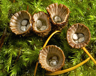
Crucibulum is a genus in the Nidulariaceae, a family of fungi whose fruiting bodies resemble tiny egg-filled bird's nests. Often called "splash cups", the fruiting bodies are adapted for spore dispersal by using the kinetic energy of falling drops of rain. The "eggs" inside the bird's nests are hard waxy shells containing spores, and tend to stick to whatever nearby herbage they land on, thus increasing the odds of being consumed and dispersed by herbivorous animals. Members of this genus are saprobic, obtaining nutrients from dead organic matter, and are typically found growing on decayed wood and wood debris. The three known Crucibulum species are distinguished from other genera of the Nidulariaceae by their relatively simple funiculus – a cord of hyphae that connects the peridiole to the exterior of the bird's nest.

<i>Nidula</i> Genus of fungi

Nidula is a genus of fungi in the family Agaricaceae. Their fruit bodies resemble tiny egg-filled birds' nests, from which they derive their common name "bird's nest fungi". Originally described in 1902, the genus differs from the related genera Cyathus and Crucibulum by the absence of a cord that attaches the eggs to the inside of the fruit body. The life cycle of this genus allows it to reproduce both sexually, with meiosis, and asexually via spores. Species in this genus produce a number of bioactive compounds, including 4-(p-hydroxyphenyl)-2-butanone, a major component of raspberry flavor and insect attractor used in pesticides.

<i>Cyathus</i> Genus of fungi in the Nidulariaceae, a family collectively known as the birds nest fungi

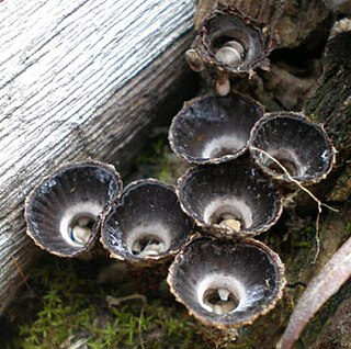
Cyathus is a genus of fungi in the Nidulariaceae, a family collectively known as the bird's nest fungi. They are given this name since they resemble tiny bird's nests filled with "eggs", structures large enough to have been mistaken in the past for seeds. However, these are now known to be reproductive structures containing spores. The "eggs", or peridioles, are firmly attached to the inner surface of this fruit body by an elastic cord of mycelia known as a funiculus. The 45 species are widely distributed throughout the world and some are found in most countries, although a few exist in only one or two locales. Cyathus stercoreus is considered endangered in a number of European countries. Species of Cyathus are also known as splash cups, which refers to the fact that falling raindrops can knock the peridioles out of the open-cup fruit body. The internal and external surfaces of this cup may be ridged longitudinally ; this is one example of a taxonomic characteristic that has traditionally served to distinguish between species.

<i>Cyathus olla</i> Species of fungus

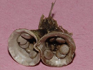
Cyathus olla also known as the field bird's nest is a species of saprobic fungus in the genus Cyathus of the family Nidulariaceae. The fruit bodies resemble tiny bird's nests filled with "eggs" – spore-containing structures called peridioles. Like other bird's nest fungi, C. olla relies on the force of falling water to dislodge peridioles from fruiting bodies to eject and disperse their spores. The life cycle of this fungus allows it to reproduce both sexually, with meiosis, and asexually via spores. C. olla is a relatively common fungus, with a worldwide distribution. It is the subject of agricultural research to determine its potential as a means to accelerate the breakdown of crop residue, and reduce the population of plant pathogens. The specific epithet is derived from the Latin word olla, meaning "pot".

<i>Cyathus stercoreus</i> Species of fungus

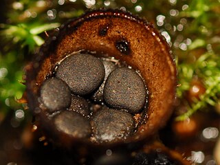
Cyathus stercoreus, commonly known as the dung-loving bird's nest or the dung bird's nest, is a species of fungus in the genus Cyathus, family Nidulariaceae. Like other species in the Nidulariaceae, the fruiting bodies of C. stercoreus resemble tiny bird's nests filled with eggs. The fruiting bodies are referred to as splash cups, because they are developed to use the force of falling drops of water to dislodge and disperse their spores. The species has a worldwide distribution, and prefers growing on dung, or soil containing dung; the specific epithet is derived from the Latin word stercorarius, meaning "of dung".

<i>Cyathus helenae</i> Species of fungus

Cyathus helenae or Helena's bird's nest is a species of fungus in the genus Cyathus, family Nidulariaceae. Like other members of the Nidulariaceae, C. helenae resembles a tiny bird's nest filled with 'eggs'—spore-containing structures known as peridioles. It was initially described by mycologist Harold Brodie in 1965, who found it growing on mountain scree in Alberta, Canada. C. helenae's life cycle allows it to reproduce both sexually and asexually. One of the smaller species of Cyathus, C. helenae produces a number of chemically unique diterpenoid molecules known as cyathins. The specific epithet of this species was given by Brodie in tribute to his late wife Helen.

<i>Astraeus hygrometricus</i> Cosmopolitan species of fungus in the family Diplocystaceae.

Astraeus hygrometricus, commonly known as the hygroscopic earthstar, the barometer earthstar, or the false earthstar, is a species of fungus in the family Diplocystaceae. Young specimens resemble a puffball when unopened. In maturity, the mushroom displays the characteristic earthstar shape that is a result of the outer layer of fruit body tissue splitting open in a star-like manner. The false earthstar is an ectomycorrhizal species that grows in association with various trees, especially in sandy soils. A. hygrometricus was previously thought to have a cosmopolitan distribution, though it is now thought to be restricted to Southern Europe, and Astraeus are common in temperate and tropical regions. Its common names refer to the fact that it is hygroscopic (water-absorbing), and can open up its rays to expose the spore sac in response to increased humidity, and close them up again in drier conditions. The rays have an irregularly cracked surface, while the spore case is pale brown and smooth with an irregular slit or tear at the top. The gleba is white initially, but turns brown and powdery when the spores mature. The spores are reddish-brown, roughly spherical with minute warts, measuring 7.5–11 micrometers in diameter.

<i>Polyozellus</i> Genus of fungus in the family Thelephoraceae, with the single species Polyozellus multiplex, found in North America and eastern Asia

Polyozellus is a fungal genus in the family Thelephoraceae, a grouping of mushrooms known collectively as the leathery earthfans. A monotypic genus, it contains the single species Polyozellus multiplex, first described in 1899, and commonly known as the blue chanterelle, the clustered blue chanterelle, or, in Alaska, the black chanterelle. The distinctive fruit body of this species comprises blue- to purple-colored clusters of vase- or spoon-shaped caps with veiny wrinkles on the undersurface that run down the length of the stem.

Limnoperdon is a fungal genus in the monotypic family Limnoperdaceae. The genus is also monotypic, as it contains a single species, the aquatic fungus Limnoperdon incarnatum. The species, described as new to science in 1976, produces fruit bodies that lack specialized structures such as a stem, cap and gills common in mushrooms. Rather, the fruit bodies—described as aquatic or floating puffballs—are small balls of loosely interwoven hyphae. The balls float on the surface of the water above submerged twigs. Experimental observations on the development of the fruit body, based on the growth on the fungus in pure culture, suggest that a thin strand of mycelium tethers the ball above water while it matures. Fruit bodies start out as a tuft of hyphae, then become cup-shaped, and eventually enclose around a single chamber that contains reddish spores. Initially discovered in a marsh in the state of Washington, the fungus has since been collected in Japan, South Africa, and Canada.
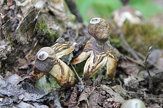
<i>Panellus stipticus</i> Species of fungus in the family Mycenaceae found in Asia, Australia, Europe, and North America

Panellus stipticus, commonly known as the bitter oyster, the astringent panus, the luminescent panellus, or the stiptic fungus, is a species of fungus in the family Mycenaceae, and the type species of the genus Panellus. A common and widely distributed species, it is found in Asia, Australia, Europe, and North America, where it grows in groups or dense overlapping clusters on the logs, stumps, and trunks of deciduous trees, especially beech, oak, and birch. During the development of the fruit bodies, the mushrooms start out as tiny white knobs, which, over a period of one to three months, develop into fan- or kidney-shaped caps that measure up to 3 cm (1.2 in) broad. The caps are orange-yellow to brownish, and attached to the decaying wood by short stubby stalks that are connected off-center or on the side of the caps. The fungus was given its current scientific name in 1879, but has been known by many names since French mycologist Jean Bulliard first described it as Agaricus stypticus in 1783. Molecular phylogenetic analysis revealed P. stipticus to have a close genetic relationship with members of the genus Mycena.

<i>Mycena aurantiomarginata</i> Species of fungus in the family Mycenaceae common in Europe and North America

Mycena aurantiomarginata, commonly known as the golden-edge bonnet, is a species of agaric fungus in the family Mycenaceae. First formally described in 1803, it was given its current name in 1872. Widely distributed, it is common in Europe and North America, and has also been collected in North Africa, Central America, and Japan. The fungus is saprobic, and produces fruit bodies (mushrooms) that grow on the floor of coniferous forests. The mushrooms have a bell-shaped to conical cap up to 2 cm (0.8 in) in diameter, set atop a slender stipe up to 6 cm (2.4 in) long with yellow to orange hairs at the base. The fungus is named after its characteristic bright orange gill edges. A microscopic characteristic is the club-shaped cystidia that are covered with numerous spiky projections, resembling a mace. The edibility of the mushroom has not been determined. M. aurantiomarginata can be distinguished from similar Mycena species by differences in size, color, and substrate. A 2010 publication reported the discovery and characterization of a novel pigment named mycenaaurin A, isolated from the mushroom. The pigment is responsible for its color, and it has antibiotic activity that may function to prevent certain bacteria from growing on the mushroom.

<i>Auriscalpium vulgare</i> Inedible European fungi

Auriscalpium vulgare, commonly known as the pinecone mushroom, the cone tooth, or the ear-pick fungus, is a species of fungus in the family Auriscalpiaceae of the order Russulales. It was first described in 1753 by Carl Linnaeus, who included it as a member of the tooth fungi genus Hydnum, but British mycologist Samuel Frederick Gray recognized its uniqueness and in 1821 transferred it to the genus Auriscalpium that he created to contain it. The fungus is widely distributed in Europe, Central America, North America, and temperate Asia. Although common, its small size and nondescript colors lead it to be easily overlooked in the pine woods where it grows. A. vulgare is not generally considered edible because of its tough texture, but some historical literature says it used to be consumed in France and Italy.

Nidulariaceae Family of fungi

The Nidulariaceae are a family of fungi in the order Agaricales. Commonly known as the bird's nest fungi, their fruiting bodies resemble tiny egg-filled birds' nests. As they are saprobic, feeding on decomposing organic matter, they are often seen growing on decaying wood and in soils enriched with wood chips or bark mulch; they have a widespread distribution in most ecological regions. The five genera within the family, namely, Crucibulum, Cyathus, Mycocalia, Nidula, and Nidularia, are distinguished from each other by differences in morphology and peridiole structure; more recently, phylogenetic analysis and comparison of DNA sequences is guiding new decisions in the taxonomic organization of this family.

References

  1. "GSD Species Synonymy: Cyathus striatus". Index Fungorum. CAB International. Retrieved 2014-07-01.
  2. Bulliard P. (1791). Histoire des champignons de la France. I (in French). Paris, France. p. 166 (figure 40A).
  3. de Brotero F. (1804). Flora Lusitanica. 2. Lisbon, Portugal. p. 474.
  4. Berkeley MJ (1839). "Descriptions of exotic fungi in the collection of Sir W.J. Hooker, from memoirs and notes of J.F. Klotzsch, with additions and corrections". Annals and Magazine of Natural History. I. 3: 375–401. doi:10.1080/03745483909443251.
  5. Phillips R. "Cyathus striatus". Roger's Plants. Archived from the original on 2008-05-16. Retrieved 2009-03-17.
  6. "Standardized Common Names for Wild Species in Canada". National General Status Working Group. 2020.
  7. 1 2 3 Lincoff GH (1981). National Audubon Society Field Guide to North American Mushrooms. New York: Random House. pp. 828–9. ISBN   978-0-394-51992-0.
  8. 1 2 3 Kuo M. (February 2014). "Cyathus striatus". MushroomExpert.Com.
  9. Brodie, The Bird's Nest Fungi, p. 173.
  10. Hudson W. (1778). Flora anglica (2nd ed.). London, UK: Impensis auctoris: Prostant Venales apud J. Nourse. p. 634.
  11. von Willdenow CL. (1787). Florae Berolinensis Prodromus. Berlin, Germany: Wilhelm Vieweg. p. 399.
  12. Gymme-Vaughan HCI, Barnes B (1927). The Structure and Development of the Fungi . Cambridge at the University Press. p.  315, figure 281.
  13. 1 2 Gates CL (1906). "The Nidulariaceae". Mycological Writings. 2: 1–30.
  14. Brodie, The Bird's Nest Fungi, p. 68.
  15. Phillips, Roger (2010). Mushrooms and Other Fungi of North America. Buffalo, NY: Firefly Books. p. 341. ISBN   978-1-55407-651-2.
  16. 1 2 Brodie, The Bird's Nest Fungi, p. 175.
  17. 1 2 Martin GW (1927). "Basidia and spores of the Nidulariaceae". Mycologia. 19 (5): 239–47. doi:10.2307/3753710. JSTOR   3753710.
  18. Healy RA, Huffman DR, Tiffany LH, Knaphaus G (2008). Mushrooms and Other Fungi of the Midcontinental United States (Bur Oak Guide). Iowa City: University of Iowa Press. p. 241. ISBN   978-1-58729-627-7.
  19. Brodie, The Bird's Nest Fungi, p. 101.
  20. Emberger G. "Cyathus striatus". Messiah College. Retrieved 2009-03-17.
  21. Roberts P, Evans S (2011). The Book of Fungi. Chicago, Illinois: University of Chicago Press. p. 533. ISBN   978-0-226-72117-0.
  22. Deacon J. (2005). Fungal Biology. Cambridge, Massachusetts: Blackwell Publishers. pp. 31–2. ISBN   978-1-4051-3066-0.
  23. Lu B, Brodie HJ (1964). "Preliminary observations of meiosis in the fungus Cyathus". Canadian Journal of Botany. 42 (3): 307–10. doi:10.1139/b64-026.
  24. Brodie, The Bird's Nest Fungi, pp. 80–92.
  25. Johri BN, Brodie HJ (1971). "Extracellular production of indolics by the fungus Cyathus". Mycologia. 63 (4): 736–44. doi:10.2307/3758043. JSTOR   3758043. PMID   5111071.
  26. Allbutt AD, Ayer WA, Brodie HJ, Johri BN, Taube H (1971). "Cyathin, a new antibiotic complex produced by Cyathus helenae". Canadian Journal of Microbiology. 17 (11): 1401–7. doi:10.1139/m71-223. PMID   5156938.
  27. Anke T, Oberwinkler F (1977). "The striatins—new antibiotics from the basidiomycete Cyathus striatus (Huds. ex Pers.) Willd". The Journal of Antibiotics. 30 (3): 221–5. doi: 10.7164/antibiotics.30.221 . PMID   863783.
  28. Ayer WA, Reffstrup T (1982). "Metabolites of bird's nest fungi. Part 18. New oxygenated cadinane derivatives from Cyathus striatus". Tetrahedron. 38 (10): 1409–12. doi:10.1016/0040-4020(82)80221-4.
  29. Ayer AW, Flanagan RJ, Reffstrup T (1984). "Metabolites of bird's nest fungi-19 New triterpenoid carboxylic acids from Cyathus striatus and Cyathus pygmaeus". Tetrahedron. 40 (11): 2069–82. doi:10.1016/S0040-4020(01)88448-9.

Cited text

Brodie HJ (1975). The Bird's Nest Fungi. Toronto, Canada: University of Toronto Press. ISBN   978-0-8020-5307-7.

Commons-logo.svg Media related to Cyathus striatus at Wikimedia Commons