Gongylonema pulchrum infections are due to humans acting as accidental hosts for the parasite. There are seven genera of spirudia nematodes that infect human hosts accidentally: Gnathostoma, Thelazia, Gongylonema, Physaloptera, Spirocerca, Rictularia. The G. pulchrum parasite is a nematode worm of the order Spirurida. It is a relatively thin nematode, and like other worms within its class, it has no circulatory or respiratory system. Most other Gongylonema species infect birds and mammals: there are 25 species found in mammals and 10 species found in birds.[citation needed]
This parasite is multi-cellular, and capable of movement. They have numerous rear mucosal projections, which assumedly assist propulsion through the thin layer of skin on the inside of the human host's mouth. They also have an excretory system possessing lateral canals. This parasite eats epithelial cells. Also, very often the canals are a place of inflammation, with accumulation of exudates in them. Gongylonema also swallows these exudates.
History of discovery
Gongylonema pulchrum was first named and presented with its own species by Molin in 1857. The first reported case was in 1850 by Dr. Joseph Leidy, when he identified a worm "obtained from the mouth of a child" from the Philadelphia Academy (however, an earlier case may have been treated in patient Elizabeth Livingstone in the seventeenth century[2]). He originally described it as Filariae hominis oris, and initially considered the worm was a guinea worm (Dracunculus medinensis), but because of the unique location of the worm (buccal cavity), and the relatively short size compared to the guinea worm, the hypothesis was disregarded. There have only been around 50 reported human cases of G. pulchrum worldwide since 1864, and these infections have been widespread and globally ubiquitous. G. pulchrum infections have been notoriously and historically hard to diagnose due to symptom complaints by patients (see "Symptoms" and "Diagnosis" below). Also, morphological diagnosis of the parasite is also somewhat complicated because of the variable size of adult worms, and the tendency of the worm to be different lengths depending on what host the worm is recovered from (see "Morphology" below).[citation needed]
Transmission
Transmission to humans is due mostly to unsanitary conditions and the ingestion of infected coprophagous insects, mostly dung beetles and cockroaches. Beyond direct ingestion of infected intermediate hosts (insects), foods can become contaminated if unsanitary conditions pervade in the production of the food- coprophagous insects are found in the food, or in the production chain. Also, contaminated water sources, again with the intermediate hosts or the infective third stage larva, can lead to transmission to humans. The infection usually occurs when someone drinks contaminated water, or consumes an infected beetle. The buccal mucosa, which is the ideal environment for the parasite, is the mucous membrane of the inside of the cheek. It is non-keratinizedstratified squamous epithelium, and is continuous with the mucosae of the soft palate, the undersurface of the tongue and the floor of the mouth.[citation needed]
Reservoir and vectors
Gongylonema pulchrum, along with most other Gongylonema nematodes, has a broad natural host range. This includes hedgehogs, cattle, dogs, cats, ruminants, rabbits, and skunks. The vector and intermediate host for Gongylonema pulchrum infections are coprophagous insects (dung beetles and cockroaches).[citation needed]
Incubation
In humans, there can be an up to six week incubation period for worm development and symptoms may not appear until the second molting of the worm, in which the young adult worms begin migration from the esophagus to the buccal and oral palate tissue. It is this movement through the mucosa of the mouth and lips that causes patients to complain of symptoms. Gongylonema pulchrum burrows in the mucosal lining of the esophagus and other parts of the buccal cavity. There the 14cm (5.5in) females lay their thick shelled eggs containing first stage larvae. The larvae all possess a cephalic hook and rows of tiny spines around a blunt anterior end, so when they hatch they may further infest their hosts.[citation needed]
Morphology
The morphology of the worm is as follows, from a 2000 Veterinary Medicine study: "The anterior end in both sexes was covered by numerous cuticular platelets. There was a pair of lateral cervical papillae. The buccal opening was small and extended in the dorsoventral direction. Around the mouth a cuticular elevation enclosed the labia, and eight papillae were located laterodorsally and lateroventrally. Two large lateral amphids were seen. On the lateral sides of the female's tail, phasmidal apertures were observed. The caudal end of the male was asymmetrically alate and bore 10 pairs of papillae and two phasmidal apertures."[3] The average length for male worms is 29.1mm (1.15in), while the average length for adult females is 58.7mm (2.31in). The worm is highly mobile, as observed in patients’ mouths and as evidenced by the morphological design of the worm.[citation needed]
Life cycle
Life cycle
In humans, the hypothesized life cycle is as follows: Ingestion of contaminated food, water, or infected dung beetle. Infects upper esophagus, moves around and lays eggs in buccal cavity of human host, ingested eggs locate near esophagus, develop and mature into adult worms after two subsequent molting stages, migrate into buccal cavity, no eggs are ever found in human feces, which strengthens the assumption that humans are solely incidental, accidental, and dead end hosts for the Gongylonema pulchrum parasite life cycle.[citation needed]
The G.pulchrum parasite has also been studied in vivo in rabbits. The life cycle is as follows:
Infective third stage larva from naturally infected dung beetles (intermediate hosts and vectors), were orally given to rabbits. The larvae entered the upper gastrointestinal tract of the rabbits (esophagus and upper stomach), and then migrated upward into the buccal cavity- pharyngeal mucosa and tongue. A third molt took place 11 days after primary infection, and the final molt took place at 36 days after primary infection. Worms reached sexual maturity at about 8 weeks, and were found mostly in the esophagus of the rabbit. 72–81 days post primary infection, embryonated eggs appeared in the feces of the rabbits.[citation needed]
Symptoms
With initial infection, some patients have reported remembering a mild fever and flu-like symptoms about a month previous to extraction or identification of worm. The most common symptom is the complaint of sensation of a worm moving around the mouth, near the lips, and in the soft palate area. This movement is normally engendered by immature adult female worms. Symptoms, once noted, may continue from a month to a year if the worm is not surgically extracted. Eosinophilia is noted in some patients. Gongylonemiasis is the affliction caused by this parasite, which is simply protracted discomfort or sensation of movement in the buccal, oral or gingival areas associated with a sensation of foreign body. Subjects commonly pull worms from their gums, tongue, lips, and inner cheeks after days and even weeks of reported discomfort. In animals, this parasite quickly spreads down the esophagus, and into the upper digestive and respiratory tracts, making it more often than not, fatal. For humans, this parasite never makes it further than the oral cavity, and is often surgically or manually extracted.[citation needed]
Diagnosis
There is a danger of misdiagnosing infections of G. pulchrum as delusional parasitosis. Diagnosis is often made by visible recognition of the worm moving through the tissue of the buccal cavity by either patient or doctor. Also, recovery of worm from patient is also a diagnostic technique. Microscopic identification of worm removed from patient's mouth or tissue is another diagnostic technique for determining the parasite infection.[citation needed]
Treatment
Treatment for infections with G. pulchrum is surgical/manual extraction of the noticed worm and albendazole (400 milligrams twice daily for 21 days). Follow up measures include periodic checks of buccal cavity and esophagus to ensure parasite infection has cleared.[citation needed]
Epidemiology
Infections of G. pulchrum are not a huge public health concern. There have only been 50 recorded infections worldwide since the first reported case in 1850.[4] The infections of G. pulchrum have been widespread, and countries reporting human infections include the United States, Germany, Iran, Japan, Laos, Morocco, China, Italy, New Zealand, and Egypt, among others. Control measures for reducing infections include making sure vector and larval contamination of food and water sources does not occur- this could be included in basic sanitary practices. Another control measure is ensuring children and adults do not accidentally or purposefully ingest infected dung beetles and other coprophagous insects.[citation needed]
Case studies
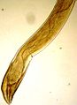
Serpentine path of Gongylonema pulchrum in the lip mucosa of a man
In 1996, the first reported case of Gongylonema pulchrum infection was reported in Japan. A 34-year-old male complaining of irritable stomatitis on his lower lip went in to see his doctor, but the pain subsided spontaneously. However, it recurred several times in the next few months. When he went in to his doctor after one of these episodes, a thread like organism was seen protruding from his ulcer. The patient also had eosinophilia, but the ulcer healed with no scar once the organism was removed. The organism was identified as a female G. pulchrum worm, and the patient needed no further treatment.[citation needed]
How the patient contracted the worm is still unknown. He didn't report eating any abnormal foods, nor had he traveled outside Japan in the past few years. He also did not report drinking any water from possibly infected wells. It is possible that he ate food that had been contaminated in an endemic country and shipped to Japan. With the globalized food market now present, this is not out of the realm of possibility, and should be considered as a possible means of transmission into countries that have no previous history of G. pulchrum infection.[4]
In 1999, a 41-year-old female resident of New York City went in to her doctor complaining of the sensation of something moving in her mouth. She said she had had the feeling for the duration of one year. Supposedly, she had removed worms from her mouth on two separate occasions- one from her lip, and one from her gums. She submitted one of the specimens for microscopic identification, and it was found to be an adult female G. pulchrum worm. She traveled frequently to visit relatives in Mississippi, so it is unknown whether she contracted the worm in New York or in the south. This was the first reported case of Gongylonema in the United States since 1963.[5]
Also in 1999, a 38-year-old woman of Cambridge, Massachusetts sought medical attention for the visible identification of a “migrating mass” in her cheek mucosa. Six months earlier, she had noted an irregular patch of mucosa on her cheek, but thought nothing of it. Previously in the year, she'd traveled to Mexico, Guatemala, and France. She didn't report ingesting any beetles, but she did eat raw foods when vacationing in Mexico. She described the foods as “raw, crunchy, and saladlike”. Approximately 12 hours after eating the food, she and five other individuals she was traveling with had an acute attack of nausea, vomiting, and dizziness. The symptoms seemed to resolve themselves with no need of further treatment. A small female Gongylonema worm was surgically removed from her cheek mucosa under local anesthesia, and follow up treatment included albendazole two times daily for three days. This was the eleventh reported case of G. pulchrum infection in the United States. Most cases reported in the US are reported from the southeastern part of the country.[6]
There was a 1916 infection reported in a 16-year-old girl from Mississippi. She presented with gastrointestinal pain, vomiting and a low fever (101.5°F (38.6°C)). She complained of a sensation of a worm moving around her lower lip, but was disregarded by her physician. As she continued to complain, the physician examined her mouth, and discovered the outline of a worm. He extracted the worm with a sewing needle, and the child's complaints stopped and she appeared to have no further symptoms of parasite infection.[citation needed]
In 2013, the first case of human gongylonemosis was reported in France.[1] The patient, a healthy 48-year-old man felt the presence of a moving, worm-like organism in his mouth. Initially, the patient would occasionally feel, but not see, this mass at different sites: cheek, palate, gums and internal surface of the lower lip. The sensation would subside after several hours without leaving any visible lesions and without being accompanied by any associated localized or generalized symptoms. The patient had no medical history. He was a resident of Alsace, France, and had not travelled abroad. He reported not to have changed his lifestyle, especially not his diet, in the recent past. He also had no knowledge of having accidentally ingested an intermediate insect host. He consulted a doctor and all results of the clinical examination fell within the normal range. Haematology investigation revealed no abnormalities, particularly no elevated eosinophil count, and no microfilariae were seen using stained blood films; the filariasis serology was negative. No medical treatment was initiated. After 3 weeks of migration, the thread-like worm installed itself on the inner surface of the lower lip, allowing the patient to extract it by tongue pressure firstly, then using his fingers. He placed the parasite in alcohol and submitted it to a medical laboratory.[citation needed]
The parasite was the subject of a 2017 episode of the science documentary series Monsters Inside Me. Jonathan Allen, a biology professor and researcher at the College of William & Mary,[7] extracted the parasite alive from his own mouth and sent it for genetic testing after recognizing that it was not a parasite common to the United States; he is only the thirteenth known infected human in the country. Once he realized the lack of documentation of gongylonema infections in scientific literature, he and a colleague conducted further tests together with scientists from the Eastern Virginia Medical School and co-authored a paper published in the American Journal of Tropical Medicine and Hygiene documenting the study.[8] Such is the rarity of human gongylonemosis that Allen's doctor failed to recognize the parasite, which Allen noted was due to so few documented cases in medical literature worldwide and the difficulty in diagnosing the infection due to its mild symptoms.[9]
Gallery
These are all pictures from a single Gongylonema pulchrum male extracted from a man in France.[1]
Complete plate (click on image for detailed caption)
Related Research Articles
Trichuriasis, also known as whipworm infection, is an infection by the parasitic worm Trichuris trichiura (whipworm). If the infection is only with a few worms, there are often no symptoms. In those who are infected with many worms, there may be abdominal pain, fatigue and diarrhea. The diarrhea sometimes contains blood. Infections in children may cause poor intellectual and physical development. Low red blood cell levels may occur due to loss of blood.
Ascariasis is a disease caused by the parasitic roundworm Ascaris lumbricoides. Infections have no symptoms in more than 85% of cases, especially if the number of worms is small. Symptoms increase with the number of worms present and may include shortness of breath and fever at the beginning of the disease. These may be followed by symptoms of abdominal swelling, abdominal pain, and diarrhea. Children are most commonly affected, and in this age group the infection may also cause poor weight gain, malnutrition, and learning problems.
Toxocariasis is an illness of humans caused by the dog roundworm and, less frequently, the cat roundworm. These are the most common intestinal roundworms of dogs, coyotes, wolves and foxes and domestic cats, respectively. Humans are among the many "accidental" or paratenic hosts of these roundworms.
Clonorchiasis is an infectious disease caused by the Chinese liver fluke and two related species. Clonorchiasis is a known risk factor for the development of cholangiocarcinoma, a neoplasm of the biliary system.
Hymenolepiasis is infestation by one of two species of tapeworm: Hymenolepis nana or H. diminuta. Alternative names are dwarf tapeworm infection and rat tapeworm infection. The disease is a type of helminthiasis which is classified as a neglected tropical disease.
Gnathostomiasis, also known as larva migrans profundus, is the human infection caused by the nematode Gnathostoma spinigerum and/or Gnathostoma hispidum, which infects vertebrates.
Hookworm infection is an infection by a type of intestinal parasite known as a hookworm. Initially, itching and a rash may occur at the site of infection. Those only affected by a few worms may show no symptoms. Those infected by many worms may experience abdominal pain, diarrhea, weight loss, and tiredness. The mental and physical development of children may be affected. Anemia may result.
Anisakis is a genus of parasitic nematodes that have life cycles involving fish and marine mammals. They are infective to humans and cause anisakiasis. People who produce immunoglobulin E in response to this parasite may subsequently have an allergic reaction, including anaphylaxis, after eating fish infected with Anisakis species.
Dipylidium caninum, also called the flea tapeworm, double-pored tapeworm, or cucumber tapeworm is a cyclophyllid cestode that infects organisms afflicted with fleas and canine chewing lice, including dogs, cats, and sometimes human pet-owners, especially children.
The oral mucosa is the mucous membrane lining the inside of the mouth. It comprises stratified squamous epithelium, termed "oral epithelium", and an underlying connective tissue termed lamina propria. The oral cavity has sometimes been described as a mirror that reflects the health of the individual. Changes indicative of disease are seen as alterations in the oral mucosa lining the mouth, which can reveal systemic conditions, such as diabetes or vitamin deficiency, or the local effects of chronic tobacco or alcohol use. The oral mucosa tends to heal faster and with less scar formation compared to the skin. The underlying mechanism remains unknown, but research suggests that extracellular vesicles might be involved.
Sparganosis is a parasitic infection caused by the plerocercoid larvae of the genus Spirometra including S. mansoni, S. ranarum, S. mansonoides and S. erinacei. It was first described by Patrick Manson in 1882, and the first human case was reported by Charles Wardell Stiles from Florida in 1908. The infection is transmitted by ingestion of contaminated water, ingestion of a second intermediate host such as a frog or snake, or contact between a second intermediate host and an open wound or mucous membrane. Humans are the accidental hosts in the life cycle, while dogs, cats, and other mammals are definitive hosts. Copepods are the first intermediate hosts, and various amphibians and reptiles are second intermediate hosts.
Mammomonogamus is a genus of parasitic nematodes of the family Syngamidae that parasitise the respiratory tracts of cattle, sheep, goats, deer, cats, orangutans, and elephants. The nematodes can also infect humans and cause the disease called mammomonogamiasis. Several known species fall under the genus Mammomonogamus, but the most common species found to infest humans is M. laryngeus. Infection in humans is very rare, with only about 100 reported cases worldwide, and is assumed to be largely accidental. Cases have been reported from the Caribbean, China, Korea, Thailand, and Philippines.
Gongylonema is a genus of thread-like nematode that was described by Molin in 1857. It is the only currently valid genus in the family Gongylonematidae, though the mysterious Spiruroides – usually placed in the Subuluridae, which are not closely related to Gongylonema among the Spiruria – might actually belong here. They are parasites of birds and mammals, transmitted by insects. Some 38 species are known, about 12 of which have been recorded in Europe.
Angiostrongyliasis is an infection by a roundworm of the Angiostrongylus type. Symptoms may vary from none to mild, to meningitis.
Mouth infections, also known as oral infections, are a group of infections that occur around the oral cavity. They include dental infection, dental abscess, and Ludwig's angina. Mouth infections typically originate from dental caries at the root of molars and premolars that spread to adjacent structures. In otherwise healthy patients, removing the offending tooth to allow drainage will usually resolve the infection. In cases that spread to adjacent structures or in immunocompromised patients, surgical drainage and systemic antibiotics may be required in addition to tooth extraction. Since bacteria that normally reside in the oral cavity cause mouth infections, proper dental hygiene can prevent most cases of infection. As such, mouth infections are more common in populations with poor access to dental care or populations with health-related behaviors that damage one's teeth and oral mucosa. This is a common problem, representing nearly 36% of all encounters within the emergency department related to dental conditions.
Ancylostoma caninum is a species of nematode known as a hookworm, which principally infects the small intestine of dogs. The result of A. caninum infection ranges from asymptomatic cases to death of the dog; better nourishment, increasing age, prior A. caninum exposure, or vaccination are all linked to improved survival. Other hosts include carnivores such as wolves, foxes, and cats, with a small number of cases having been reported in humans.
Nanophyetus salmincola is a food-borne intestinal trematode parasite prevalent on the Pacific Northwest coast. The species may be the most common trematode endemic to the United States.
Trichomonas gallinae is a cosmopolitan parasite of birds including finches, pigeons, doves, turkeys, chickens, parrots, and raptors. The condition in birds of prey is called frounce. It is believed to be an ancient pathogen causing frounce-like symptoms in theropod dinosaurs. The same condition in pigeons is commonly called canker.
Gongylonema ingluvicola is a species in the genus Gongylonema. It is one of the spirurid nematodes that infect a vast array of animals throughout the world. Infections have been noted in both humans and in animals ranging from cows to birds. As of recent, there have been 50+ human cases reported in countries such as New Zealand, Australia, Middle East, China, and United States.
Anisakis simplex, known as the herring worm, is a species of nematode in the genus Anisakis. Like other nematodes, it infects and settles in the organs of marine animals, such as salmon, mackerels and squids. It is commonly found in cold marine waters, such as the Pacific Ocean and Atlantic Ocean.
1 2 Haruki, K., Furuya, H., Saito, S., Kamiya, S. & Kagei, N. 2005: Gongylonema infection in man: A first case of gongylonemosis in Japan. Helminthologia, 42, 63-66. Free PDF
Lichtenfels, JR (1971). "Morphological variation in the gullet nematode, Gongylonema pulchrum Molin, 1857, from eight species of definitive hosts with a consideration of Gongylonema from Macaca spp". Journal of Parasitology. 57 (2): 348–355. doi:10.2307/3278041. JSTOR3278041. PMID5553452.
This page is based on this Wikipedia article Text is available under the CC BY-SA 4.0 license; additional terms may apply. Images, videos and audio are available under their respective licenses.