Taenia saginata

Last updated

Taenia saginata
Taenia saginata adult 5260 lores.jpg
Scientific classification OOjs UI icon edit-ltr.svg
Domain: Eukaryota
Kingdom: Animalia
Phylum: Platyhelminthes
Class: Cestoda
Order: Cyclophyllidea
Family: Taeniidae
Genus: Taenia
Species:
T. saginata
Binomial name
Taenia saginata
Goeze, 1782

Taenia saginata (synonym Taeniarhynchus saginatus), commonly known as the beef tapeworm, is a zoonotic tapeworm belonging to the order Cyclophyllidea and genus Taenia. It is an intestinal parasite in humans causing taeniasis (a type of helminthiasis) and cysticercosis in cattle. Cattle are the intermediate hosts, where larval development occurs, while humans are definitive hosts harbouring the adult worms. It is found globally and most prevalently where cattle are raised and beef is consumed. It is relatively common in Africa, Europe, Southeast Asia, South Asia, and Latin America. [1] Humans are generally infected as a result of eating raw or undercooked beef which contains the infective larvae, called cysticerci. As hermaphrodites, each body segment called proglottid has complete sets of both male and female reproductive systems. Thus, reproduction is by self-fertilisation. From humans, embryonated eggs, called oncospheres, are released with faeces and are transmitted to cattle through contaminated fodder. Oncospheres develop inside muscle, liver, and lungs of cattle into infective cysticerci. [2]

Contents

T. saginata has a strong resemblance to the other human tapeworms, such as Taenia asiatica and Taenia solium , in structure and biology, except for few details. It is typically larger and longer, with more proglottids, more testes, and higher branching of the uteri. It also lacks an armed scolex unlike other Taenia. Like the other tapeworms, it causes taeniasis inside the human intestine, but does not cause cysticercosis. Its infection is relatively harmless and clinically asymptomatic. [3] [4]

Description

Taenia saginata proglottid stained to show uterine branches: The pore on the side identifies it as a cyclophyllid cestode. Taenia saginataD.JPG
Taenia saginata proglottid stained to show uterine branches: The pore on the side identifies it as a cyclophyllid cestode.

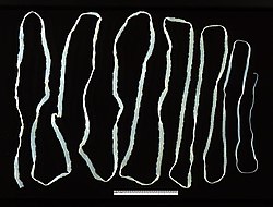
T. saginata is the largest of species in the genus Taenia . An adult worm is normally 4 to 10 m in length, but can become very large; specimens over 22 m long are reported. Typical of cestodes, its body is flattened dorsoventrally and heavily segmented. It is entirely covered by a tegument. The body is white in colour and consists of three portions: scolex, neck, and strobila. The scolex has four suckers, but they have no hooks. Lack of hooks and a rostellum is an identifying feature from other Taenia species. [4] The rest of the body proper, the strobila, is basically a chain of numerous body segments called proglottids. The neck is the shortest part of the body, and consists of immature proglottids. The midstrobila is made of mature proglottids that eventually lead to the gravid proglottids, which are at the posterior end. An individual can have as many as 1000 to 2000 proglottids. [5]

T. saginata does not have a digestive system, mouth, anus, or digestive tract. It derives nutrients from the host through its tegument, as the tegument is completely covered with absorptive hair-like microtriches. It is also an acoelomate, having no body cavity. The inside of each mature proglottid is filled with muscular layers and complete male and female reproductive systems, including the tubular unbranched uterus, ovary, genital pore, testes, and vitelline gland. In the gravid proglottid, the uterus contains up to 15 side branches filled with eggs. [3] [6]

Life cycle

Intermediate host

Cattle acquire the embryonated eggs, the oncospheres, when they eat contaminated food. Oncospheres enter the duodenum, the anterior portion of the small intestine, and hatch there under the influence of gastric juices. The embryonic membranes are removed, liberating free hexacanth ("six-hooked") larvae. With their hooks, they attach to the intestinal wall and penetrate the intestinal mucosa into the blood vessels. The larvae can move to all parts of the body by the general circulatory system, and finally settle in skeletal muscles within 70 days. Inside the tissue, they cast off their hooks and instead develop a protective cuticular shell, called the cyst. Thus, they become fluid-filled cysticerci. Cysterci can also form in lungs and liver. The inner membrane of the cysticercus soon develops numerous protoscolices (small scolices) that are invertedly attached to the inner surface. The cysticercus of T. saginata is specifically named cysticercus bovis to differentiate from that of T. solium, cysticercus cellulosae. [3] [6]

Definitive host

Taenia saginata - oncosphere T.saginata-egg.jpg
Taenia saginata - oncosphere

Humans contract infective cysticerci by eating raw or undercooked meat. Once reaching the jejunum, the inverted scolex becomes evaginated to the exterior under stimuli from the digestive enzymes of the host. Using the scolex, it attaches to the intestinal wall. The larva mature into adults about 5 to 12 weeks later. Adult worms can live about 25 years in the host. [3] Usually, only a single worm is present at a time, but multiple worms are also reported. In each mature proglottid, self-fertilisation produces zygotes, which divide and differentiate into embryonated eggs called oncospheres. With thousands of oncospheres, the oldest gravid proglottids detach. Unlike in other Taenia, gravid proglottids are shed individually. In some cases, the proglottid ruptures inside the intestine, and the eggs are released. The free proglottids and liberated eggs are removed by peristalsis into the environment. On the ground, the proglottids are motile and shed eggs as they move. These oncospheres in an external environment can remain viable for several days to weeks in sewage, rivers, and pastures. [3] [6] [7]

Genome

Epidemiology

The disease is relatively common in Africa, some parts of Eastern Europe, the Philippines, and Latin America. [2] There is also a widespread occurrence of the parasite throughout East, Southeast, and South Asia. [8] This parasite is found anywhere where beef is eaten, including countries such as the United States, with strict federal sanitation policies. In the US, the incidence of infection is low, but 25% of cattle sold are still infected. [6] However, not all slaughterhouses are federally inspected. [9] The total global infection is estimated to be between 40 and 60 million. [1] It is most prevalent in Sub-Saharan Africa and the Middle East. [7]

Religious beliefs may also play a role in infection rates. In India, Muslims can have a higher rate of infection compared to Hindus, who do not eat beef. [9]

Infection

Symptoms

T. saginata infection is usually asymptomatic, but heavy infection often results in weight loss, dizziness, abdominal pain, diarrhea, headaches, nausea, constipation, chronic indigestion, and loss of appetite. Intestinal obstruction in humans can be alleviated by surgery. The tapeworm can also expel antigens that can cause an allergic reaction in the individual. [10] It is also a rare cause of ileus, pancreatitis, cholecystitis, and cholangitis. [11] Taenia saginata has been reported as a cause of gallbladder perforation if left untreated in some cases. [12] Taenia saginata adult worms can live in the host for up to 25 years and most infections will last 2 to 3 years without treatment. [13]

The Taenia saginata remains asymptomatic due to the fact the organism does not present cysticerci in humans. Therefore, there is no presence of cysticercosis in humans either. Typically, cysticercosis is a parasitical tissue infection which infect the brain and muscle tissues. However the Taenia saginata can cause taeniasis, which is an infection. Taeniasis causes weight loss, pain and blockages in the intestines which can potentially become life-threatening. [14]

Diagnosis

The basic diagnosis is done from a stool sample. Feces are examined to find parasite eggs. The eggs look like other eggs from the family Taeniidae, so it is only possible to identify the eggs to the family, not to the species level. Since it is difficult to diagnose using eggs alone, looking at the scolex or the gravid proglottids can help identify it as Taenia saginata. [6] Proglottids sometimes trickle down the thighs of infected humans and are visible with unaided eye, so can aid with identification. Observation of scolex help distinguish between T. saginata, T. solium and T. asiatica . When the uterus is injected with India ink, its branches become visible. Counting the uterine branches enables some identification (T. saginata uteri have 12 or more branches on each side, while other species such as T. solium only have five to 10). [2]

Differentiation of the species of Taenia , such as T. solium and T. asiatica, is notoriously difficult because of their close morphological resemblance, and their eggs are more or less identical. Identification often requires histological observation of the uterine branches and PCR detection of ribosomal 5.8S gene. [15] The uteri of T. saginata stem out from the center to form 12 to 20 branches, but in contrast to its closely related Taenia species, the branches are much less in number and comparatively thicker; in addition, the ovaries are bilobed and testes are twice as many. [16]

Eosinophilia and elevated IgE levels are chief hematological findings. Also Ziehl–Neelsen stain can be used to differentiate between mature T. saginata and T. solium, in most cases T. saginata will stain while T. solium will not, but the method is not strictly reliable. [17]

Prevention

Adequate cooking at 56 °C (133 °F) for 5 minutes of beef viscera destroys cysticerci. Refrigeration, freezing at −10 °C (14 °F) for 9 days or long periods of salting is lethal to cysticerci. Inspection of beef and proper disposal of human excreta are also important measures. [10]

Treatment

Taeniasis is easily treated with praziquantel (5–10 mg/kg, single-administration) or niclosamide (adults and children over 6 years: 2 g, single-administration after a light breakfast, followed after 2 hours by a laxative; children aged 2–6 years: 1 g; children under 2 years: 500 mg). [10] Albendazole is also highly effective for treatment of cattle infection. [18] During the 1940s, the preferred treatment was oleoresin of aspidium, which would be introduced into the duodenum via a Rehfuss tube. [19]

See also

Related Research Articles

<span class="mw-page-title-main">Cysticercosis</span> Tissue infection caused by the young form of the pork tapeworm

Cysticercosis is a tissue infection caused by the young form of the pork tapeworm. People may have few or no symptoms for years. In some cases, particularly in Asia, solid lumps of between one and two centimetres may develop under the skin. After months or years these lumps can become painful and swollen and then resolve. A specific form called neurocysticercosis, which affects the brain, can cause neurological symptoms. In developing countries this is one of the most common causes of seizures.

<i>Diphyllobothrium</i> Genus of flatworms

Diphyllobothrium is a genus of tapeworms which can cause diphyllobothriasis in humans through consumption of raw or undercooked fish. The principal species causing diphyllobothriasis is D. latum, known as the broad or fish tapeworm, or broad fish tapeworm. D. latum is a pseudophyllid cestode that infects fish and mammals. D. latum is native to Scandinavia, western Russia, and the Baltics, though it is now also present in North America, especially the Pacific Northwest. In Far East Russia, D. klebanovskii, having Pacific salmon as its second intermediate host, was identified.

Hymenolepiasis is infestation by one of two species of tapeworm: Hymenolepis nana or H. diminuta. Alternative names are dwarf tapeworm infection and rat tapeworm infection. The disease is a type of helminthiasis which is classified as a neglected tropical disease.

<i>Taenia solium</i> Species of Cestoda

Taenia solium, the pork tapeworm, belongs to the cyclophyllid cestode family Taeniidae. It is found throughout the world and is most common in countries where pork is eaten. It is a tapeworm that uses humans as its definitive host and pigs as the intermediate or secondary hosts. It is transmitted to pigs through human feces that contain the parasite eggs and contaminate their fodder. Pigs ingest the eggs, which develop into larvae, then into oncospheres, and ultimately into infective tapeworm cysts, called cysticercus. Humans acquire the cysts through consumption of uncooked or under-cooked pork and the cysts grow into an adult worms in the small intestine.

<i>Taenia</i> (tapeworm) Genus of flatworms

Taenia is the type genus of the Taeniidae family of tapeworms. It includes some important parasites of livestock. Members of the genus are responsible for taeniasis and cysticercosis in humans, which are types of helminthiasis belonging to the group of neglected tropical diseases. More than 100 species are recorded. They are morphologically characterized by a ribbon-like body composed of a series of segments called proglottids; hence the name Taenia. The anterior end of the body is the scolex. Some members of the genus Taenia have an armed scolex ; of the two major human parasites, Taenia saginata has an unarmed scolex, while Taenia solium has an armed scolex.

<i>Hymenolepis nana</i> Species of flatworm

Dwarf tapeworm is a cosmopolitan species though most common in temperate zones, and is one of the most common cestodes infecting humans, especially children.

<i>Taenia crassiceps</i> Species of Cestoda

Taenia crassiceps is a tapeworm in the family Taeniidae. It is a parasitic organism whose adult form infects the intestine of carnivores, like canids. It is related to Taenia solium, the pork tapeworm, and to Taenia saginata, the beef tapeworm. It is commonly found in the Northern Hemisphere, especially throughout Canada and the northern United States.

<span class="mw-page-title-main">Taeniidae</span> Family of flatworms

The Taeniidae are a family of tapeworms. It is the largest family representing the order Cyclophyllidea. It includes many species of medical and veterinary importance, as Taenia solium, Taenia saginata, and Echinococcus granulosus. The Taeniidae are parasites of mammals and many are infectious to humans.

<i>Taenia pisiformis</i> Species of flatworm

Taenia pisiformis, commonly called the rabbit tapeworm, is an endoparasitic tapeworm which causes infection in lagomorphs, rodents, and carnivores. Adult T. pisiformis typically occur within the small intestines of the definitive hosts, the carnivores. Lagomorphs, the intermediate hosts, are infected by fecal contamination of grasses and other food sources by the definitive hosts. The larval stage is often referred to as Cysticercus pisiformis and is found on the livers and peritoneal cavities of the intermediate hosts. T. pisiformis can be found worldwide.

<span class="mw-page-title-main">Taeniasis</span> Parasitic disease due to infection with tapeworms belonging to the genus Taenia

Taeniasis is an infection within the intestines by adult tapeworms belonging to the genus Taenia. There are generally no or only mild symptoms. Symptoms may occasionally include weight loss or abdominal pain. Segments of tapeworm may be seen in the stool. Complications of pork tapeworm may include cysticercosis.

<span class="mw-page-title-main">Eucestoda</span> Subclass of flatworms

Eucestoda, commonly referred to as tapeworms, is the larger of the two subclasses of flatworms in the class Cestoda. Larvae have six posterior hooks on the scolex (head), in contrast to the ten-hooked Cestodaria. All tapeworms are endoparasites of vertebrates, living in the digestive tract or related ducts. Examples are the pork tapeworm with a human definitive host, and pigs as the secondary host, and Moniezia expansa, the definitive hosts of which are ruminants.

<i>Hymenolepis</i> (tapeworm) Genus of worms

Hymenolepis is a genus of cyclophyllid tapeworms that cause hymenolepiasis. They parasitise mammals, including humans. Some notable species are:

<span class="mw-page-title-main">Cestoda</span> Class of flatworms

Cestoda is a class of parasitic worms in the flatworm phylum (Platyhelminthes). Most of the species—and the best-known—are those in the subclass Eucestoda; they are ribbon-like worms as adults, known as tapeworms. Their bodies consist of many similar units known as proglottids—essentially packages of eggs which are regularly shed into the environment to infect other organisms. Species of the other subclass, Cestodaria, are mainly fish infecting parasites.

Diphyllobothrium mansonoides is a species of tapeworm (cestodes) that is endemic to North America. Infection with D. mansonoides in humans can result in sparganosis. Justus F. Mueller first reported this organism in 1935. D. mansonoides is similar to D. latum and Spirometra erinacei. When the organism was discovered, scientist did not know if D. mansonoides and S. erinacei were separate species. PCR analysis of the two worms has shown the two to be separate but closely related organisms.

Taenia asiatica, commonly known as Asian taenia or Asian tapeworm, is a parasitic tapeworm of humans and pigs. It is one of the three species of Taenia infecting humans and causes taeniasis. Discovered only in 1980s from Taiwan and other East Asian countries as an unusual species, it is so notoriously similar to Taenia saginata, the beef tapeworm, that it was for a time regarded as a slightly different strain. But anomaly arose as the tapeworm is not of cattle origin, but of pigs. Morphological details also showed significant variations, such as presence of rostellar hooks, shorter body, and fewer body segments. The scientific name designated was then Asian T. saginata. But the taxonomic consensus turns out to be that it is a unique species. It was in 1993 that two Korean parasitologists, Keeseon S. Eom and Han Jong Rim, provided the biological bases for classifying it into a separate species. The use of mitochondrial genome sequence and molecular phylogeny in the late 2000s established the taxonomic status.

Moniezia expansa is commonly known as sheep tapeworm or double-pored ruminant tapeworm. It is a large tapeworm inhabiting the small intestines of ruminants such as sheep, goats and cattle. It has been reported from Peru that pigs are also infected. There is an unusual report of human infection in an Egyptian. It is characterized by unarmed scolex, presence of two sets of reproductive systems in each proglottid, and each proglottid being very short but very broad.

Taenia serialis, also known as a canid tapeworm, is found within canines such as foxes and dogs. Adult T. serialis are parasites of carnivores, particularly dogs, with herbivorous lagomorph mammals such as rabbits and hares, serving as intermediate hosts. In definitive hosts, T. serialis is acquired by eating tissues from a variety of intermediate hosts. Accidental infection of humans though, can occur when eggs are ingested from food or water contaminated with dog feces and the human then becomes the T. serialis intermediate host.

<i>Raillietina tetragona</i> Species of flatworm

Raillietina tetragona is a parasitic tapeworm belonging to the class Cestoda. It is a cosmopolitan helminth of the small intestine of pigeon, chicken and guinea fowl, and is found throughout the world.

<i>Taenia hydatigena</i> Species of flatworm

Taenia hydatigena is one of the adult forms of the canine and feline tapeworm. This infection has a worldwide geographic distribution. Humans with taeniasis can infect other humans or animal intermediate hosts by eggs and gravid proglottids passed in the feces.

<span class="mw-page-title-main">Cysticercus</span> Larval tapeworm

Cysticercus is a scientific name given to the young tapeworms (larvae) belonging to the genus Taenia. It is a small, sac-like vesicle resembling a bladder; hence, it is also known as bladder worm. It is filled with fluid, in which the main body of the larva, called scolex, resides. It normally develops from the eggs, which are ingested by the intermediate hosts, such as pigs and cattle. The tissue infection is called cysticercosis. Inside such hosts, they settle in the muscles. When humans eat raw or undercooked pork or beef that is contaminated with cysticerci, the larvae grow into adult worms inside the intestine. Under certain circumstances, specifically for the pork tapeworm, the eggs can be accidentally eaten by humans through contaminated foodstuffs. In such case, the eggs hatch inside the body, generally moving to muscles as well as inside the brain. Such brain infection can lead to a serious medical condition called neurocysticercosis. This disease is the leading cause of acquired epilepsy.

References

  1. 1 2 Eckert J. (2005). "Helminths". In Kayser, F.H.; Bienz, K.A.; Eckert, J.; Zinkernagel, R.M. (eds.). Medical Microbiology. Stuttgart: Thieme. pp. 560–562. ISBN   9781588902450.
  2. 1 2 3 Somers, Kenneth D.; Morse, Stephen A. (2010). Lange Microbiology and Infectious Diseases Flash Cards (2nd ed.). New York: Lange Medical Books/ McGraw-Hill. pp. 184–186. ISBN   9780071628792.
  3. 1 2 3 4 5 Bogitsh, Burton J.; Carter, Clint E. (2013). Human Parasitology (4th ed.). Amsterdam: Academic Press. pp. 244–245. ISBN   9780124159150.
  4. 1 2 Winn Jr. Washington; Allen, Stephen; Janda, William; Koneman, Elmer; Procop, Gary; Schreckenberger, Paul; Woods, Gail (2006). Koneman's Color Atlas and Textbook of Diagnostic Microbiology (6th ed.). Philadelphia: Lippincott Williams & Wilkins. pp. 1282–1284. ISBN   9780781730143.
  5. Patel, Nayan M.; Tatar, Eric L. (2007). "Unusual colonoscopy finding: Taenia saginata proglottid". World Journal of Gastroenterology. 13 (41): 5540–5541. doi: 10.3748/wjg.v13.i41.5540 . PMC   4171297 . PMID   17907306.
  6. 1 2 3 4 5 Roberts, Larry S.; Janovy, John Jr. (2009). Gerald D. Schmidt & Larry S. Roberts' Foundations of parasitology (8th ed.). Boston: McGraw-Hill. ISBN   978-0-07-128458-5.
  7. 1 2 Ortega, Ynes R. (2006). Foodborne parasites. New York: Springer. pp. 207–210. ISBN   9780387311975.
  8. Eichenberger, Ramon M.; Thomas, Lian F.; Gabriël, Sarah; Bobić, Branco; Devleesschauwer, Brecht; Robertson, Lucy J.; Saratsis, Anastasios; Torgerson, Paul R.; Braae, Uffe C. (May 7, 2020). Dermauw, Veronique; Dorny, Pierre (eds.). "Epidemiology of Taenia saginata taeniosis/cysticercosis: a systematic review of the distribution in East, Southeast and South Asia". Parasites & Vectors. 13 (1): 234. doi: 10.1186/s13071-020-04095-1 . PMC   7206752 . PMID   32381027.
  9. 1 2 Roberts, Larry S.; Janovy, JR, John; Nadler, Steve (2013). Gerald D. Schmidt & Larry S. Roberts' Foundations of Parasitology (9th ed.). New York, NY: McGrawHill. pp. 330–332. ISBN   978-0-07-352419-1.
  10. 1 2 3 "Taeniasis/Cysticercosis". WHO Fact sheet no. 376. World Health Organization. 2013. Retrieved 7 February 2014.
  11. Uygur-Bayramiçli, O; Ak, O; Dabak, R; Demirhan, G; Ozer, S (2012). "Taenia saginata a rare cause of acute cholangitis: a case report". Acta Clinica Belgica. 67 (6): 436–7. doi:10.1179/ACB.67.6.2062709. PMID   23340150.
  12. Hendrickx, Emilie, et al. "Epidemiology of Taenia Saginata Taeniosis/Cysticercosis: A Systematic Review of the Distribution in West and Central Africa." Parasites & Vectors, vol. 12, no. 1, 2019, doi:10.1186/s13071-019-3584-7.
  13. "Taeniasis/Cysticercosis." World Health Organization, World Health Organization, www.who.int/news-room/fact-sheets/detail/taeniasis-cysticercosis.
  14. Mehlhorn, Heinz (2016). Encyclopedia of parasitology (Fourth ed.). Berlin, Germany: Berlin, Heidelberg: Springer Berlin Heidelberg. pp. 184–186. ISBN   978-3662439777.
  15. González LM, Montero E, Harrison LJ, Parkhouse RM, Garate T (2000). "Differential diagnosis of Taenia saginata and Taenia solium infection by PCR". J Clin Microbiol. 38 (2): 737–744. doi:10.1128/JCM.38.2.737-744.2000. PMC   86191 . PMID   10655377.
  16. Zarlenga DS. (1991). "The differentiation of a newly described Asian taeniid from Taenia saginata using enzymatically amplified non-transcribed ribosomal DNA repeat sequences". Southeast Asian J Trop Med Public Health. 22 (suppl): 251–255. PMID   1822899.
  17. Juan A. Jimenez; Silvia Rodriguez; Luz M. Moyano; Yesenia Castillo; Héctor H. García (2010). "Differentiating Taenia eggs found in human stools - Does Ziehl Neelsen staining help?". Tropical Medicine & International Health. 15 (9): 1077–1081. doi:10.1111/j.1365-3156.2010.02579.x. PMC   3428859 . PMID   20579318.
  18. Lopes, Welber Daniel Zanetti; Cruz Breno Cayeiro; Soares, Vando Edésio; Nunes, Jorge Luis N.; Teixeira, Weslen Fabricio Pires; Maciel, Willian Giquelin; Buzzulini, Carolina; Pereira, João Carlos Melo; Felippelli, Gustavo; Soccol, Vanette Thomaz; de Oliveira, Gilson Pereira; da Costa, Alvimar José (2014). "Historic of therapeutic efficacy of albendazol sulphoxide administered in different routes, dosages and treatment schemes, against Taenia saginata cysticercus in cattle experimentally infected". Experimental Parasitology. 137 (1): 14–20. doi:10.1016/j.exppara.2013.11.007. PMID   24309372.
  19. "Clinical Aspects and Treatment of the More Common Intestinal Parasites of Man (TB-33)". Veterans Administration Technical Bulletin 1946 & 1947. 10: 1–14. 1948.