Thelazia

Last updated

Thelazia
Thelazia callipaeda female.jpg
Thelazia callipaeda female [1]
Scientific classification
Kingdom:
Phylum:
Class:
Order:
Family:
Genus:
Thelazia

Bosc, 1819
Species

See text

Thelazia is a genus of nematode worms which parasitize the eyes and associated tissues of various bird and mammal hosts, including humans. [2] [3] They are often called "eyeworms", and infestation with Thelazia species is referred to as "thelaziasis" (occasionally spelled "thelaziosis"). Adults are usually found in the eyelids, tear glands, tear ducts, or the so-called "third eyelid" (nictitating membrane). Occasionally, they are found in the eyeball itself, either under the conjunctiva (the membrane that covers the white part of the eye) or in the vitreous cavity of the eyeball. [4] All species of Thelazia for which the life cycle has been studied are transmitted by species of Diptera (flies) which do not bite, but which feed on tears.

Contents

Representative species

Human infections/cases

Thelazia anolabiata

Thelazia anolabiata is a spirurid nematode living in the orbits of birds which can cause lacrimation, keratitis, conjunctivitis, and corneal ulcers. This species is reported for the first time from an Andean Cock of the Rock, Rupicola peruviana (Passeriformes: Cotingidae), from a zoo in Lima, Peru. Clinical signs of keratoconjunctivitis were resolved with the treatment of ivermectin, ciprofloxacin, and an epithelium regenerator, and the host is presently in good health. The nematodes were identified using the primary characteristics used to differentiate the species of this genus as lengths of spicules and other morphologic characteristics. [7]

Thelazia californiensis

The eyeworm, Thelazia californiensis is a spiruroid nematode. It was first described by Price in 1930, as a parasite of dogs in the western United States. T. Californiensis can infest dog, cat, sheep, deer and other mammals and causes a disease known as thelaziasis. This is not a common disease. This eyeworm commonly parasitizes in the tear ducts and conjunctival sac of its hosts, and appears as a creamy, thin-white thread measuring slightly over a half an inch in length. The adult worms may be found in the conjunctival sac or tear ducts. The eggs or larvae can be seen when the tears or secretions are examined under the microscope. The eyeworms are most common on the Pacific Coast. Human can become infested, but is extremely rare and thought to be accidental. There are only few reports of Thelazia californiensis infestation in humans. [8]

Thelazia callipaeda

Thelazia callipaeda eye worm is a nematode transmitted by drosophilid flies not only primarily to carnivores and lagomorphs but also to humans. Only a few cases have been reported in Europe (Italy, France, and Portugal). Here, we report the first eye infection in a German patient. [9]

Thelazia gulosa

Thelazia gulosa, the cattle eyeworm, was found in the left eye of a 26-year-old woman from Oregon in 2018. This case was the first-ever reported human infection from this genus. [10] The ten previous cases reported from the United States were caused by Thelazia californiensis. Human infections caused by Thelazia callipaeda have also been reported in Japan, China, India, Taiwan, Burma, Thailand, Korea, Indonesia, Italy, Russia, and France.

The 26-year-old woman had been practicing horsemanship in Gold Beach, Oregon, which is a region where cattle farming is common. After a few weeks, her left eye started to feel irritated followed by a sensation of a foreign object. On the eighth day of her symptoms, she found and removed a small, translucent worm from her eye. She went to a local physician, who found and removed two more worms. These worms were sent to Northwest Pathology for analysis and identification. It was then forwarded to the Center for Disease Control and Prevention (CDC) Parasitic Diseases Reference Laboratory for identification.

The next day, the woman went to an optometrist where three more worms were removed. She was sent to see an infectious disease specialist, who was able to remove a partial worm. The patient was diagnosed with parasitic infiltration of the left periocular tissues and a secondary bilateral papillary reaction of the upper and lower palpebral conjunctive. Even after multiple washouts by ophthalmologists, no more worms were seen, but the patient continued to remove additional worms from her eye. Over 20 days, a total of 14 worms were removed. Since then, the patient no longer had any symptoms and no more worms were detected.

Thelazia lacrymalis

Equine thelaziosis is a neglected vector-borne parasitic disease in modern veterinary medicine, lacking recent reports. It is transmitted by Musca autumnalis, and potentially other Muscidae species, by ingesting the lachrymal secretions of its equine host. The distribution of both Thelazia lacrymalis and its intermediate hosts remains largely unknown throughout Europe, with most studies dating back 20 years. The aim of this study was to assess the presence, prevalence and distribution of T. lacrymalis in horses from Romania. [11]

Thelazia skrjabini

Examination of 122 eyes and 162 nasolachrymal ducts of cattle over 8 years old in Denmark revealed Thelazia skrjabini in 2 cows. This is the first record of Thelazia in Denmark. [12]

See also

Related Research Articles

<i>Loa loa</i> Species of roundworm

Loa loa is a filarial (arthropod-borne) nematode (roundworm) that causes Loa loa filariasis. Loa loa actually means "worm worm", but is commonly known as the "eye worm", as it localizes to the conjunctiva of the eye. Loa loa is commonly found in Africa. It mainly inhabits rain forests in West Africa and has native origins in Ethiopia. The disease caused by Loa loa is called loiasis and is one of the neglected tropical diseases.

Gnathostomiasis, also known as larva migrans profundus, is the human infection caused by the nematode Gnathostoma spinigerum and/or Gnathostoma hispidum, which infects vertebrates.

<i>Echinococcus multilocularis</i> Species of flatworm

Echinococcus multilocularis, the fox tapeworm, is a small cyclophyllid tapeworm found extensively in the northern hemisphere. E. multilocularis, along with other members of the Echinococcus genus, produce diseases known as echinococcosis. Unlike E. granulosus,E. multilocularis produces many small cysts that spread throughout the internal organs of the infected animal. The resultant disease is called Alveolar echinococcosis, and is caused by ingesting the eggs of E. multilocularis.

Elaeophora is a genus of parasitic nematodes which live attached to the interior surfaces of major arteries, veins and/or heart chambers in various large mammal hosts. Infestation with Elaeophora species is referred to as elaeophorosis. The species of Elaeophora have been found in Africa, Asia, Europe, and North America. Despite the fact that they produce aneurysms in the arteries and heart of their hosts which measure up to 2 cm in diameter, overt clinical symptoms of infestation are seldom reported, with the notable exception of E. schneideri infestation in sheep, elk, and moose.

Mammomonogamus is a genus of parasitic nematodes of the family Syngamidae that parasitise the respiratory tracts of cattle, sheep, goats, deer, cats, orangutans, and elephants. The nematodes can also infect humans and cause the disease called mammomonogamiasis. Several known species fall under the genus Mammomonogamus, but the most common species found to infest humans is M. laryngeus. Infection in humans is very rare, with only about 100 reported cases worldwide, and is assumed to be largely accidental. Cases have been reported from the Caribbean, China, Korea, Thailand, and Philippines.

Elaeophora schneideri is a nematode which infests several mammalian hosts in North America. It is transmitted by horse-flies. Infection in the normal definitive hosts, mule deer or black-tailed deer, seldom produces clinical symptoms. In other hosts, such as sheep, elk, moose, and goats, infection with E. schneideri leads to elaeophorosis. Symptoms of elaeophorosis include necrosis of the muzzle, ears, and optic nerves; lack of coordination (ataxia); facial or lower limb dermatitis; horn deformities; blindness; and death.

<i>Musca autumnalis</i> Species of fly

Musca autumnalis, the face fly or autumn housefly, is a pest of cattle and horses.

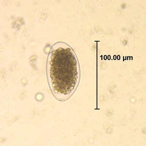
Capillaria philippinensis is a parasitic nematode which causes intestinal capillariasis. This sometimes fatal disease was first discovered in Northern Luzon, Philippines, in 1964. Cases have also been reported from China, Egypt, Indonesia, Iran, Japan, Korea, Lao PDR, Taiwan and Thailand. Cases diagnosed in Italy and Spain were believed to be acquired abroad, with one case possibly contracted in Colombia. The natural life cycle of C. philippinensis is believed to involve fish as intermediate hosts, and fish-eating birds as definitive hosts. Humans acquire C. philippinensis by eating small species of infested fish whole and raw.

<i>Gongylonema pulchrum</i> Species of human parasite

Gongylonema pulchrum is the only parasite of the genus Gongylonema capable of infecting humans.

<i>Angiostrongylus cantonensis</i> Species of roundworm

Angiostrongylus cantonensis is a nematode (roundworm) parasite that causes angiostrongyliasis, an infection that is the most common cause of eosinophilic meningitis in Southeast Asia and the Pacific Basin. The nematode commonly resides in the pulmonary arteries of rats, giving it the common name rat lungworm. Snails are the primary intermediate hosts, where larvae develop until they are infectious.
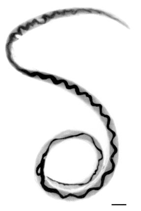
Capillaria plica is a parasitic nematode which is most often found in the urinary bladder, and occasionally in the kidneys, of dogs and foxes. It has also been found in the domestic cat, and various wild mammals. Its presence usually produces no clinical symptoms, but in some cases, it leads to hematuria, cystitis, or difficulty in urination.

<i>Thelazia callipaeda</i> Species of worm

Thelazia callipaeda is a parasitic nematode, and the most common cause of thelaziasis in humans, dogs and cats. It was first discovered in the eyes of a dog in China in 1910. By 2000, over 250 human cases had been reported in the medical literature.

<span class="mw-page-title-main">Thelaziasis</span> Medical condition

Thelaziasis is the term for infestation with parasitic nematodes of the genus Thelazia. The adults of all Thelazia species discovered so far inhabit the eyes and associated tissues of various mammal and bird hosts, including humans. Thelazia nematodes are often referred to as "eyeworms".

<i>Trichostrongylus</i> Genus of roundworms

Trichostrongylus species are nematodes, which are ubiquitous among herbivores worldwide, including cattle, sheep, donkeys, goats, deer, and rabbits. At least 10 Trichostrongylus species have been associated with human infections. Infections occur via ingestion of infective larvae from contaminated vegetables or water. Epidemiological studies indicate a worldwide distribution of Trichostrongylus infections in humans, with the highest prevalence rates observed in individuals from regions with poor sanitary conditions, in rural areas, or who are farmers / herders. Human infections are most prevalent in the Middle East and Asia, with a worldwide estimated prevalence of 5.5 million people.

<i>Moniliformis moniliformis</i> Species of thorny-headed worm

Moniliformis moniliformis is a parasite of the Acanthocephala phylum in the family Moniliformidae. The adult worms are usually found in intestines of rodents or carnivores such as cats and dogs. The species can also infest humans, though this is rare.

Thelazia californiensis is a nematode that originates in the genus Thelazia, which comes from phylum Nematoda. This worm has been known to cause Thelaziasis in hosts.

<span class="mw-page-title-main">John Stoffolano</span> American entomologist

John G. Stoffolano, Jr. is an American entomologist specializing in non-biting fly behavior, physiology, and veterinary issues of flies as vectors of pathogens.

Phortica variegata is a species of vinegar fly in the family Drosophilidae. Phortica and related fly species are perhaps best known for their behaviour of feeding on the lacrimal secretions of mammals. As a consequence of this behaviour, P. variegata can serve as a vector of Thelazia callipaeda roundworms.

<span class="mw-page-title-main">Cat worm infections</span> Worm infections in cats

Cat worm infections, the infection of cats (Felidae) with parasitic worms, occur frequently. Most worm species occur worldwide in both domestic and other cats, but there are regional, species and lifestyle differences in the frequency of infestation. According to the classification of the corresponding parasites in the zoological system, infections can be divided into those caused by nematode and flatworms - in the case of the latter, mainly cestoda and trematoda - while other strains are of no veterinary significance. While threadworms usually do not require an intermediate host for their reproduction, the development cycle of flatworms always proceeds via alternate hosts.

<span class="mw-page-title-main">Nematode infection in dogs</span> Threadworm infections of dogs are frequent

Nematode infection in dogs - the infection of dogs with parasitic nemamotodes - are, along with tapeworm infections and infections with protozoa, frequent parasitoses in veterinary practice. Nematodes, as so-called endoparasites, colonize various internal organs - most of them the digestive tract - and the skin. To date, about 30 different species of nematode have been identified in domestic dogs; they are essentially also found in wild dog species. However, the majority of them often cause no or only minor symptoms of disease in adult animals. The infection therefore does not necessarily have to manifest itself in a worm disease (helminthosis). For most nematodes, an infection can be detected by examining the feces for eggs or larvae. Roundworm infection in dogs and the hookworm in dogs is of particular health significance in Central Europe, as they can also be transmitted to humans (zoonosis). Regular deworming can significantly reduce the frequency of infection and thus the risk of infection for humans and dogs.

References

  1. This image is from Otranto, Domenico; Dutto, Moreno (2008). "Human Thelaziasis, Europe". Emerging Infectious Diseases. 14 (4): 647–649. doi:10.3201/eid1404.071205. PMC   2570937 . PMID   18394285.
  2. Otranto, D.; Traversa, D. (2005). "Thelazia eyeworm: An original endo- and ecto-parasitic nematode". Trends in Parasitology. 21 (1): 1–4. doi:10.1016/j.pt.2004.10.008. PMID   15639731.
  3. do Vale, Beatriz; Lopes, Ana Patrícia; da Conceição Fontes, Maria; Silvestre, Mário; Cardoso, Luís; Coelho, Ana Cláudia (2020). "Systematic review on infection and disease caused by Thelazia callipaeda in Europe: 2001–2020". Parasite. 27: 52. doi: 10.1051/parasite/2020048 . ISSN   1776-1042. PMC   7526429 . PMID   32996882.
  4. Xue, Chunyan; Tian, Nong; Huang, Zhenping (2007). "Thelazia callipaeda in human vitreous". Canadian Journal of Ophthalmology. 42 (6): 884–885. doi: 10.3129/i07-167 . PMID   18059522.
  5. de Oliveira Rodrigues, H. (1992) "Thelazia anolabiata (Molin, 1860) Railliet & Henry, 1910 (Nematoda: Thelazioidea), a new host record and systematic considerations." Memórias do Instituto Oswaldo Cruz 87(Suppl 1):217-222.
  6. KOYAMA Y; OHIRA A; KONO T; YONEYAMA T; SHIWAKU K (2000). "Five cases of thelaziasis". The British Journal of Ophthalmology. 84 (4): 439. doi:10.1136/bjo.84.4.439c. PMC   1723424 . PMID   10777285.
  7. Elias, R.; Mamani, J.; Hermoza, C.; Kinsella, J. (2008-12-20). "First report of thelaziosis (Thelazia anolabiata) in an Andean Cock of the Rock (Rupicola peruviana) from Peru". Veterinary Parasitology. 158 (4): 382–383. doi:10.1016/j.vetpar.2008.09.016. ISSN   0304-4017. PMID   18980808.
  8. "Case 279 --Clinical Microbiology Case". path.upmc.edu. Retrieved 2023-12-14.
  9. Dolff, S.; Kehrmann, J.; Eisermann, P.; Dalbah, S.; Tappe, D.; Rating, P. (2020). "Case Report: Thelazia callipaeda Eye Infection: The First Human Case in Germany". The American Journal of Tropical Medicine and Hygiene. 102 (2): 350–351. doi:10.4269/ajtmh.19-0483. PMC   7008349 . PMID   31912777.
  10. Bradbury, R. S.; Breen, K. V.; Bonura, E. M.; Hoyt, J. W.; Bishop, H. S. (2018). "Case Report: Conjunctival Infestation with Thelazia gulosa: A Novel Agent of Human Thelaziasis in the United States". The American Journal of Tropical Medicine and Hygiene. 98 (4): 1171–1174. doi:10.4269/ajtmh.17-0870. PMC   5928835 . PMID   29436343.
  11. Cotuțiu, Vlad-Dan; Ionică, Angela Monica; Lefkaditis, Menelaos; Cazan, Cristina Daniela; Hașaș, Alina Diana; Mihalca, Andrei Daniel (2022-11-14). "Thelazia lacrymalis in horses from Romania: epidemiology, morphology and phylogenetic analysis". Parasites & Vectors. 15 (1): 425. doi: 10.1186/s13071-022-05532-z . ISSN   1756-3305. PMC   9664606 . PMID   36376981.
  12. "How to access research remotely". www.cabdirect.org. Retrieved 2023-12-14.