List of biophysically important macromolecular crystal structures

Last updated

Nucleosome X-ray crystal structure 1aoi nucleosome rib round.tif
Nucleosome X-ray crystal structure

Crystal structures of protein and nucleic acid molecules and their complexes are central to the practice of most parts of biophysics, and have shaped much of what we understand scientifically at the atomic-detail level of biology. Their importance is underlined by the United Nations declaring 2014 as the International Year of Crystallography, as the 100th anniversary of Max von Laue's 1914 Nobel prize for discovering the diffraction of X-rays by crystals. This chronological list of biophysically notable protein and nucleic acid structures is loosely based on a review in the Biophysical Journal . [1] The list includes all the first dozen distinct structures, those that broke new ground in subject or method, and those that became model systems for work in future biophysical areas of research.

Contents

Myoglobin

Myoglobin sketch Myoglobin helix cylinder sketch.jpg
Myoglobin sketch
Alpha helix Helix electron density myoglobin 2nrl 17-32.jpg
Alpha helix

1958  Myoglobin was the very first crystal structure of a protein molecule. [2] Myoglobin cradles an iron-containing heme group that reversibly binds oxygen for use in powering muscle fibers, and those first crystals were of myoglobin from the sperm whale, whose muscles need copious oxygen storage for deep dives. The myoglobin 3-dimensional structure is made up of 8 alpha-helices, and the crystal structure showed that their conformation was right-handed and very closely matched the geometry proposed by Linus Pauling, with 3.6 residues per turn and backbone hydrogen bonds from the peptide NH of one residue to the peptide CO of residue i+4. Myoglobin is a model system for many types of biophysical studies, [3] especially involving the binding process of small ligands such as oxygen and carbon monoxide.
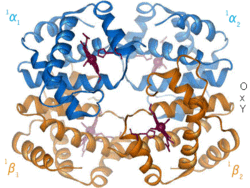
Hemoglobin

Hemoglobin beta chain Hemoglobin beta red whBkg.tif
Hemoglobin beta chain
Hemoglobin oxy/deoxy transition Hemoglobin t-r state ani.gif
Hemoglobin oxy/deoxy transition

1960  The hemoglobin crystal structure [4] showed a tetramer of two related chain types and was solved at much lower resolution than the monomeric myoglobin, but it clearly had the same basic 8-helix architecture (now called the "globin fold"). Further hemoglobin crystal structures at higher resolution (PDB 1MHB, 1DHB) soon showed the coupled change of both local and quaternary conformation between the oxy and deoxy states of hemoglobin, [5] which explains the cooperativity of oxygen binding in the blood and the allosteric effect of factors such as pH and DPG. For decades hemoglobin was the primary teaching example for the concept of allostery, as well as being an intensive focus of research and discussion on allostery. In 1909, hemoglobin crystals from >100 species were used to relate taxonomy to molecular properties. [6] That book was cited by Perutz in the 1938 report [7] of horse hemoglobin crystals that began his long saga to solve the crystal structure. Hemoglobin crystals are pleochroic dark red in two directions and pale red in the third [6] because of the orientation of the hemes, and the bright Soret band of the heme porphyrin groups is used in spectroscopic analysis of hemoglobin ligand binding.

HEW lysozyme ribbon HEW lysozyme rib bw.tif
HEW lysozyme ribbon

Hen-egg-white lysozyme

1965  Hen-egg-white lysozyme (PDB file 1lyz). [8] was the first crystal structure of an enzyme (it cleaves small carbohydrates into simple sugars), used for early studies of enzyme mechanism. [9] It contained beta sheet (antiparallel) as well as helices, and was also the first macromolecular structure to have its atomic coordinates refined (in real space). [10] The starting material for preparation can be bought at the grocery store, and hen-egg lysozyme crystallizes very readily in many different space groups; it is the favorite test case for new crystallographic experiments and instruments. Recent examples are nanocrystals of lysozyme for free-electron laser data collection [11] and microcrystals for micro electron diffraction. [12]

Ribonuclease A ribbon drawing RibonucleaseA SS paleRib.png
Ribonuclease A ribbon drawing

Ribonuclease

1967  Ribonuclease A (PDB file 2RSA) [13] is an RNA-cleaving enzyme stabilized by 4 disulfide bonds. It was used in Anfinsen's seminal research on protein folding which led to the concept that a protein's 3-dimensional structure was determined by its amino-acid sequence. Ribonuclease S, the cleaved, two-component form studied by Fred Richards, was also enzymatically active, had a nearly identical crystal structure (PDB file 1RNS), [14] and was shown to be catalytically active even in the crystal, [15] helping dispel doubts about the relevance of protein crystal structures to biological function.

Two domains of elastase Elastase ribbon BW.jpg
Two domains of elastase

Serine proteases

1967  The serine proteases are a historically very important group of enzyme structures, because collectively they illuminated catalytic mechanism (in their case, by the Ser-His-Asp "catalytic triad"), the basis of differing substrate specificities, and the activation mechanism by which a controlled enzymatic cleavage buries the new chain end to properly rearrange the active site. [16] The early crystal structures included chymotrypsin (PDB file 2CHA), [17] chymotrypsinogen (PDB file 1CHG), [18] trypsin (PDB file 1PTN), [19] and elastase (PDB file 1EST). [20] They also were the first protein structures that showed two near-identical domains, presumably related by gene duplication. One reason for their wide use as textbook and classroom examples was the insertion-code numbering system, which made Ser195 and His57 consistent and memorable despite the protein-specific sequence differences.[ citation needed ]

Papain

1968  Papain

Carboxypeptidase, with Zn Carboxypeptidase-A rib bw.tif
Carboxypeptidase, with Zn
CPA inhibitor Potato CPA inhibitor ribbon.jpg
CPA inhibitor

Carboxypeptidase

1969  Carboxypeptidase A is a zinc metalloprotease. Its crystal structure (PDB file 1CPA) [21] showed the first parallel beta structure: a large, twisted, central sheet of 8 strands with the active-site Zn located at the C-terminal end of the middle strands and the sheet flanked on both sides with alpha helices. It is an exopeptidase that cleaves peptides or proteins from the carboxy-terminal end rather than internal to the sequence. Later a small protein inhibitor of carboxypeptidase was solved (PDB file 4CPA) [22] that mechanically stops the catalysis by presenting its C-terminal end just sticking out from between a ring of disulfide bonds with tight structure behind it, preventing the enzyme from sucking in the chain past the first residue.

Subtilisin ribbon Subtilisin rib bw.tif
Subtilisin ribbon

Subtilisin

1969  Subtilisin (PDB file 1sbt [23] ) was a second type of serine protease with a near-identical active site to the trypsin family of enzymes, but with a completely different overall fold. This gave the first view of convergent evolution at the atomic level. Later, an intensive mutational study on subtilisin documented the effects of all 19 other amino acids at each individual position. [24]

Lactate dehydrogenase

1970  Lactate dehydrogenase

Commputer ribbon, with 3 SS BPTI ribbon 1bpi.jpg
Commputer ribbon, with 3 SS

Trypsin inhibitor

1970  Basic pancreatic trypsin inhibitor, or BPTI (PDB file 2pti [25] ), is a small, very stable protein that has been a highly productive model system for study of super-tight binding, disulfide bond (SS) formation, protein folding, molecular stability by amino-acid mutations or hydrogen-deuterium exchange, and fast local dynamics by NMR. Biologically, BPTI binds and inhibits trypsin while stored in the pancreas, allowing activation of protein digestion only after trypsin is released into the stomach.

Rubredoxin with nn-heme iron Rubredoxin Fe 2rxn rib.tif
Rubredoxin with nn-heme iron

Rubredoxin

1970  Rubredoxin (PDB file 2rxn [26] ) was the first redox structure solved, a minimalist protein with the iron bound by 4 Cys sidechains from 2 loops at the top of β hairpins. It diffracted to 1.2Å, enabling the first reciprocal-space refinement of a protein (4,5rxn [27] ). (NB: note that 4rxn was done without geometry restraints.) Archaeal rubredoxins account for many of the highest-resolution small structures in the PDB.

SS-linked insulin monomer Insulin worm bw.jpg
SS-linked insulin monomer
Space-grown insulin crystals Insulincrystals.jpg
Space-grown insulin crystals

Insulin

1971  Insulin (PDB file 1INS) [28] is a hormone central to the metabolism of sugar and fat storage, and important in human diseases such as obesity and diabetes. It is biophysically notable for its Zn binding, its equilibrium between monomer, dimer, and hexamer states, its ability to form crystals in vivo, and its synthesis as a longer "pro" form which is then cleaved to fold up as the active 2-chain, SS-linked monomer. Insulin was a success of NASA's crystal-growth program on the Space Shuttle, producing bulk preparations of very uniform tiny crystals for controlled dosage.

Staphylococcal nuclease

1971  Staphylococcal nuclease

Cytochrome C

1971  Cytochrome C

T4 phage lysozyme

1974  T4 phage lysozyme

Immunoglobulins

1974  Immunoglobulins

Superoxide dismutase

1975  Cu,Zn Superoxide dismutase

Transfer RNA

1976  Transfer RNA

TIM ribbon drawing, PDB 1tim TriosePhosphateIsomerase Ribbon pastel photo mat.png
TIM ribbon drawing, PDB 1tim

Triose phosphate isomerase

1976  Triose phosphate isomerase

Pepsin-like aspartic proteases

Later structures (1978 onwards)

Photosynthetic reaction center in membrane Photosynthetic Reaction Center Drawing.png
Photosynthetic reaction center in membrane
CAP protein dimer on DNA, PDB 1cgp CAP-DNA 1cgp rib.tiff
CAP protein dimer on DNA, PDB 1cgp
Kinesin dimer, PDB 3kin 064-Kinesin-3kin-composite.png
Kinesin dimer, PDB 3kin
Topoisomerase I on DNA (by David Goodsell) 073-Topoisomerases Topo I-1a36.png
Topoisomerase I on DNA (by David Goodsell)
tRNA 3-step progression through a ribosome 121- RCSB Molecule-of-Month 70S 3-step Ribosome elongation.tif
tRNA 3-step progression through a ribosome
beta-adrenergic receptor & G protein 3sn6 G-coupled ribbon.png
beta-adrenergic receptor & G protein
Half of a vault particle Half-vault front.png
Half of a vault particle
Photosystem II 4fby spline het.png
Photosystem II

Related Research Articles

<span class="mw-page-title-main">Chymotrypsin</span> Digestive enzyme

Chymotrypsin (EC 3.4.21.1, chymotrypsins A and B, alpha-chymar ophth, avazyme, chymar, chymotest, enzeon, quimar, quimotrase, alpha-chymar, alpha-chymotrypsin A, alpha-chymotrypsin) is a digestive enzyme component of pancreatic juice acting in the duodenum, where it performs proteolysis, the breakdown of proteins and polypeptides. Chymotrypsin preferentially cleaves peptide amide bonds where the side chain of the amino acid N-terminal to the scissile amide bond (the P1 position) is a large hydrophobic amino acid (tyrosine, tryptophan, and phenylalanine). These amino acids contain an aromatic ring in their side chain that fits into a hydrophobic pocket (the S1 position) of the enzyme. It is activated in the presence of trypsin. The hydrophobic and shape complementarity between the peptide substrate P1 side chain and the enzyme S1 binding cavity accounts for the substrate specificity of this enzyme. Chymotrypsin also hydrolyzes other amide bonds in peptides at slower rates, particularly those containing leucine at the P1 position.

<span class="mw-page-title-main">Proteolysis</span> Breakdown of proteins into smaller polypeptides or amino acids

Proteolysis is the breakdown of proteins into smaller polypeptides or amino acids. Uncatalysed, the hydrolysis of peptide bonds is extremely slow, taking hundreds of years. Proteolysis is typically catalysed by cellular enzymes called proteases, but may also occur by intra-molecular digestion.

<span class="mw-page-title-main">Trypsin</span> Family of digestive enzymes

Trypsin is an enzyme in the first section of the small intestine that starts the digestion of protein molecules by cutting long chains of amino acids into smaller pieces. It is a serine protease from the PA clan superfamily, found in the digestive system of many vertebrates, where it hydrolyzes proteins. Trypsin is formed in the small intestine when its proenzyme form, the trypsinogen produced by the pancreas, is activated. Trypsin cuts peptide chains mainly at the carboxyl side of the amino acids lysine or arginine. It is used for numerous biotechnological processes. The process is commonly referred to as trypsinogen proteolysis or trypsinization, and proteins that have been digested/treated with trypsin are said to have been trypsinized. Trypsin was discovered in 1876 by Wilhelm Kühne and was named from the Ancient Greek word for rubbing since it was first isolated by rubbing the pancreas with glycerin.

<span class="mw-page-title-main">Lysozyme</span> Antimicrobial enzyme produced by animals

Lysozyme is an antimicrobial enzyme produced by animals that forms part of the innate immune system. It is a glycoside hydrolase that catalyzes the following process:

<span class="mw-page-title-main">Serine protease</span> Class of enzymes

Serine proteases are enzymes that cleave peptide bonds in proteins. Serine serves as the nucleophilic amino acid at the (enzyme's) active site. They are found ubiquitously in both eukaryotes and prokaryotes. Serine proteases fall into two broad categories based on their structure: chymotrypsin-like (trypsin-like) or subtilisin-like.

<span class="mw-page-title-main">Structural Classification of Proteins database</span> Biological database of proteins

The Structural Classification of Proteins (SCOP) database is a largely manual classification of protein structural domains based on similarities of their structures and amino acid sequences. A motivation for this classification is to determine the evolutionary relationship between proteins. Proteins with the same shapes but having little sequence or functional similarity are placed in different superfamilies, and are assumed to have only a very distant common ancestor. Proteins having the same shape and some similarity of sequence and/or function are placed in "families", and are assumed to have a closer common ancestor.

<span class="mw-page-title-main">Ramachandran plot</span> Visual representation of allowable protein conformations

In biochemistry, a Ramachandran plot, originally developed in 1963 by G. N. Ramachandran, C. Ramakrishnan, and V. Sasisekharan, is a way to visualize energetically allowed regions for backbone dihedral angles ψ against φ of amino acid residues in protein structure. The figure on the left illustrates the definition of the φ and ψ backbone dihedral angles. The ω angle at the peptide bond is normally 180°, since the partial-double-bond character keeps the peptide bond planar. The figure in the top right shows the allowed φ,ψ backbone conformational regions from the Ramachandran et al. 1963 and 1968 hard-sphere calculations: full radius in solid outline, reduced radius in dashed, and relaxed tau (N-Cα-C) angle in dotted lines. Because dihedral angle values are circular and 0° is the same as 360°, the edges of the Ramachandran plot "wrap" right-to-left and bottom-to-top. For instance, the small strip of allowed values along the lower-left edge of the plot are a continuation of the large, extended-chain region at upper left.

<span class="mw-page-title-main">Robert Huber</span> German biochemist and Nobel laureate (born 1937)

Robert Huber is a German biochemist and Nobel laureate. known for his work crystallizing an intramembrane protein important in photosynthesis and subsequently applying X-ray crystallography to elucidate the protein's structure.

<span class="mw-page-title-main">Catalytic triad</span> Set of three coordinated amino acids

A catalytic triad is a set of three coordinated amino acids that can be found in the active site of some enzymes. Catalytic triads are most commonly found in hydrolase and transferase enzymes. An acid-base-nucleophile triad is a common motif for generating a nucleophilic residue for covalent catalysis. The residues form a charge-relay network to polarise and activate the nucleophile, which attacks the substrate, forming a covalent intermediate which is then hydrolysed to release the product and regenerate free enzyme. The nucleophile is most commonly a serine or cysteine amino acid, but occasionally threonine or even selenocysteine. The 3D structure of the enzyme brings together the triad residues in a precise orientation, even though they may be far apart in the sequence.

α-Bungarotoxin Chemical compound

α-Bungarotoxin is one of the bungarotoxins, components of the venom of the elapid Taiwanese banded krait snake. It is a type of α-neurotoxin, a neurotoxic protein that is known to bind competitively and in a relatively irreversible manner to the nicotinic acetylcholine receptor found at the neuromuscular junction, causing paralysis, respiratory failure, and death in the victim. It has also been shown to play an antagonistic role in the binding of the α7 nicotinic acetylcholine receptor in the brain, and as such has numerous applications in neuroscience research.

<span class="mw-page-title-main">Barnase</span> Bacterial ribonuclease protein

Barnase (a portmanteau of "BActerial" "RiboNucleASE") is a bacterial protein that consists of 110 amino acids and has ribonuclease activity. It is synthesized and secreted by the bacterium Bacillus amyloliquefaciens, but is lethal to the cell when expressed without its inhibitor barstar. The inhibitor binds to and occludes the ribonuclease active site, preventing barnase from damaging the cell's RNA after it has been synthesized but before it has been secreted. The barnase/barstar complex is noted for its extraordinarily tight protein-protein binding, with an on-rate of 108s−1M−1.

<span class="mw-page-title-main">Subtilisin</span> Proteolytic enzyme found in Bacillus subtilis

Subtilisin is a protease initially obtained from Bacillus subtilis.

<span class="mw-page-title-main">Frederic M. Richards</span> American biochemist and biophysicist (1925–2009)

Frederic Middlebrook Richards, commonly referred to as Fred Richards, was an American biochemist and biophysicist known for solving the pioneering crystal structure of the ribonuclease S enzyme in 1967 and for defining the concept of solvent-accessible surface. He contributed many key experimental and theoretical results and developed new methods, garnering over 20,000 journal citations in several quite distinct research areas. In addition to the protein crystallography and biochemistry of ribonuclease S, these included solvent accessibility and internal packing of proteins, the first side-chain rotamer library, high-pressure crystallography, new types of chemical tags such as biotin/avidin, the nuclear magnetic resonance (NMR) chemical shift index, and structural and biophysical characterization of the effects of mutations.

<span class="mw-page-title-main">Protein domain</span> Self-stable region of a proteins chain that folds independently from the rest

In molecular biology, a protein domain is a region of a protein's polypeptide chain that is self-stabilizing and that folds independently from the rest. Each domain forms a compact folded three-dimensional structure. Many proteins consist of several domains, and a domain may appear in a variety of different proteins. Molecular evolution uses domains as building blocks and these may be recombined in different arrangements to create proteins with different functions. In general, domains vary in length from between about 50 amino acids up to 250 amino acids in length. The shortest domains, such as zinc fingers, are stabilized by metal ions or disulfide bridges. Domains often form functional units, such as the calcium-binding EF hand domain of calmodulin. Because they are independently stable, domains can be "swapped" by genetic engineering between one protein and another to make chimeric proteins.

<span class="mw-page-title-main">Louise Johnson</span> British biochemist and protein crystallographer 1940–2012

Dame Louise Napier Johnson,, was a British biochemist and protein crystallographer. She was David Phillips Professor of Molecular Biophysics at the University of Oxford from 1990 to 2007, and later an emeritus professor.

<span class="mw-page-title-main">Subtilase</span>

Subtilases are a family of subtilisin-like serine proteases. They appear to have independently and convergently evolved an Asp/Ser/His catalytic triad, like in the trypsin serine proteases. The structure of proteins in this family shows that they have an alpha/beta fold containing a 7-stranded parallel beta sheet.

<span class="mw-page-title-main">SSI protease inhibitor</span>

In molecular biology the protein SSI is a Subtilisin inhibitor-like which stands for Streptomyces subtilisin inhibitor. This is a protease inhibitor. These are often synthesised as part of a larger precursor protein, either as a prepropeptide. The function of this protein domain is to prevent access of the substrate to the active site. It is found only in bacteria.

Streptogrisin B is an enzyme. This enzyme catalyses the following chemical reaction

<span class="mw-page-title-main">PA clan of proteases</span>

The PA clan is the largest group of proteases with common ancestry as identified by structural homology. Members have a chymotrypsin-like fold and similar proteolysis mechanisms but can have identity of <10%. The clan contains both cysteine and serine proteases. PA clan proteases can be found in plants, animals, fungi, eubacteria, archaea and viruses.

<span class="mw-page-title-main">Structure validation</span> Process of evaluating 3-dimensional atomic models of biomacromolecules

Macromolecular structure validation is the process of evaluating reliability for 3-dimensional atomic models of large biological molecules such as proteins and nucleic acids. These models, which provide 3D coordinates for each atom in the molecule, come from structural biology experiments such as x-ray crystallography or nuclear magnetic resonance (NMR). The validation has three aspects: 1) checking on the validity of the thousands to millions of measurements in the experiment; 2) checking how consistent the atomic model is with those experimental data; and 3) checking consistency of the model with known physical and chemical properties.

References

  1. Richardson JS, Richardson DC (2014). "Biophysical highlights from 54 years of macromolecular crystallography". Biophysical Journal. 106 (3): 510–525. Bibcode:2014BpJ...106..510R. doi:10.1016/j.bpj.2014.01.001. PMC   3945011 . PMID   24507592.
  2. Kendrew JC, Bodo G, Dintzis HM, Parrish RG, Wyckoff H, Phillips DC (1958). "A Three-Dimensional Model of the Myoglobin Molecule Obtained by X-Ray Analysis". Nature. 181 (4610): 662–666. doi:10.1038/181662a0.
  3. Frauenfelder H, McMahon BH, Fenimore PW (2003). "Myoglobin: the hydrogen atom of biology and a paradigm of complexity". Proceedings of the National Academy of Sciences USA. 100 (15): 8615–8617. Bibcode:2003PNAS..100.8615F. doi: 10.1073/pnas.1633688100 . PMC   166357 . PMID   12861080.
  4. Perutz MF, Rossmann MG, Cullis AF, Muirhead H, North AC (1960). "Structure of haemoglobin: A three-dimensional Fourier synthesis at 5.5-Å resolution, obtained by X-ray analysis". Nature. 185 (4711): 416–422. Bibcode:1960Natur.185..416P. doi:10.1038/185416a0. PMID   18990801. S2CID   4208282.
  5. Perutz MF (1970). "Stereochemistry of Cooperative Effects in Haemoglobin: Haem–Haem Interaction and the Problem of Allostery". Nature. 228 (5273): 726–734. Bibcode:1970Natur.228..726P. doi:10.1038/228726a0. PMID   5528785. S2CID   43624891.
  6. 1 2 Edward Tyson Reichert & Amos Peaslee Brown (1909). The Differentiation and Specificity of Corresponding Proteins and other Vital Substances in Relation to Biological Classification and Organic Evolution: The Crystallography of Hemoglobins. Washington, DC: Carnegie Institution.
  7. Bernal JD, Fankuchen I, Perutz M (1938). "X-ray study of chymotrypsin and hemoglobin". Nature. 141 (3568): 523–524. doi:10.1038/141523a0. S2CID   4063748.
  8. Blake CC, Koenig DF, Mair GA, North AC, Phillips DC, Sarma VR (1965). "Structure of hen egg-white lysozyme: A three-dimensional Fourier synthesis at 2Å resolution". Nature. 206 (4986): 757–761. Bibcode:1965Natur.206..757B. doi:10.1038/206757a0. PMID   5891407. S2CID   4161467.
  9. Warshel A, Levitt M (1976). "Theoretical studies of enzymic reactions: Dielectric, electrostatic, and steric stabilization of the carbonium ion in the reaction of lysozyme". J Mol Biol. 103 (2): 227–49. doi:10.1016/0022-2836(76)90311-9. PMID   985660.
  10. Diamond R (1974). "Real-space refinement of the structure of hen egg-white lysozyme". Journal of Molecular Biology. 82 (3): 371–374. doi:10.1016/0022-2836(74)90598-1. PMID   4856347.
  11. Boutet S, Lomb L, Williams GJ, et al. (2012). "High-resolution protein structure determination by serial femtosecond crystallography" (PDF). Science. 337 (6092): 362–364. Bibcode:2012Sci...337..362B. doi:10.1126/science.1217737. PMC   3788707 . PMID   22653729.
  12. Shi D, Nannenga BL, Iadenza MG, Gonen T (2013). "Three-dimensional electron crystallography of protein microcrystals". eLife. 2: e01345. doi: 10.7554/elife.01345 . PMC   3831942 . PMID   24252878.
  13. Kartha G, Bello J, Harker D (1967). "Tertiary structure of ribonuclease". Nature. 213 (5079): 862–865. Bibcode:1967Natur.213..862K. doi:10.1038/213862a0. PMID   6043657. S2CID   4183721.
  14. Wyckoff HW, Hardman KD, Allewell NM, Inagami T, Johnson LN, Richards FM (1967). "The structure of ribonuclease-S at 3.5 Å resolution". Journal of Biological Chemistry. 242 (17): 3984–3988. doi: 10.1016/S0021-9258(18)95844-8 .
  15. Doscher MS, Richards FM (1963). "The activity of an enzyme in the crystalline state: Ribonuclease-S". Journal of Biological Chemistry. 238 (7): 2399–2406. doi: 10.1016/S0021-9258(19)67984-6 .
  16. Dickerson RE, Geis I (1969). Structure and Action of Proteins. New York: Harper.
  17. Matthews BW, Sigler PB, Henderson R, Blow DM (1967). "Three-dimensional structure of tosyl-α-chymotrypsin". Nature. 214 (5089): 652–656. Bibcode:1967Natur.214..652M. doi:10.1038/214652a0. PMID   6049071. S2CID   4273406.
  18. Freer ST, Kraut J, Robertus JD, Tonle H, Wright HT (1970). "Chymotrypsinogen: 2.5-Å crystal structure, comparison with α-chymotrypsin, and implications for zymogen activation". Biochemistry. 9 (9): 1997–2009. doi:10.1021/bi00811a022. PMID   5442169.
  19. Fehlhammer H, Bode W (1975). "The refined crystal structure of bovine beta-trypsin at 1.8Å resolution, II. Crystallographic refinement, calcium binding site and active site at pH 7.0". Journal of Molecular Biology. 98 (4): 693–697. doi:10.1016/s0022-2836(75)80005-2. PMID   512.
  20. Sawyer L, Shotton DM, Campbell JW, Ladner RC (1978). "The atomic structure of crystalline porcine pancreatic elastase at 2.5Å resolution: comparison with the structure of alpha-chymotrypsin". Journal of Molecular Biology. 118 (2): 137–208. doi:10.1016/0022-2836(78)90412-6. PMID   628010.
  21. Lipscomb WN; Hartsuck JA; Reeke GN; Quiocho FA; Bethge PH; Ludwif=g ML; Steitz TA; Muirhead H; Coppola JC (1969). "The structure of carboxypeptidase A, VII. The 2.0-Å resolution studies of the enzyme and of its complex with glycyltyrosine, and mechanistic deductions". Brookhaven Symposia in Biology. 21 (1): 24–90. PMID   5719196.
  22. Rees DC, Lipscomb WN (1982). "Refined crystal structure of the potato inhibitor complex of carboxypeptidase A at 2.5 A resolution". Journal of Molecular Biology. 160 (3): 475–498. doi:10.1016/0022-2836(82)90309-6. PMID   7154070.
  23. Alden RA, Birktoft JJ, Kraut J, Robertus JD, Wright CS (1971). "Atomic coordinates for subtilisin BPN' (or novo)". Biochem Biophys Res Commun. 45 (2): 337–44. doi:10.1016/0006-291X(71)90823-0. PMID   5160720.
  24. Wells J, Estell D (1988). "Subtilisin - an enzyme designed to be engineered". Trends Biochem Sci. 13 (8): 291–297. doi:10.1016/0968-0004(88)90121-1. PMID   3154281.
  25. Huber R, Kukla D, Ruhmann A, Epp O, Formanek H (1970). "The basic trypsin inhibitor of bovine pancreas. I. Structure analysis and conformation of the polypeptide chain". Naturwissenschaften. 57 (5304): 389–392. doi:10.1038/231506a0. PMID   4932997. S2CID   4158731.
  26. Herriott JR, Sieker LC, Jensen LH, Lovenberg W (1970). "Structure of rubredoxin: An X-ray study to 2.5Å resolution". Journal of Molecular Biology. 50 (2): 391–402. doi:10.1016/0022-2836(70)90200-7. PMID   5476919.
  27. Watenpaugh KD, Sieker LC, Jensen LH (1980). "Crystallographic refinement of rubredoxin at 1.2Å resolution". Journal of Molecular Biology. 138 (3): 615–633. doi:10.1016/s0022-2836(80)80020-9. PMID   7411618.
  28. Blundell TL, Cutfield JF, Cutfield SM, Dodson EJ, Dodson GG, Hodgkin DC, Mercola DA, Vijayan M (1971). "Atomic positions in rhombohedral 2-zinc insulin crystals". Nature. 231 (5304): 506–511. Bibcode:1971Natur.231..506B. doi:10.1038/231506a0. PMID   4932997. S2CID   4158731.
  29. Tanaka H, Kato K, Yamashita E, Sumizawa T, Zhou Y, Yau M, Iwasaki K, Yoshimura M, Tsukihara T (2009). "The Structure of Rat Liver Vault at 3.5 Angstrom Resolution". Science. 323 (5912): 384–388. Bibcode:2009Sci...323..384T. doi:10.1126/science.1164975. PMID   19150846. S2CID   2072790.
  30. Casanas A, Querol-Audi J, Guerra P, Pous J, Tanaka H, Tsukihara T, Verdaguer N, Fita I (2013). "New features of vault architecture and dynamics revealed by novel refinement using the deformable elastic network approach" (PDF). Acta Crystallographica. D69 (Pt 6): 1054–1061. doi:10.1107/S0907444913004472. hdl: 10261/88208 . PMID   23695250.