Schmitt Gillenwater Kelly syndrome

Last updated
Schmitt Gillenwater Kelly syndrome
Other namesRadial hypoplasia-triphalangeal thumbs-hypospadias-maxillary diastema syndrome
Autosomal dominant - en.svg
Schmitt Gillenwater Kelly syndrome has an autosomal dominant pattern of inheritance.

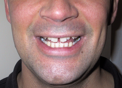
Schmitt Gillenwater Kelly syndrome is a rare autosomal dominant [1] congenital disorder consisting of radial hypoplasia, triphalangeal thumbs, hypospadias, and maxillary diastema. [1] [2]

Contents

Discovery

It was first identified by Edward Schmitt, Jay Y. Gillenwater, Thadeus E. Kelly, and John M. Opitz in 1962, where they published their results in a case study. [1] The family was found to be minimally restricted in normal functions, and lived relatively normal lives. The symptoms were consistent throughout all the members of the family, with the exception of all three of the boys having hypospadias. [1]

Radial Hypoplasia

Signs and Symptoms

The family had hypoplastic radii, which resulted in approximately 50% shorter radii. Because of this, the ulna was bowed outwards with outermost part of the ulna being pushed towards the skin. This resulted in a shorter reach.

Causes

Although more research is needed, the genetic cause of radial hypoplasia is believed to come from a rare allele of the Sonic hedgehog (Shh) gene. [3] This gene produces the Shh protein that induces development of the ulna, and the index, middle, ring, and pinky fingers while increasing the expression of fibroblast growth factor (FGF), another signaling molecule, which induces development of the radius and thumb. Both Shh and FGF are widely expressed during early embryo development. [4] [5] When this rare allele of Shh gene is expressed, the result is reduced Shh protein production, which hampers FGF expression, potentially leading to radial hypoplasia. [6]

Treatment and Prognosis

Treatment usually begins after birth and minor cases involve stretching, manipulation, and splinting. The goal of surgery is to increase length and straighten forearm and thumb reconstruction. [3]

Triphalangeal thumbs

Signs and Symptoms

Triphalangeal thumbs TPT five-fingered hand.jpeg
Triphalangeal thumbs

Another symptom of the individuals with the syndrome was Bilaterally symmetrical triphalangeal thumbs had three phalanges rather than two, and a longer finger like appearance. The thumbs were non-opposable.

Causes

The cause of this condition is understood to be genetic in nature, but the exact mechanism is unknown. However, research has shown that the gene of interest is located in chromosome 7, with potential candidate genes including EN2 and the human homologs of mouse genes Hx and Hm. [7]

Treatment and Prognosis

Surgery is done to correct any variations in the thumb and improve appearance; methods would vary on a case by case basis [8]

Hypospadia

Signs and Symptoms

Examples of different types of hypospadias Hypospadias-lg.jpg
Examples of different types of hypospadias

In the males, Hypospadias was seen, which is the opening of the urethra was at the underside of the penis rather than at the tip.

Causes

Hypospadias can come about as a result of imbalances in the Wnt, Shh, Hox, and BMP pathways during fetal development. The Wnt, Shh, Hox, and BMP families are widely expressed throughout development. [4] [9] [10] [11] During the development of male external genitalia, Shh acts as a central cue, indirectly activating Hoxa13 and Hoxd13 by binding to the Patched receptor, and directly activating BMP2, Fgf10, Wnt5a, and BMP4. Fgf10 induces further Shh expression, while BMP4 represses Wnt5a expression. Rare allelic expression of any one of these genes can result in hypospadias. [12]

Treatment and Prognosis

Minor forms do not require reconstructive surgery; Interventions include: correcting the location of urethral opening, repairing skin near the urethra opening, and straightening penile shaft. [13]

Maxillary diastema

Signs and Symptoms

Diastema of the upper (maxillary) teeth Brian diastema.png
Diastema of the upper (maxillary) teeth

The family also had Anterior Maxillary Diastema, a space between the upper incisors.

Causes

It highly likely this condition is caused by a genetic factor, but the exact gene is unknown.[ citation needed ]

Treatment and Prognosis

Treatment is centered around closing the gap between the incisors: either by veneers, braces, implants, or boding to conceal the gap. [14]

Similar diseases

Marfan syndrome - Is another autosomal dominant congenital condition. Symptoms consist of curved spine, thumb abnormalities, heart disease. Like Schmitt Gillenwater Kelly syndrome, surgery is done for cosmetic and reconstructive purposes. [15] Unlike Schmitt GIllenwater Kelly Syndrome, Marfan syndrome has a higher likelihood of developing life-threatening complications. [16]

Related Research Articles

<span class="mw-page-title-main">Marfan syndrome</span> Genetic disorder involving connective tissue

Marfan syndrome (MFS) is a multi-systemic genetic disorder that affects the connective tissue. Those with the condition tend to be tall and thin, with long arms, legs, fingers, and toes. They also typically have exceptionally flexible joints and abnormally curved spines. The most serious complications involve the heart and aorta, with an increased risk of mitral valve prolapse and aortic aneurysm. The lungs, eyes, bones, and the covering of the spinal cord are also commonly affected. The severity of the symptoms is variable.

<span class="mw-page-title-main">Polydactyly</span> Physical anomaly involving extra fingers or toes

Polydactyly or polydactylism, also known as hyperdactyly, is an anomaly in humans and animals resulting in supernumerary fingers and/or toes. Polydactyly is the opposite of oligodactyly.

<span class="mw-page-title-main">Jackson–Weiss syndrome</span> Medical condition

Jackson–Weiss syndrome (JWS) is a genetic disorder characterized by foot abnormalities and the premature fusion of certain bones of the skull (craniosynostosis), which prevents further growth of the skull and affects the shape of the head and face. This genetic disorder can also sometimes cause intellectual disability and crossed eyes. It was characterized in 1976.

Abruzzo–Erickson syndrome is an extremely rare disorder characterized by deafness, protruding ears, coloboma, a cleft palate or palatal rugosity, radial synostosis, and short stature. It was first characterized by Abruzzo and Erickson in 1977 as a CHARGE like syndrome as variably expressed among a family of two brothers, their mother, and their maternal uncle. Members of this family exhibited many of the CHARGE symptoms, but notably did not have choanal atresia and the brothers experienced typical genital development. Due to the recent discovery of this disorder, its etiology is not fully known but it is understood that it arises from mutations on the TBX22 gene on the X-chromosome. The disorder is inherited in an X-linked recessive manner. There is currently no known cure but its symptoms can be treated.

<span class="mw-page-title-main">Duane-radial ray syndrome</span> Medical condition

Duane-radial ray syndrome, also known as Okihiro Syndrome, is a rare autosomal dominant disorder that primarily affects the eyes and causes abnormalities of bones in the arms and hands. This disorder is considered to be a SALL4-related disorder due to the SALL4 gene mutations leading to these abnormalities. It is diagnosed by clinical findings on a physical exam as well as genetic testing and imaging. After being diagnosed, there are other evaluations that one may go through in order to determine the extent of the disease. There are various treatments for the symptoms of this disorder.

<span class="mw-page-title-main">Apical ectodermal ridge</span>

The apical ectodermal ridge (AER) is a structure that forms from the ectodermal cells at the distal end of each limb bud and acts as a major signaling center to ensure proper development of a limb. After the limb bud induces AER formation, the AER and limb mesenchyme—including the zone of polarizing activity (ZPA)—continue to communicate with each other to direct further limb development.

<span class="mw-page-title-main">Limb development</span>

Limb development in vertebrates is an area of active research in both developmental and evolutionary biology, with much of the latter work focused on the transition from fin to limb.

The limb bud is a structure formed early in vertebrate limb development. As a result of interactions between the ectoderm and underlying mesoderm, formation occurs roughly around the fourth week of development. In the development of the human embryo the upper limb bud appears in the third week and the lower limb bud appears four days later.

Congenital contractural arachnodactyly (CCA), also known as Beals-Hecht syndrome, is a rare autosomal dominant congenital connective tissue disorder. As with Marfan syndrome, people with CCA typically have an arm span that is greater than their height and very long fingers and toes. However, Beals and Hecht discovered in 1972 that, unlike Marfan's, CCA is caused by mutations to the fibrillin-2 (FBN2) gene rather than the fibrillin-1 (FBN1) gene.

<span class="mw-page-title-main">WNT4</span> Protein-coding gene in the species Homo sapiens

WNT4 is a secreted protein that, in humans, is encoded by the WNT4 gene, found on chromosome 1. It promotes female sex development and represses male sex development. Loss of function may have consequences, such as female to male sex reversal.

<span class="mw-page-title-main">TBX2</span> Protein-coding gene in the species Homo sapiens

T-box transcription factor 2 Tbx2 is a transcription factor that is encoded by the Tbx2 gene on chromosome 17q21-22 in humans. This gene is a member of a phylogenetically conserved family of genes that share a common DNA-binding domain, the T-box. Tbx2 and Tbx3 are the only T-box transcription factors that act as transcriptional repressors rather than transcriptional activators, and are closely related in terms of development and tumorigenesis. This gene plays a significant role in embryonic and fetal development through control of gene expression, and also has implications in various cancers. Tbx2 is associated with numerous signaling pathways, BMP, TGFβ, Wnt, and FGF, which allow for patterning and proliferation during organogenesis in fetal development.

<span class="mw-page-title-main">Zone of polarizing activity</span>

The zone of polarizing activity (ZPA) is an area of mesenchyme that contains signals which instruct the developing limb bud to form along the anterior/posterior axis. Limb bud is undifferentiated mesenchyme enclosed by an ectoderm covering. Eventually, the limb bud develops into bones, tendons, muscles and joints. Limb bud development relies not only on the ZPA, but also many different genes, signals, and a unique region of ectoderm called the apical ectodermal ridge (AER). Research by Saunders and Gasseling in 1948 identified the AER and its subsequent involvement in proximal distal outgrowth. Twenty years later, the same group did transplantation studies in chick limb bud and identified the ZPA. It wasn't until 1993 that Todt and Fallon showed that the AER and ZPA are dependent on each other.

<span class="mw-page-title-main">Acropectoral syndrome</span> Medical condition

Acropectoral syndrome is an autosomal dominant skeletal dysplasia syndrome affecting the hands, feet, sternum, and lumbosacral spine. A recently proposed candidate gene for preaxial polydactyly is LMBR1, encoding a novel transmembrane receptor, which may be an upstream regulator of SHH. The LMBR1 gene is on human chromosome 7q36.

<span class="mw-page-title-main">Maxillary hypoplasia</span> Medical condition

Maxillary hypoplasia, or maxillary deficiency, is an underdevelopment of the bones of the upper jaw. It is associated with Crouzon syndrome, Angelman syndrome, as well as Fetal alcohol syndrome. It can also be associated with Cleft lip and cleft palate. Some people could develop it due to poor dental extractions.

<span class="mw-page-title-main">Bat wing development</span>

The order Chiroptera, comprising all bats, has evolved the unique mammalian adaptation of flight. Bat wings are modified tetrapod forelimbs. Because bats are mammals, the skeletal structures in their wings are morphologically homologous to the skeletal components found in other tetrapod forelimbs. Through adaptive evolution these structures in bats have undergone many morphological changes, such as webbed digits, elongation of the forelimb, and reduction in bone thickness. Recently, there have been comparative studies of mouse and bat forelimb development to understand the genetic basis of morphological evolution. Consequently, the bat wing is a valuable evo-devo model for studying the evolution of vertebrate limb diversity.

<span class="mw-page-title-main">Triphalangeal thumb</span> Medical condition

Triphalangeal thumb (TPT) is a congenital malformation where the thumb has three phalanges instead of two. The extra phalangeal bone can vary in size from that of a small pebble to a size comparable to the phalanges in non-thumb digits. The true incidence of the condition is unknown, but is estimated at 1:25,000 live births. In about two-thirds of the patients with triphalangeal thumbs, there is a hereditary component. Besides the three phalanges, there can also be other malformations. It was first described by Columbi in 1559.

<span class="mw-page-title-main">Polysyndactyly</span> Medical condition

Polysyndactyly is a congenital anomaly, combining polydactyly and syndactyly, in which affected individuals have an extra finger or toe that is connected, via fusing or webbing, to an adjacent digit.

T-box transcription factor Tbx4 is a transcription factor that belongs to T-box gene family that is involved in the regulation of embryonic developmental processes. The transcription factor is encoded by the TBX4 gene located on human chromosome 17. Tbx4 is known mostly for its role in the development of the hindlimb, but it also plays a critical role in the formation of the umbilicus. Tbx4 has been shown to be expressed in the allantois, hindlimb, lung and proctodeum.

<span class="mw-page-title-main">Familial opposable triphalangeal thumbs duplication</span> Medical condition

Familial opposable triphalangeal thumb duplication is a limb malformation syndrome and a type of pre-axial polydactyly, characterized by having duplicated opposable triphalangeal thumbs. This condition can be a symptom of other genetic disorders, such as Holt–Oram syndrome and Fanconi anemia. This trait is autosomal dominant and often runs in families. Sometimes big toe duplication, post-axial polydactyly, and syndactyly of the hand and feet can occur alongside this malformation Approximately 20 families with the condition have been described in medical literature.

<span class="mw-page-title-main">Tibial hemimelia-polysyndactyly-triphalangeal thumb syndrome</span> Medical condition

Tibial hemimelia-polysyndactyly-triphalangeal thumb syndrome is a rare genetic limb malformation syndrome which is characterized by thumb triphalangy, polysyndactyly of the hand and foot, and hypoplasia/aplasia of the tibia bone. Additional features include short stature, radio-ulnar synostosis, ectrodactyly and abnormalities of the carpals and metatarsals. Only 19 affected families worldwide have been recorded in medical literature. It is associated with a heterozygous base pair substitution of A to G in position 404–406, located on intron 5 in the LMBR1 gene.

References

  1. 1 2 3 4 Schmitt E, Gillenwater JY, Kelly TE (1982). "An autosomal dominant syndrome of radial hypoplasia, triphalangeal thumbs, hypospadias, and maxillary diastema". Am. J. Med. Genet. 13 (1): 63–69. doi:10.1002/ajmg.1320130111. PMID   7137222.
  2. Schmitt Gillenwater Kelly syndrome; Radial hypoplasia triphalangeal thumbs hypospadias maxillary diastema at NIH's Office of Rare Diseases
  3. 1 2 Olson, Nathanael; Hosseinzadeh, Shayan (2023), "Radial Dysplasia", StatPearls, Treasure Island (FL): StatPearls Publishing, PMID   32491425 , retrieved 2023-12-05
  4. 1 2 "SHH sonic hedgehog signaling molecule [Homo sapiens (human)] - Gene - NCBI". www.ncbi.nlm.nih.gov. Retrieved 2023-12-05.
  5. Beenken, Andrew; Mohammadi, Moosa (March 2009). "The FGF family: biology, pathophysiology and therapy". Nature Reviews. Drug Discovery. 8 (3): 235–253. doi:10.1038/nrd2792. ISSN   1474-1784. PMC   3684054 . PMID   19247306.
  6. Elmakky, Amira; Stanghellini, Ilaria; Landi, Antonio; Percesepe, Antonio (August 2015). "Role of Genetic Factors in the Pathogenesis of Radial Deficiencies in Humans". Current Genomics. 16 (4): 264–278. doi:10.2174/1389202916666150528000412. ISSN   1389-2029. PMC   4765521 . PMID   26962299.
  7. Heutink, Peter; Zguricas, Julia; Oosterhout, Linda van; Breedveld, Guido J.; Testers, Leon; Sandkuijl, Lodewijk A.; Snijders, Pieter J. L. M.; Weissenbach, Jean; Lindhout, Dick; Hovius, Steven E. R.; Oostra, Ben A. (March 1994). "The gene for triphalangeal thumb maps to the subtelomeric region of chromosome 7q". Nature Genetics. 6 (3): 287–292. doi:10.1038/ng0394-287. hdl: 1765/57316 . ISSN   1546-1718. PMID   8012392. S2CID   5711857.
  8. Hovius, Steven E. R.; Zuidam, J. Michiel; de Wit, Thijs (December 2004). "Treatment of the Triphalangeal Thumb". Techniques in Hand & Upper Extremity Surgery. 8 (4): 247–256. doi:10.1097/00130911-200412000-00008. ISSN   1531-6572. PMID   16518099.
  9. Komiya, Yuko; Habas, Raymond (April 2008). "Wnt signal transduction pathways". Organogenesis. 4 (2): 68–75. doi:10.4161/org.4.2.5851. ISSN   1547-6278. PMC   2634250 . PMID   19279717.
  10. "Hox Genes in Development: The Hox Code | Learn Science at Scitable". www.nature.com. Retrieved 2023-12-05.
  11. Katagiri, Takenobu; Watabe, Tetsuro (June 2016). "Bone Morphogenetic Proteins". Cold Spring Harbor Perspectives in Biology. 8 (6): a021899. doi:10.1101/cshperspect.a021899. ISSN   1943-0264. PMC   4888821 . PMID   27252362.
  12. Bouty, Aurore; Ayers, Katie L.; Pask, Andrew; Heloury, Yves; Sinclair, Andrew H. (2015). "The Genetic and Environmental Factors Underlying Hypospadias". Sexual Development. 9 (5): 239–259. doi:10.1159/000441988. PMC   5012964 . PMID   26613581 . Retrieved 2023-12-05.
  13. Keays, Melise A.; Dave, Sumit (February 2016). "Current hypospadias management: Diagnosis, surgical management, and long-term patient-centred outcomes". Canadian Urological Association Journal. 11 (1–2S): S48–53. doi:10.5489/cuaj.4386. ISSN   1920-1214. PMC   5332236 . PMID   28265319.
  14. Vijaya, Sheetal; Vijaya, Shilpa; J Shetty, Meghan (July 2023). "Management of Midline Diastema in a Young Adult With Minimal-Thickness Porcelain Laminate Veneers". Cureus. 15 (7). e41904. doi: 10.7759/cureus.41904 . ISSN   2168-8184. PMC   10425606 . PMID   37588337.
  15. CDC (December 2019). "Marfan Syndrome | cdc.gov". Centers for Disease Control and Prevention. Retrieved 2023-10-23.
  16. Salik, Irim; Rawla, Prashanth (2023), "Marfan Syndrome", StatPearls, Treasure Island (FL): StatPearls Publishing, PMID   30726024 , retrieved 2023-12-04