Spinal locomotion

Last updated
Simple Walk-Cycle Blender3D SimpleWalkCycle.gif
Simple Walk-Cycle

Spinal locomotion results from intricate dynamic interactions between a central program in lower thoracolumbar spine and proprioceptive feedback from body in the absence of central control by brain as in complete spinal cord injury (SCI). [1] [2] [3] [4] Following SCI, the spinal circuitry below the lesion site does not become silent; rather, it continues to maintain active and functional neuronal properties, although in a modified manner. [5] [6]

Contents

Components of spinal locomotion

Reflex pathway: a pain receptor (sensory neuron) sends signals via the posterior horn, followed by a muscle activation (motor neuron) response via the anterior horn. Anatomy and physiology of animals Relation btw sensory, relay & motor neurons.jpg
Reflex pathway: a pain receptor (sensory neuron) sends signals via the posterior horn, followed by a muscle activation (motor neuron) response via the anterior horn.

Centrally generated patterns

The spinal cord executes rhythmical and sequential activation of muscles in locomotion. The central pattern generator (CPG) provides the basic locomotor rhythm and synergies by integrating commands from various sources that serve to initiate or modulate its output to meet the requirements of the environment. CPG within the lumbosacral spinal cord segments represent an important component of the total circuitry that generates and controls posture and locomotion. [7] This spinal circuitry can function independently in the absence of descending input from the brain to generate stable posture and locomotion and even modulate activity to match changing conditions (e.g., stepping over obstacles). [8] This capability improve with training (spinal plasticity) [7] and therefore it is believed that spinal cord has the capability to learn and memorize. [9] [10]

Sensory feedback

The sensory feedback originates from muscles, joints, tendons and skin afferents as well as from special senses and dynamically adapts the locomotor pattern of spinal cord to the requirements of the environment. These afferent sensory receptors perceive deformation of tissue, the amount of pressure (stretch or simply, placement), direction of movement, speed and velocity at which movement is occurring.

Sensory modulation of CPG

Simplified schema of basic nervous system function: signals are picked up by sensory receptors and sent to the spinal cord and brain, where processing occurs AT EACH LEVEL AND results in MODULATION OF signals sent FROM the spinal cord and out to motor neurons Nervous system organization en.svg
Simplified schema of basic nervous system function: signals are picked up by sensory receptors and sent to the spinal cord and brain, where processing occurs AT EACH LEVEL AND results in MODULATION OF signals sent FROM the spinal cord and out to motor neurons

The dynamic interactions between Spinal cord and sensory input are ensured by modulating transmission in locomotor pathways in a state- and phase-dependent manner. For instance, proprioceptive inputs from extensors can, during stance, adjust the timing and amplitude of muscle activities of the limbs to the speed of locomotion but be silenced during the swing phase of the cycle. Similarly, skin afferents participate predominantly in the correction of limb and foot placement during stance on uneven terrain, but skin stimuli can evoke different types of responses depending on when they occur within the step cycle. [11] It is important to note that inputs from the hip appear to play a critical role in spinal locomotion. Experiments on spinal animals showed that when one limb is held with the hip flexed, locomotion on that side stops while the other limb continues walking. However, when the stopped limb is extended at the hip joint to a point normally reached at the end of stance during walking, it suddenly flexes and starts walking again provided that the contralateral limb is a position to accept the weight of the hindquarters. [12] Other work confirmed the importance of hip afferents for locomotor rhythm generation since flexion of the hip will abolish the rhythm whereas extension will enhance it. [13]

The spinal cord processes and interprets proprioception in a manner similar to how our visual system processes information. [14] When a painting is viewed, the brain interprets the total visual field, as opposed to processing each individual pixel of information independently, and then derives an image. At any instant the spinal cord receives an ensemble of information from all receptors throughout the body that signals a proprioceptive “image” that represents time and space, and it computes which neurons to excite next based on the most recently perceived “images.” The importance of the CPG is not simply its ability to generate repetitive cycles, but also to receive, interpret, and predict the appropriate sequences of actions during any part of the step cycle, i.e., state dependence. The peripheral input then provides important information from which the probabilities of a given set of neurons being active at any given time can be finely tuned to a given situation during a specific phase of a step cycle. An excellent example of this is when a mechanical stimulus is applied to the dorsum of the paw of a cat. When the stimulus is applied during the swing phase, the flexor muscles of that limb are excited, and the result is enhanced flexion in order to step over the obstacle that created the stimulus. [15] However, when the same stimulus is applied during stance, the extensors are excited. Thus, the functional connectivity between mechanoreceptors and specific interneuronal populations within the spinal cord varies according to the physiological state. Even the efficacy of the monosynaptic input from muscle spindles to the motor neuron changes readily from one part of the step cycle to another, according to whether a subject is running or walking. [16]

In the absence of CPG, control by brain as it happens in complete spinal cord injury, sensory feedback is very important in generating rhythmic locomotion. Firstly, locomotor movements can be initiated or blocked by some proprioceptive afferent inputs. [12] Other work confirmed the importance of hip afferents for locomotor rhythm generation since flexion of the hip will abolish the rhythm whereas extension will enhance it. [13] Secondly, proprioceptive afferents may participate in adapting walking speed, in determining overall cycle duration, and in regulating the structure of the step cycle’s subphases (i.e., swing, stance), which is required for speed adaptation and interlimb coupling. [16] [17] Thirdly, proprioceptive afferents are involved in setting the level of muscle activity through various reflex pathways. [18]

Developmental evidence

Ultrasound recordings have captured in utero images of human fetuses at 13–14 gestational weeks "creeping and climbing" and producing alternating steps. [19] Onset of stepping in the fetus precedes development and myelination of most descending brain pathways strongly suggesting human spinal cord locomotor CPG and sensory feedback coordination and plasticity. Collectively, studies across the first postnatal year indicates that a locomotor continuum extends from neonatal stepping to the onset of independent walking further suggesting human locomotion is controlled by CPG and sensory input interaction.

Rehabilitation

The injured spinal cord is an “altered” spinal cord. After a SCI, supraspinal and spinal sources of control of movement differ substantially from that which existed prior to the injury, [20] thus resulting in an altered spinal cord. The automaticity of posture and locomotion emerge from the interactions between peripheral nervous system (PNS) and central nervous system (CNS) to work in synergy, each system having intrinsic activation and inhibition patterns that can generate coordinated motor outputs.

Electrical stimulation

Numerous experiments have demonstrated that electrical stimulation (ES) of the lumbosacral enlargement and dorsal root can induce locomotor EMG patterns and even hindlimb stepping in acute and chronic low-spinal animals and humans. [21] [22] Increased stimulation amplitude resulted in increased EMG amplitudes and an increased frequency of rhythmic activity. High frequencies of stimulation (>70 Hz) produced tonic activity in the leg musculature, which suggests that the upper lumbar stimulation may activate neuronal structures that then recruit interneurons involved in CPG. [23]

Treadmill training

Treadmill training (more commonly known as body weight supported treadmill training) can be applied via manual (therapist) or robotic assistance. In manual treadmill training the therapists provide assistance to facilitate an upright posture and a normal stepping pattern. [24] Therapist assistance may be provided at the patient’s pelvis, leg and foot, and a third therapist controlling the treadmill settings. [25] In robotic-assisted treadmill training, a device replaces the need for therapists to assist the patient in generating a normal stepping pattern. Currently, there are three different models available: Hocoma's Lokomat, the HealthSouth AutoAmbulator, and the Mechanized Gait Trainer II. [25] The Lokomat is a driven gait orthosis that consists of a computer -controlled exoskeleton that secured to the patient’s legs while being supported over a treadmill. [24] In addition to a belt driven treadmill and an overhead lift, the HealthSouth AutoAmbulator also includes a pair of articulated arms (that drives the hip and knee joints) and two upright structures that house the computer controls and body-weight unloading mechanism. [25] Unlike the first two, the Mechanized Gait Trainer II does not work in conjunction with a treadmill; instead it is based on a crank and rocker gear system which provides limb motion similar to an elliptical trainer. [25] Robotic-assisted treadmill training was developed with three goals in mind: 1. to decrease therapist physical demand and time, 2. to improve repeatability of step kinematics, and 3. to increase volume of locomotor training. [25]

In Humans with clinically complete SCI, there is evidence that treadmill training can improve several aspects of walking with some weight support assistance. Dietz and colleagues reported that after several weeks of treadmill training, the levels of weight bearing that can be imposed on the legs of clinically complete SCI subjects during treadmill walking significantly increases. [26] When stepping on a treadmill with body-weight support, rhythmic leg muscle activation patterns can be elicited in clinically complete subjects who are otherwise unable to voluntarily produce muscle activity in their legs. [27] A recent study has demonstrated that the levels of leg extensor muscle activity recorded in clinically complete SCI subjects significantly improved over course of several weeks of step training. [28] the stepping ability of clinically complete SCI subjects can improve in response to step training, but the level of improvement has not reached a level that allows complete independence from assistance during full weight-bearing. Also in humans with complete or motor-complete SCI, a novel approach using a CPG-activating drug treatment called Spinalon was shown to acutely induce episodes of rhythmic locomotor-like leg movements or corresponding electromyographic activity. [29] Largely due to the knowledge gained from studies on spinalized animals, two general principles have emerged for exciting the spinal circuits that generate stepping:

See also

Related Research Articles

<span class="mw-page-title-main">Muscle spindle</span> Innervated muscle structure involved in reflex actions and proprioception

Muscle spindles are stretch receptors within the body of a skeletal muscle that primarily detect changes in the length of the muscle. They convey length information to the central nervous system via afferent nerve fibers. This information can be processed by the brain as proprioception. The responses of muscle spindles to changes in length also play an important role in regulating the contraction of muscles, for example, by activating motor neurons via the stretch reflex to resist muscle stretch.

<span class="mw-page-title-main">Gait (human)</span> A pattern of limb movements made during locomotion

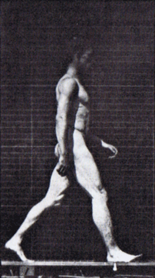
A gait is a manner of limb movements made during locomotion. Human gaits are the various ways in which humans can move, either naturally or as a result of specialized training. Human gait is defined as bipedal forward propulsion of the center of gravity of the human body, in which there are sinuous movements of different segments of the body with little energy spent. Varied gaits are characterized by differences such as limb movement patterns, overall velocity, forces, kinetic and potential energy cycles, and changes in contact with the ground.

<span class="mw-page-title-main">Type Ia sensory fiber</span> Type of afferent nerve fiber

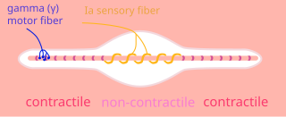
A type Ia sensory fiber, or a primary afferent fiber is a type of afferent nerve fiber. It is the sensory fiber of a stretch receptor called the muscle spindle found in muscles, which constantly monitors the rate at which a muscle stretch changes. The information carried by type Ia fibers contributes to the sense of proprioception.

<span class="mw-page-title-main">Spinocerebellar tract</span> Nerve tract in humans

The spinocerebellar tract is a nerve tract originating in the spinal cord and terminating in the same side (ipsilateral) of the cerebellum.

Central pattern generators (CPGs) are self-organizing biological neural circuits that produce rhythmic outputs in the absence of rhythmic input. They are the source of the tightly-coupled patterns of neural activity that drive rhythmic and stereotyped motor behaviors like walking, swimming, breathing, or chewing. The ability to function without input from higher brain areas still requires modulatory inputs, and their outputs are not fixed. Flexibility in response to sensory input is a fundamental quality of CPG-driven behavior. To be classified as a rhythmic generator, a CPG requires:

  1. "two or more processes that interact such that each process sequentially increases and decreases, and
  2. that, as a result of this interaction, the system repeatedly returns to its starting condition."
<span class="mw-page-title-main">Scratch reflex</span> Response to activation of sensory neurons

The scratch reflex is a response to activation of sensory neurons whose peripheral terminals are located on the surface of the body. Some sensory neurons can be activated by stimulation with an external object such as a parasite on the body surface. Alternatively, some sensory neurons can respond to a chemical stimulus that produces an itch sensation. During a scratch reflex, a nearby limb reaches toward and rubs against the site on the body surface that has been stimulated. The scratch reflex has been extensively studied to understand the functioning of neural networks in vertebrates. Despite decades of research, key aspects of the scratch reflex are still unknown, such as the neural mechanisms by which the reflex is terminated.

<span class="mw-page-title-main">Proprioception</span> Sense of self-movement, force, and body position

Proprioception, also called kinaesthesia, is the sense of self-movement, force, and body position.

<span class="mw-page-title-main">Undulatory locomotion</span>

Undulatory locomotion is the type of motion characterized by wave-like movement patterns that act to propel an animal forward. Examples of this type of gait include crawling in snakes, or swimming in the lamprey. Although this is typically the type of gait utilized by limbless animals, some creatures with limbs, such as the salamander, forgo use of their legs in certain environments and exhibit undulatory locomotion. In robotics this movement strategy is studied in order to create novel robotic devices capable of traversing a variety of environments.

<span class="mw-page-title-main">Locomotor effects of shoes</span>

Locomotor effects of shoes are the way in which the physical characteristics or components of shoes influence the locomotion neuromechanics of a person. Depending on the characteristics of the shoes, the effects are various, ranging from alteration in balance and posture, muscle activity of different muscles as measured by electromyography (EMG), and the impact force. There are many different types of shoes that exist, such as running, walking, loafers, high heels, sandals, slippers, work boots, dress shoes, and many more. However, a typical shoe will be composed of an insole, midsole, outsole, and heels, if any. In an unshod condition, where one is without any shoes, the locomotor effects are primarily observed in the heel strike patterns and resulting impact forces generated on the ground.

A gait trainer is a wheeled device that assists a person who is unable to walk independently to learn or relearn to walk safely and efficiently as part of gait training. Gait trainers are intended for children or adults with physical disabilities, to provide the opportunity to improve walking ability. A gait trainer offers both unweighting support and postural alignment to enable gait practice. It functions as a support walker and provides more assistance for balance and weight-bearing, than does a traditional rollator walker, or a walker with platform attachments. It also provides opportunities to stand and to bear weight in a safe, supported position.

<span class="mw-page-title-main">Neural substrate of locomotor central pattern generators in mammals</span>

Central pattern generators are biological neural networks organized to produce any rhythmic output without requiring a rhythmic input. In mammals, locomotor CPGs are organized in the lumbar and cervical segments of the spinal cord, and are used to control rhythmic muscle output in the arms and legs. Certain areas of the brain initiate the descending neural pathways that ultimately control and modulate the CPG signals. In addition to this direct control, there exist different feedback loops that coordinate the limbs for efficient locomotion and allow for the switching of gaits under appropriate circumstances.

When treating a person with a spinal cord injury, repairing the damage created by injury is the ultimate goal. By using a variety of treatments, greater improvements are achieved, and, therefore, treatment should not be limited to one method. Furthermore, increasing activity will increase his/her chances of recovery.

<span class="mw-page-title-main">Spinal interneuron</span> Interneuron relaying signals between sensory and motor neurons in the spinal cord

A spinal interneuron, found in the spinal cord, relays signals between (afferent) sensory neurons, and (efferent) motor neurons. Different classes of spinal interneurons are involved in the process of sensory-motor integration. Most interneurons are found in the grey column, a region of grey matter in the spinal cord.

<span class="mw-page-title-main">Proportional myoelectric control</span>

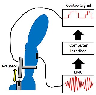
Proportional myoelectric control can be used to activate robotic lower limb exoskeletons. A proportional myoelectric control system utilizes a microcontroller or computer that inputs electromyography (EMG) signals from sensors on the leg muscle(s) and then activates the corresponding joint actuator(s) proportionally to the EMG signal.

<span class="mw-page-title-main">Cutaneous reflex in human locomotion</span>

Cutaneous, superficial, or skin reflexes, are activated by skin receptors and play a valuable role in locomotion, providing quick responses to unexpected environmental challenges. They have been shown to be important in responses to obstacles or stumbling, in preparing for visually challenging terrain, and for assistance in making adjustments when instability is introduced. In addition to the role in normal locomotion, cutaneous reflexes are being studied for their potential in enhancing rehabilitation therapy (physiotherapy) for people with gait abnormalities.

<span class="mw-page-title-main">Golgi tendon organ</span> Proprioceptive sensory receptor organ that senses changes in muscle tension

The Golgi tendon organ (GTO) is a proprioceptor – a type of sensory receptor that senses changes in muscle tension. It lies at the interface between a muscle and its tendon known as the musculotendinous junction also known as the myotendinous junction. It provides the sensory component of the Golgi tendon reflex.

<span class="mw-page-title-main">Mesencephalic locomotor region</span>

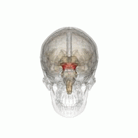
The mesencephalic locomotor region (MLR) is a functionally defined area of the midbrain that is associated with the initiation and control of locomotor movements in vertebrate species.

Postural control refers to the maintenance of body posture in space. The central nervous system interprets sensory input to produce motor output that maintains upright posture. Sensory information used for postural control largely comes from visual, proprioceptive, and vestibular systems. While the ability to regulate posture in vertebrates was previously thought to be a mostly automatic task, controlled by circuits in the spinal cord and brainstem, it is now clear that cortical areas are also involved, updating motor commands based on the state of the body and environment.

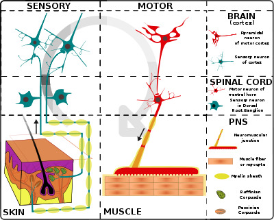
Proprioception refers to the sensory information relayed from muscles, tendons, and skin that allows for the perception of the body in space. This feedback allows for more fine control of movement. In the brain, proprioceptive integration occurs in the somatosensory cortex, and motor commands are generated in the motor cortex. In the spinal cord, sensory and motor signals are integrated and modulated by motor neuron pools called central pattern generators (CPGs). At the base level, sensory input is relayed by muscle spindles in the muscle and Golgi tendon organs (GTOs) in tendons, alongside cutaneous sensors in the skin.

<span class="mw-page-title-main">Interlimb coordination</span> Coordination of the left and right limbs

Interlimb coordination is the coordination of the left and right limbs. It could be classified into two types of action: bimanual coordination and hands or feet coordination. Such coordination involves various parts of the nervous system and requires a sensory feedback mechanism for the neural control of the limbs. A model can be used to visualize the basic features, the control centre of locomotor movements, and the neural control of interlimb coordination. This coordination mechanism can be altered and adapted for better performance during locomotion in adults and for the development of motor skills in infants. The adaptive feature of interlimb coordination can also be applied to the treatment for CNS damage from stroke and the Parkinson's disease in the future.

References

  1. Edgerton et al, 1998a. Locomotor capacity attributable to step training versus spontaneous recovery after spinalization in adult cats. Journal of Neurophysiol. 79:1329–1340.
  2. Edgerton et al, 1999a. Retention of hindlimb stepping ability in adult spinal cats after the cessation of step training. Journal of Neurophysiology. 81:85–94.
  3. Edgerton et al, 2002. Use of robotics in assessing the adaptive capacity of the rat lumbar spinal cord. Prog. Brain Res. 137:141–149.
  4. Guertin PA (December 2009). "The mammalian central pattern generator for locomotion". Brain Research Reviews. 62 (4): 345–56. doi:10.1016/j.brainresrev.2009.08.002. PMID   1972008. S2CID   9374670.
  5. Edgerton, V.R., Harkema, S.J., Dobkin, B.H. 2003. Retraining the Human Spinal Cord In: Spinal Cord Medicine: Principles and Practices. Demos Medical Publishing, Chapter 60, 817-826.
  6. de Leon, R.D., Roy, R.R., and Edgerton, V.R.2001 Is the recovery of stepping following spinal cord injury mediated by modifying existing neural pathways or by generating new pathways? Physical Therapy. 81(12): 1904-1911.
  7. 1 2 Dietz 2003. Spinal cord pattern generators for locomotion. Clin. Neurophysiol. 114:1379–89.
  8. Forssberg H, Grillner S, Rossignol S. 1975 Phase dependent reflex reversal during walking in chronic spinal cats. Brain Research. 85:103–107.
  9. Garraway SM, Hochman S. 2001. Serotonin increases the incidence of primary afferentevoked long-term depression in rat deep dorsal horn neurons. Journal of Neurophysiology 85:1864–1872.
  10. Rygh LJ, Tjolsen A, Hole K, Svendsen F. 2002. Cellular memory in spinal nociceptive circuitry. Scandinavian Journal of Psychology. 43:153–159.
  11. Simonsen EB, Dyhre-Poulsen P. 1999. Amplitude of the human soleus H reflex during walking and running. Journal of Physiology. 515:929–939.
  12. 1 2 Grillner, S., Rossignol, S., 1978. On the initiation of the swing phase of locomotion in chronic spinal cats. Brain Research. 146, 269–277.
  13. 1 2 Pearson, K.G., Rossignol, S., 1991. Fictive motor patterns in chronic spinal cats. Journal of Neurophysiology. 66, 1874–1887.
  14. Reggie Edgerton 2004. Plasticity of the spinal neural circuitry after injury. Annual Review of Neuroscience. 27:145–167.
  15. Forssberg H.1979 Stumbling corrective reaction: a phase-dependent compensatory reaction during locomotion. Journal of Neurophysiology, 42:936–953.
  16. 1 2 Lovely RG, Gregor RJ, Roy RR, Edgerton VR.1990. Weight-bearing hindlimb stepping in treadmill-exercised adult spinal cats. Brain Research. 514:206–218
  17. Zehret al,2003 Neural control of rhythmic human arm movement: phase dependence and task modulation of Hoffmann reflexes in forearm muscles. Journal of Neurophysiology. 89:12–21.
  18. Duysens J, Pearson KG. 1980. Inhibition of flexor burst generation by loading ankle extensor muscles in walking cats. Brain Res.187:321–332.
  19. Ianniruberto and Tajani Ultrasonographic study of fetal movements. Seminars in Perinatology 5: 175–181, 1981. [Web of Science][Medline].
  20. Dietz et al, 1998b. Locomotor pattern in paraplegic patients: training effects and recovery of spinal cord function. Spinal Cord. 36:380–390.
  21. Grillner S, Zangger P. 1984. The effect of dorsal root transection on the efferent motor pattern in the cat’s hindlimb during locomotion. Acta Physiologica Scandinavia. 120:393–405.
  22. Gerasimenko YP, Avelev VD, Nikitin OA, Lavrov IA. 2003. Initiation of locomotor activity in spinal cats by epidural stimulation of the spinal cord. Neuroscience and Behavioral Physiology. 33:247–254.
  23. Dimitrijevic MR, Gerasimenko Y, Pinter MM.1998. Evidence for a spinal central pattern generator in humans. Annals of the New York Academy of Science. 860:360–376.
  24. 1 2 Hornby, George T., Zemon, David H., & Campbell, Donielle. 2005. Robotic-assisted body weight supported treadmill training in individuals following motor incomplete spinal cord injury. Physical Therapy, 85(1), 52-66.
  25. 1 2 3 4 5 Winchester, Patricia & Querry, Ross. 2006. Robotic-orthoses for body weight supported treadmill training. Physical Medicine and Rehabilitation Clinics of North America, 17(1), 159-172.
  26. Dietz V, Colombo G, Jensen L, Baumgartner L.1995. Locomotor capacity of spinal cord in paraplegic patients. Annals of Neurology. 37:574–582.
  27. Maegele M, Muller S, Wernig A, Edgerton VR, Harkema SJ. 2002. Recruitment of spinal motor pools during voluntary movements versus stepping after human spinal cord injury. Journal of Neurotrauma. 19:1217–1229.
  28. Wirz M, Colombo G, Dietz V. 2001. Long term effects of locomotor training in spinal man. Journal of Neurology Neurosurgery and Psychiatry. 71:93–96.
  29. Radhakrishna M, Steuer I, Prince F, Roberts M, Mongeon D, Kia M, Dyck S, Matte G, Vaillancourt M, Guertin PA (December 2017). "Double-blind, placebo-controlled, randomized phase I/IIa study (safety and efficacy) with buspirone/levodopa/carbidopa (Spinalon) in subjects with complete AIS A or motor-complete AIS B spinal cord injury". Current Pharmaceutical Design. 23 (12): 1789–1804. doi:10.2174/1381612822666161227152200. PMID   28025945.