Related Research Articles

Coccolithophores, or coccolithophorids, are single-celled organisms which are part of the phytoplankton, the autotrophic (self-feeding) component of the plankton community. They form a group of about 200 species, and belong either to the kingdom Protista, according to Robert Whittaker's five-kingdom system, or clade Hacrobia, according to a newer biological classification system. Within the Hacrobia, the coccolithophores are in the phylum or division Haptophyta, class Prymnesiophyceae. Coccolithophores are almost exclusively marine, are photosynthetic, and exist in large numbers throughout the sunlight zone of the ocean.

An epiphysis is one of the rounded ends or tips of a long bone that ossify from a secondary center of ossification. Between the epiphysis and diaphysis lies the metaphysis, including the epiphyseal plate. At the joint, the epiphysis is covered with articular cartilage; below that covering is a zone similar to the epiphyseal plate, known as subchondral bone.

The long bones are those that are longer than they are wide. They are one of five types of bones: long, short, flat, irregular and sesamoid. Long bones, especially the femur and tibia, are subjected to most of the load during daily activities and they are crucial for skeletal mobility. They grow primarily by elongation of the diaphysis, with an epiphysis at each end of the growing bone. The ends of epiphyses are covered with hyaline cartilage. The longitudinal growth of long bones is a result of endochondral ossification at the epiphyseal plate. Bone growth in length is stimulated by the production of growth hormone (GH), a secretion of the anterior lobe of the pituitary gland.

Primary familial brain calcification (PFBC), also known as familial idiopathic basal ganglia calcification (FIBGC) and Fahr's disease, is a rare, genetically dominant, inherited neurological disorder characterized by abnormal deposits of calcium in areas of the brain that control movement. Through the use of CT scans, calcifications are seen primarily in the basal ganglia and in other areas such as the cerebral cortex.

Dystrophic calcification (DC) is the calcification occurring in degenerated or necrotic tissue, as in hyalinized scars, degenerated foci in leiomyomas, and caseous nodules. This occurs as a reaction to tissue damage, including as a consequence of medical device implantation. Dystrophic calcification can occur even if the amount of calcium in the blood is not elevated, in contrast to metastatic calcification, which is a consequence of a systemic mineral imbalance, including hypercalcemia and/or hyperphosphatemia, that leads to calcium deposition in healthy tissues. In dystrophic calcification, basophilic calcium salt deposits aggregate, first in the mitochondria, then progressively throughout the cell. These calcifications are an indication of previous microscopic cell injury, occurring in areas of cell necrosis when activated phosphatases bind calcium ions to phospholipids in the membrane.
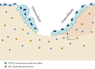
Ossification in bone remodeling is the process of laying down new bone material by cells named osteoblasts. It is synonymous with bone tissue formation. There are two processes resulting in the formation of normal, healthy bone tissue: Intramembranous ossification is the direct laying down of bone into the primitive connective tissue (mesenchyme), while endochondral ossification involves cartilage as a precursor.
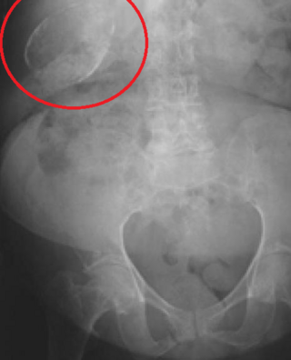
Porcelain gallbladder is a calcification of the gallbladder believed to be brought on by excessive gallstones, although the exact cause is not clear. As with gallstone disease in general, this condition occurs mostly in overweight female patients of middle age. It is a morphological variant of chronic cholecystitis. Inflammatory scarring of the wall, combined with dystrophic calcification within the wall transforms the gallbladder into a porcelain-like vessel. Removal of the gallbladder (cholecystectomy) is the recommended treatment.

Calcification is the accumulation of calcium salts in a body tissue. It normally occurs in the formation of bone, but calcium can be deposited abnormally in soft tissue, causing it to harden. Calcifications may be classified on whether there is mineral balance or not, and the location of the calcification. Calcification may also refer to the processes of normal mineral deposition in biological systems, such as the formation of stromatolites or mollusc shells.

An ossification center is a point where ossification of the hyaline cartilage begins. The first step in ossification is that the chondrocytes at this point become hypertrophic and arrange themselves in rows.

Calcifying odontogenic cyst (COC) is a rare developmental lesion that comes from odontogenic epithelium. It is also known as a calcifying cystic odontogenic tumor, which is a proliferation of odontogenic epithelium and scattered nest of ghost cells and calcifications that may form the lining of a cyst, or present as a solid mass.

Chondroblastoma is a rare, benign, locally aggressive bone tumor that typically affects the epiphyses or apophyses of long bones. It is thought to arise from an outgrowth of immature cartilage cells (chondroblasts) from secondary ossification centers, originating from the epiphyseal plate or some remnant of it.

Calcific tendinitis is a common condition where deposits of calcium phosphate form in a tendon, sometimes causing pain at the affected site. Deposits can occur in several places in the body, but are by far most common in the rotator cuff of the shoulder. Around 80% of those with deposits experience symptoms, typically chronic pain during certain shoulder movements, or sharp acute pain that worsens at night. Calcific tendinitis is typically diagnosed by physical exam and X-ray imaging. The disease often resolves completely on its own, but is typically treated with non-steroidal anti-inflammatory drugs to relieve pain, rest and physical therapy to promote healing, and in some cases various procedures to breakdown and/or remove the calcium deposits.
Fitzsimmons–Guilbert syndrome is an extremely rare genetic disease characterized by a slowly progressive spastic paraplegia, skeletal anomalies of the hands and feet with brachydactyly type E, cone-shaped epiphyses, abnormal metaphyseal–phalangeal pattern profile, sternal anomaly, dysarthria, and mild intellectual deficit.

Mönckeberg's arteriosclerosis, or Mönckeberg's sclerosis, is a form of arteriosclerosis or vessel hardening, where calcium deposits are found in the muscular middle layer of the walls of arteries. It is an example of dystrophic calcification. This condition occurs as an age-related degenerative process. However, it can occur in pseudoxanthoma elasticum and idiopathic arterial calcification of infancy as a pathological condition, as well. Its clinical significance and cause are not well understood and its relationship to atherosclerosis and other forms of vascular calcification are the subject of disagreement. Mönckeberg's arteriosclerosis is named after Johann Georg Mönckeberg, who first described it in 1903.

Hydrops-ectopic calcification-moth-eaten skeletal dysplasia is a defect in cholesterol biosynthesis. Greenberg characterized the condition in 1988.
An odontogenic tumor is a neoplasm of the cells or tissues that initiate odontogenic processes.

Calcific bursitis refers to calcium deposits within the bursae. This most occurs in the shoulder area. The most common bursa for calcific bursitis to occur is the subacromial bursa. A bursa is a small, fluid-filled sac that reduces friction, and facilitates movements between its adjacent tissues. Inflammation of the bursae is called bursitis.

Pulp stones are nodular, calcified masses appearing in either or both the coronal and root portion of the pulp organ in teeth. Pulp stones are not painful unless they impinge on nerves.

Spondyloepimetaphyseal dysplasia-short limb-abnormal calcification syndrome is a rare genetic disorder which is characterized by osseous anomalies resulting in short stature and other afflictions.
References
- ↑ Primer of Diagnostic Imaging (6 ed.). Elsevier. 2019. pp. 628–690.
| | This human musculoskeletal system article is a stub. You can help Wikipedia by expanding it. |