
A CT scan or computed tomography scan makes use of computer-processed combinations of many X-ray measurements taken from different angles to produce cross-sectional (tomographic) images of specific areas of a scanned object, allowing the user to see inside the object without cutting.

Medical ultrasound is a diagnostic imaging technique based on the application of ultrasound. It is used to create an image of internal body structures such as tendons, muscles, joints, blood vessels, and internal organs. Its aim is often to find a source of a disease or to exclude pathology. The practice of examining pregnant women using ultrasound is called obstetric ultrasound, and was an early development and application of clinical ultrasonography.
Urinary incontinence (UI), also known as involuntary urination, is any uncontrolled leakage of urine. It is a common and distressing problem, which may have a large impact on quality of life. It has been identified as an important issue in geriatric health care. The term enuresis is often used to refer to urinary incontinence primarily in children, such as nocturnal enuresis.

An intravenous pyelogram (IVP), also called an intravenous urogram (IVU), is a radiological procedure used to visualize abnormalities of the urinary system, including the kidneys, ureters, and bladder. Unlike a kidneys, ureters, and bladder x-ray (KUB), which is a plain radiograph, an IVP uses contrast to highlight the urinary tract.

Interventional radiology (IR) is a medical specialty which provides minimally invasive image-guided diagnosis and treatment of disease. Although the range of procedures performed by interventional radiologists is broad, the unifying concept behind these procedures is the application of image guidance and minimally invasive techniques in order to minimize risk to the patient.

JAMA: The Journal of the American Medical Association is a peer-reviewed medical journal published 48 times a year by the American Medical Association. It publishes original research, reviews, and editorials covering all aspects of biomedicine. The journal was established in 1883 with Nathan Smith Davis as the founding editor. The journal's editor-in-chief is Howard Bauchner of Boston University, who succeeded Catherine DeAngelis on July 1, 2011.
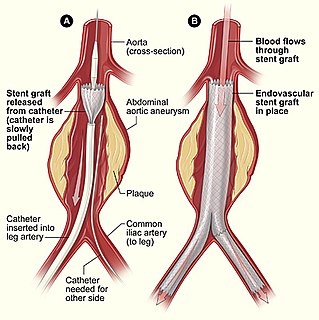
Minimally invasive procedures encompass surgical techniques that limit the size of incisions needed and so lessen wound healing time, associated pain and risk of infection. Surgery by definition is invasive and many operations requiring incisions of some size are referred to as open surgery, in which incisions made can sometimes leave large wounds that are painful and take a long time to heal. Minimally invasive procedures have been enabled by the advance of various medical technologies. An endovascular aneurysm repair as an example of minimally invasive surgery is much less invasive in that it involves much smaller incisions than the corresponding open surgery procedure of open aortic surgery. This minimally invasive surgery became the most common method of repairing abdominal aortic aneurysms in 2003 in the United States.
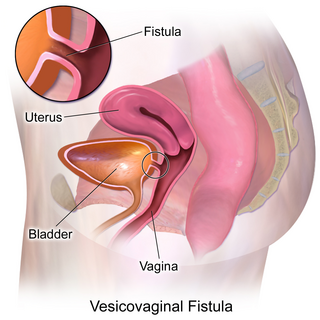
Vesicovaginal fistula (VVF) is a subtype of female urogenital fistula (UGF).

Percutaneous transhepatic cholangiography or percutaneous hepatic cholangiogram is a radiological technique used to visualize the anatomy of the biliary tract. A contrast medium is injected into a bile duct in the liver, after which X-rays are taken. It allows access to the biliary tree in cases where endoscopic retrograde cholangiopancreatography (ERCP) has been unsuccessful. Initially reported in 1937, the procedure became popular in 1952.
Urologic diseases or conditions include urinary tract infections, kidney stones, bladder control problems, and prostate problems, among others. Some urologic conditions don't affect a person for that long and some are lifetime conditions. Kidney diseases are normally investigated and treated by nephrologists, while the specialty of urology deals with problems in the other organs. Gynecologists may deal with problems of incontinence in women.
A puerperal disorder or postpartum disorder is a disorder which presents primarily during the puerperium, or postpartum period. The postpartum period can be divided into three distinct stages; the initial or acute phase, 6–12 hours after childbirth; subacute postpartum period, which lasts 2–6 weeks, and the delayed postpartum period, which can last up to six months. In the subacute postpartum period, 87% to 94% of women report at least one health problem. Long term health problems are reported by 31% of women.
VOXEL-MAN is the name of a set of a computer programs for creation and visualization of three-dimensional digital models of the human body derived from cross-sectional images of Computer Tomography, Magnetic Resonance Tomography or photography. It was developed at the University Medical Center Hamburg-Eppendorf. Applications include diagnostic imaging, digital anatomical atlases and surgery simulators. The 3D interactive atlases of anatomy and radiology for brain/skull and inner organs are available for free download. The name Voxel-Man is derived from the term Voxel, the elementary cuboid component of a digital representation of a three-dimensional object. Occasionally the name Voxel-Man is also used as a general term for a digital representation of the human body.

Cone beam computed tomography is a medical imaging technique consisting of X-ray computed tomography where the X-rays are divergent, forming a cone.
Gynoroentgenology is the abbreviation of gynecological roentgenology. It is the radiologic imaging of the gynecologic parts of the female human body in order to make a radiologic diagnosis of a gynecologic disease. The term gynecologic radiology is related to gynoroentgenology. Gynoroentgenologic imaging can detect and diagnose primary neoplasms, metastasis, therapy-related lesions, congenital lesions, inflammation, miscellaneous diseases, pseudolesions, normal variants, infection uterine arteriovenous malformations and cystic adenomyosis. An example procedure is gynography.

Tubo-ovarian abscesses are one of the late complications of pelvic inflammatory disease (PID) and can be life-threatening if the abscess ruptures and results in sepsis. It consists of an encapsulated or confined 'pocket of pus' with defined boundaries that forms during an infection of a fallopian tube and ovary. These abscesses are found most commonly in reproductive age women and typically result from upper genital tract infection. It is an inflammatory mass involving the fallopian tube, ovary and, occasionally, other adjacent pelvic organs. A TOA can also develop as a complication of a hysterectomy.
Jayaram K Udupa is an Indian-American radiologist and academic. He serves as chief professor of Radiological Science at Perlman School of Medicine at the University of Pennsylvania. Udupa has worked in the fields of medical image science, image processing, and physics analysis of medical imaging and medical diagnostic procedures since the 1980s. He is known for his contributions in image processing and its applications in various fields of science, medicine, and engineering.
A ureterovaginal fistula is an abnormal passageway existing between the ureter and the vagina. It presents as urinary incontinence. Its impact on women is to reduce the "quality of life dramatically."

Ron Kikinis is an American physician and scientist best known for his research in the fields of imaging informatics, image guided surgery, and medical image computing. Dr. Kikinis is the founding Director of the Surgical Planning Laboratory in the Department of Radiology at Brigham and Women's Hospital, in Boston, Massachusetts and is a Professor of Radiology at Harvard Medical School. Since 2014 Dr. Kikinis is also the Head of Fraunhofer MEVIS and Professor of Computer Science at University of Bremen in Germany.
The Mallinckrodt Institute of Radiology (MIR), established 1931, is an academic radiology center associated with the Washington University School of Medicine, located within the Washington University Medical Center in St. Louis, Missouri. In addition to providing diagnostic and therapeutic patient-care services, the institute is a top research and education center. It employs over 140 academic staff and is among the top recipients of National Institutes of Health funding of radiology departments. The center provides radiology services to Barnes-Jewish and St. Louis Children's hospitals, as well as multiple other hospitals and outpatient centers in the St. Louis area. The center performs 700,000 examinations and procedures annually.
A urogenital fistula is an abnormal tract that exist between the vagina and bladder, ureters, or urethra. A urogenital fistula can occur between any of the organs and structures of the pelvic region. A fistula allows urine to continually exit through and out the vagina. This can result in significant disability, interference with sexual activity, and other physical health issues, the effects of which may in turn have a negative impact on mental or emotional state, including an increase in social isolation. Urogenital fistulas vary in etiology. Fistulas are usually caused by injury or surgery, but they can also result from malignancy, infection, prolonged and obstructed labor and deliver in childbirth, hysterectomy, radiation therapy or inflammation. Of the fistulas that develop from difficult childbirth, 97 percent occur in developing countries. Congenital urogenital fistulas are rare; only ten cases have been documented. Abnormal passageways can also exist between the vagina and the organs of the gastrointestinal system, and these may also be termed fistulas.

