Cardiology is a branch of medicine that deals with the disorders of the heart as well as some parts of the circulatory system. The field includes medical diagnosis and treatment of congenital heart defects, coronary artery disease, heart failure, valvular heart disease and electrophysiology. Physicians who specialize in this field of medicine are called cardiologists, a specialty of internal medicine. Pediatric cardiologists are pediatricians who specialize in cardiology. Physicians who specialize in cardiac surgery are called cardiothoracic surgeons or cardiac surgeons, a specialty of general surgery.

Angiography or arteriography is a medical imaging technique used to visualize the inside, or lumen, of blood vessels and organs of the body, with particular interest in the arteries, veins, and the heart chambers. This is traditionally done by injecting a radio-opaque contrast agent into the blood vessel and imaging using X-ray based techniques such as fluoroscopy.

Interventional radiology (IR), is a radiology specialty which provides minimally invasive image-guided diagnosis and treatment of disease. Although the range of procedures performed by interventional radiologists is broad, the unifying concept behind these procedures is the application of image guidance and minimally invasive techniques in order to minimize risk to the patient.

A coronary catheterization is a minimally invasive procedure to access the coronary circulation and blood filled chambers of the heart using a catheter. It is performed for both diagnostic and interventional (treatment) purposes.

Interventional cardiology is a branch of cardiology that deals specifically with the catheter based treatment of structural heart diseases. Andreas Gruentzig is considered the father of interventional cardiology after the development of angioplasty by interventional radiologist Charles Dotter.
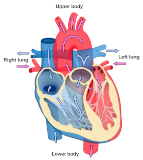
Cyanotic heart defect is a group-type of congenital heart defect (CHD) that occurs due to deoxygenated blood bypassing the lungs and entering the systemic circulation or a mixture of oxygenated and unoxygenated blood entering the systemic circulation. It is caused by structural defects of the heart, or any condition which increases pulmonary vascular resistance. The result being the development of collateral circulation.
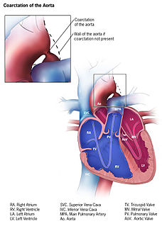
Coarctation of the aorta, also called aortic narrowing, is a congenital condition whereby the aorta is narrow, usually in the area where the ductus arteriosus inserts. The word "coarctation" means narrowing. Coarctations are most common in the aortic arch. The arch may be small in babies with coarctations. Other heart defects may also occur when coarctation is present, typically occurring on the left side of the heart. When a patient has a coarctation, the left ventricle has to work harder. Since the aorta is narrowed, the left ventricle must generate a much higher pressure than normal in order to force enough blood through the aorta to deliver blood to the lower part of the body. If the narrowing is severe enough, the left ventricle may not be strong enough to push blood through the coarctation, thus resulting in lack of blood to the lower half of the body. Physiologically its complete form is manifested as interrupted aortic arch.

Cardiac catheterization is the insertion of a catheter into a chamber or vessel of the heart. This is done both for diagnostic and interventional purposes. A common example of cardiac catheterization is coronary catheterization that involves catheterization of the coronary arteries for coronary artery disease and myocardial infarctions. Catheterization is most often performed in special laboratories with fluoroscopy and highly maneuverable tables. These "cath labs" are often equipped with cabinets of catheters, stents, balloons, etc of various sizes to increase efficiency. Monitors show the fluoroscopy imaging, EKG, pressure waves, and more.

Pulmonary atresia is a congenital malformation of the pulmonary valve in which the valve orifice fails to develop. The valve is completely closed thereby obstructing the outflow of blood from the heart to the lungs. The pulmonary valve is located on the right side of the heart between the right ventricle and pulmonary artery. In a normal functioning heart, the opening to the pulmonary valve has three flaps that open and close
Valvular heart disease is any cardiovascular disease process involving one or more of the four valves of the heart. These conditions occur largely as a consequence of aging, but may also be the result of congenital (inborn) abnormalities or specific disease or physiologic processes including rheumatic heart disease and pregnancy.

Computed tomography angiography is a computed tomography technique used to visualize arterial and venous vessels throughout the body. Using contrast injected into the blood vessels, images are created to look for blockages, aneurysms, dissections, and stenosis. CTA can be used to visualize the vessels of the heart, the aorta and other large blood vessels, the lungs, the kidneys, the head and neck, and the arms and legs.

The smallest cardiac veins are minute valveless veins in the walls of all four heart chambers. The veins are sometimes accurately referred to as vessels, but they are frequently confused with a distinct set of artery connections eponymously referred to as the "vessels of Wearn". In his 1928 publication, Wearn himself referred to the arterio-cameral connections as thebesian. However, Wearn's 1933 and 1941 publications emphatically described the thebesian veins as distinct from arterio-cameral vessels.

Pulmonary insufficiency is a condition in which the pulmonary valve is incompetent and allows backflow from the pulmonary artery to the right ventricle of the heart during diastole. While a small amount of backflow may occur ordinarily, it is usually only shown on an echocardiogram and is harmless. More pronounced regurgitation that is noticed through a routine physical examination is a medical sign of disease and warrants further investigation. If it is secondary to pulmonary hypertension it is referred to as a Graham Steell murmur.

Cardiovascular magnetic resonance imaging (CMR), also known as cardiac MRI, is a medical imaging technology for non-invasive assessment of the function and structure of the cardiovascular system. Conventional MRI sequences are adapted for cardiac imaging by using ECG gating and high temporal resolution protocols. The development of CMR is an active field of research and continues to see a rapid expansion of new and emerging techniques.
The following outline is provided as an overview of and topical guide to cardiology:

Volume overload refers to the state of one of the chambers of the heart in which too large a volume of blood exists within it for it to function efficiently. Ventricular volume overload is approximately equivalent to an excessively high preload. It is a cause of cardiac failure.
Aortography involves placement of a catheter in the aorta and injection of contrast material while taking X-rays of the aorta. The procedure is known as an aortogram. The diagnosis of aortic dissection can be made by visualization of the intimal flap and flow of contrast material in both the true lumen and the false lumen. The catheter has to be inserted through the right femoral artery, because in about two thirds of cases the aortic dissection spreads into the left common iliac artery.

Cardiac ventriculography is a medical imaging test used to determine a person's heart function in the right, or left ventricle. Cardiac ventriculography involves injecting contrast media into the heart's ventricle(s) to measure the volume of blood pumped. Cardiac ventriculography can be performed with a radionuclide in radionuclide ventriculography or with an iodine-based contrast in cardiac chamber catheterization.
Charles E. Mullins is a retired pediatric cardiologist who practiced at Baylor College of Medicine and Texas Children's Hospital. He is known for advancing cardiac catheterization techniques to treat congenital heart defects.


