Zenker's paralysis

Last updated
Peroneal nerve palsy
Slide3i.JPG
Location of peroneal nerve on foot
Classification and external resources

Zenker’s paralysis, also known as peroneal nerve paralysis or palsy, is a paralysis on common fibular nerve that affects patient’s ability to lift the foot at the ankle. The condition was named after Friedrich Albert von Zenker. Peroneal nerve paralysis usually leads to neuromuscular disorder, peroneal nerve injury, or foot drop which can be symptoms of more serious disorders such as nerve compression. The origin of peroneal nerve palsy has been reported to be associated with musculoskeletal injury or isolated nerve traction and compression. Also it has been reported to be mass lesions and metabolic syndromes. Peroneal nerve is most commonly interrupted at the knee and possibly at the joint of hip and ankle. Most studies reported that about 30% of peroneal nerve palsy is followed from knee dislocations. [1]

Friedrich Albert von Zenker German pathologist and physician

Friedrich Albert von Zenker was a German pathologist and physician, celebrated for his discovery of trichinosis.

Traction (orthopedics) set of mechanisms for straightening broken bones or relieving pressure on the spine and skeletal system; types of traction: skin traction and skeletal traction

Traction refers to the set of mechanisms for straightening broken bones or relieving pressure on the spine and skeletal system. There are two types of traction: skin traction and skeletal traction. They are used in orthopedic medicine.

Contents

Peroneal nerve injury occurs when the knee is exposed to various stress. It occurs when the posterolateral corner structure of knee is injured. Relatively tethered location around fibular head, tenuous vascular supply and epineural connective tissues are possible factors that cause damage on the common peroneal nerve. Treatment options for nerve palsy include both operative and non-operative techniques. Initial treatment includes physical therapy and ankle-foot orthosis. Physical therapy mainly focuses on preventing deformation by stretching the posterior ankle capsule. A special brace or splint worn inside the shoe (called an Ankle Foot Orthosis) holds the foot in the best position for walking. Orthosis stretches posterior ankle structures. Physical therapy can help patients to learn how to walk with a foot drop. [2]

Signs and symptoms

Slapping gait makes a slapping noise at the joint of an ankle.

Signs and symptoms of peroneal nerve palsy are related to mostly lower legs and foot which are the followings: [3]

Sensation is an animal's, including humans', detection of external or internal stimulation. It is different from perception, which is about making sense of, or describing, the stimulation.

Gait pattern of movement of the limbs of animals

Gait is the pattern of movement of the limbs of animals, including humans, during locomotion over a solid substrate. Most animals use a variety of gaits, selecting gait based on speed, terrain, the need to maneuver, and energetic efficiency. Different animal species may use different gaits due to differences in anatomy that prevent use of certain gaits, or simply due to evolved innate preferences as a result of habitat differences. While various gaits are given specific names, the complexity of biological systems and interacting with the environment make these distinctions 'fuzzy' at best. Gaits are typically classified according to footfall patterns, but recent studies often prefer definitions based on mechanics. The term typically does not refer to limb-based propulsion through fluid mediums such as water or air, but rather to propulsion across a solid substrate by generating reactive forces against it.

Patients may need pain relievers to control pain. Other medications that are used to reduce pain include gabapentin, carbamazepine, or tricyclic antidepressants such as amitriptyline. Whenever if possible, patients need to avoid or limit the use of medication to reduce the risk of side effects. If the pain is severe, a pain specialist can help patients to explore all options to relieve the pain. Physical therapy exercises may help patients to maintain muscle strength. Also, orthopedic devices may improve patient's ability to walk and prevent contractures. Orthopedic devices may include braces, splints, orthopedic shoes, or other equipment. Vocational counseling, occupational therapy, or similar programs may help patients to maximize their mobility and independence.

Gabapentin chemical compound

Gabapentin, sold under the brand name Neurontin among others, is an anticonvulsant medication used to treat partial seizures, neuropathic pain, hot flashes, and restless legs syndrome. It is recommended as one of a number of first-line medications for the treatment of neuropathic pain caused by diabetic neuropathy, postherpetic neuralgia, and central neuropathic pain. About 15% of those given gabapentin for diabetic neuropathy or postherpetic neuralgia have a measurable benefit. Gabapentin is taken by mouth.

Carbamazepine an antiepileptic drug

Carbamazepine (CBZ), sold under the trade name Tegretol, among others, is a anticonvulsant medication used primarily in the treatment of epilepsy and neuropathic pain. It is not effective for absence or myoclonic seizures. It is used in schizophrenia along with other medications and as a second-line agent in bipolar disorder. Carbamazepine appears to work as well as phenytoin and valproate.

Tricyclic Chemical compounds

Tricyclics are chemical compounds that contain three interconnected rings of atoms.

Causes

Causing factors of peroneal nerve palsy are such as musculoskeletal or peroneal nerve injuries. Usually paralysis occurs at the outside of the leg and the top of the foot. Palsy causes decrease of muscle strength to lift the foot, twist ankle outside, and move toes around. Major cause of palsy is due to dislocation of knee. Other possible causing factors are metabolic dysfunction of lower part of knee or disorientation of hip or pelvis. Damages on peroneal nerves destroy the myelin sheath that covers the axon or the whole nerve cell. There might be a loss of feeling, muscle control, muscle tone, and eventual loss of muscle mass because the nerves aren't stimulating the muscles after they are damaged. Dysfunction of a single nerve such as the common peroneal nerve is called a mononeuropathy. Mononeuropathy means the nerve damage is occurred in one area. However, certain conditions may also cause single nerve injuries. [4]

Axon The long process of a neuron that conducts nerve impulses, usually away from the cell body to the terminals and varicosities, which are sites of storage and release of neurotransmitter.

An axon, or nerve fiber, is a long, slender projection of a nerve cell, or neuron, in vertebrates, that typically conducts electrical impulses known as action potentials away from the nerve cell body. The function of the axon is to transmit information to different neurons, muscles, and glands. In certain sensory neurons, such as those for touch and warmth, the axons are called afferent nerve fibers and the electrical impulse travels along these from the periphery to the cell body, and from the cell body to the spinal cord along another branch of the same axon. Axon dysfunction has caused many inherited and acquired neurological disorders which can affect both the peripheral and central neurons. Nerve fibers are classed into three types – group A nerve fibers, group B nerve fibers, and group C nerve fibers. Groups A and B are myelinated, and group C are unmyelinated. These groups include both sensory fibers and motor fibers. Another classification groups only the sensory fibers as Type I, Type II, Type III, and Type IV.

Common causes of damage to the peroneal nerve include the following:

Common peroneal nerve injury is more common in people:

Prolonged pressure on nerve may occur because of:

Diagnosis

For partial nerve palsy, more than 80% of patients will recover completely. For complete nerve palsy, less than 40% of patients will have complete recovery. Peroneal nerve in continuity arises from defined cause will be recovered better than those arise from unknown causes. [7]

Examinations are required for following reasons:

Electromyography

Electromyography is used to observe peroneal nerve palsy within one month of injuries. And if it is partial peroneal nerve palsy, patients have higher chance to recover fully from the palsy. More than 70 to 80 percent of patients with partial paralysis recovered completely, but those with complete paralysis have chances less than 30 percent to recover completely. If the symptom does not get any better in few months, surgery is required to decompress the nerve compression. [8]

Nerve conduction velocity

Nerve conduction velocity is an important aspect of nerve conduction studies. It is the speed at which an electrochemical impulse propagates down a neural pathway. Conduction velocities are affected by a wide array of factors, including age, sex, and various medical conditions. Studies allow for better diagnoses of various neuropathies, especially demyelinating conditions as these conditions result in reduced or non-existent conduction velocities. To perform nerve conduction velocity, surface electrodes are placed onto the skin over nerves at various locations. Each patch sends electrical impulses which stimulate the nerve. Resulting electrical activity of nerve is recorded by the other electrodes. The distance between electrodes and the time it takes for electrical impulses to travel between electrodes are used to determine the velocity of the nerve signals. [9]

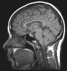
Image of brain MRI. MRI brain.jpg
Image of brain MRI.

MRI

An MRI (magnetic resonance imaging) scan is an imaging test that uses powerful magnets and radio waves to create pictures of the body. It does not use radiation. Single MRI images are called slices. The images can be stored on a computer or printed on film. One exam produces dozens or sometimes hundreds of images. To locate nerve palsy, MRI is used by physicians to detect the position and location of damaged peroneal nerve. [10]

Prevention

Avoid putting long-term pressure on the back or side of the knee. Treat injuries to the leg or knee right away. If a cast, splint, dressing, or other pressure on the lower leg causes a tight feeling or numbness, call the health care provider. [11]

Treatments

Precise knowledge about the length and exact localization of a damaged nerve segment is essential for surgical intervention. On one hand, certain preoperative information about the overall state of an injured nerve (state of the neural and perineural tissue) is important because exploratory inspection of a nerve itself may lead to additional inadvertent damage. If, on the other hand, the surgeon, during genicular ligament reconstruction, inspects a nerve at the site of most probable injury only (limited neurolysis), he or she may sometimes by chance expose an unaffected section of a nerve. Because of the mechanism of nerve injury during traction, however, a more proximal or distal segment of the nerve may be severely damaged. A limited nerve inspection without preoperative knowledge about the site of nerve injury may thus give the false impression of an unimpaired nerve and wrongly lead to conservative treatment of the nerve lesion. If no neurologic improvement is shown after 2–3 months from injuries, then operative decompression is indicated. Surgical operations such as grafting and tendon transfer are necessarily required. [12]

Tendon transfer

Many different conditions can be treated by tendon transfer surgery. Tendon transfer surgery is necessary when a certain muscle function is lost because of a nerve injury. If a nerve is injured and cannot be repaired, then the nerve no longer sends signals to certain muscles. Those muscles are paralyzed and their muscle function is lost. Tendon transfer surgery can be used to attempt to replace that function. Common nerve injuries that are treated with tendon transfer surgery are spinal cord, radial nerve, ulnar nerve, or median nerve injury. Tendon transfers have higher chance to treat nerve palsy, and such transfers include posterior, anterior, and anteroposterior tibial tendon transfer. Peroneal nerve and its nerve branches need to be fixed from adherence to proximal fibula, which proximal fibula is about 3~5 cm. [13]

Tendon graft

Grafting is a surgical procedure to move tissue from one site to another on the body, or from another person, without bringing its own blood supply with it. Instead, a new blood supply grows in after it is placed. A similar technique where tissue is transferred with the blood supply intact is called a flap. In some instances a grafting can be an artificially manufactured device. Examples of this are a tube to carry blood flow across a defect or from an artery to a vein for use in hemodialysis. [14]

Arthroplasty

Arthroplasty on knee has been broadly used to treat knee and musculoskeletal joint dislocation. It is an elective procedure that is done to relieve pain and restore function to the joint after damage by arthritis or some other type of trauma. However, there has been series of reports arthroplasty worsens condition of peroneal nerve, causing paralysis. Other forms of arthroplasty include resection (al) arthroplasty, resurfacing arthroplasty, mold arthroplasty, cup arthroplasty, silicone replacement arthroplasty, etc. [15]

History

Friedrich Albert von Zenker FAZenker.png
Friedrich Albert von Zenker

Friedrich Albert von Zenker (1825–1898) was a German pathologist and physician, celebrated for his discovery of trichinosis. He was born in Dresden, and was educated in Leipzig and Heidelberg. Attached to the city hospital of Dresden in 1851, he added, in 1855, the duties of professor of pathological anatomy and general pathology in the surgico-medical academy of that city. In 1862 he became professor of pathological anatomy and pharmacology at Erlangen. Three years afterwards he assumed with Ziemssen the editorship of the Deutsches Archiv für klinische Medizin. In 1895 he retired from active service. His important discovery of the danger of trichine dates from 1860. In that year he published "Ueber die Trichinenkrankheit des Menschen" (in volume xviii of Virchow's Archiv). Beside, Zenker also found Zenker's degeneration and Zenker's diverticulum. [16]

See also

Related Research Articles

Human leg lower extremity or limb of the human body (foot, lower leg, thigh and hip)

The human leg, in the general meaning, is the entire lower limb of the human body, including the foot, thigh and even the hip or gluteal region. However, the definition in human anatomy refers only to the section of the lower limb extending from the knee to the ankle, also known as the crus. Legs are used for standing, and all forms of locomotion including recreational such as dancing, and constitute a significant portion of a person's mass. Female legs generally have greater hip anteversion and tibiofemoral angles, but shorter femur and tibial lengths than those in males.

Sciatic nerve large nerve in humans and other animals

The sciatic nerve is a large nerve in humans and other animals. It begins in the lower back and runs through the buttock and down the lower limb. It is the longest and widest single nerve in the human body, going from the top of the leg to the foot on the posterior aspect. The sciatic nerve provides the connection to the nervous system for nearly the whole of the skin of the leg, the muscles of the back of the thigh, and those of the leg and foot. It is derived from spinal nerves L4 to S3. It contains fibers from both the anterior and posterior divisions of the lumbosacral plexus.

Peroneus longus

In human anatomy, the peroneus longus is a superficial muscle in the lateral compartment of the leg, and acts to evert and plantarflex the ankle.
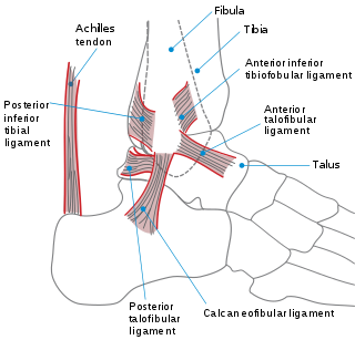
Ankle region where the foot and the leg meet

The ankle, or the talocrural region, is the region where the foot and the leg meet. The ankle includes three joints: the ankle joint proper or talocrural joint, the subtalar joint, and the inferior tibiofibular joint. The movements produced at this joint are dorsiflexion and plantarflexion of the foot. In common usage, the term ankle refers exclusively to the ankle region. In medical terminology, "ankle" can refer broadly to the region or specifically to the talocrural joint.

Extensor hallucis longus muscle

The extensor hallucis longus is a thin muscle, situated between the tibialis anterior and the extensor digitorum longus, that functions to extend the big toe and dorsiflects the foot, and assists with foot eversion and inversion.

Popliteal artery

The popliteal artery is a deeply placed continuation of the femoral artery after it passes through the adductor hiatus, or opening in the distal portion of the adductor magnus muscle. It courses through the popliteal fossa and ends at the lower border of the popliteus muscle, where it branches into the anterior and posterior tibial arteries.

A soft tissue injury (STI) is the damage of muscles, ligaments and tendons throughout the body. Common soft tissue injuries usually occur from a sprain, strain, a one off blow resulting in a contusion or overuse of a particular part of the body. Soft tissue injuries can result in pain, swelling, bruising and loss of function.

Tibialis anterior muscle

The tibialis anterior is a muscle in humans that originates in the upper two-thirds of the lateral (outside) surface of the tibia and inserts into the medial cuneiform and first metatarsal bones of the foot. It acts to dorsiflex and invert the foot. This muscle is mostly located near the shin.

Tarsal tunnel syndrome compression neuropathy and painful foot condition in which the tibial nerve is compressed as it travels through the tarsal tunnel

Tarsal tunnel syndrome (TTS), also known as posterior tibial neuralgia, is a compression neuropathy and painful foot condition in which the tibial nerve is compressed as it travels through the tarsal tunnel. This tunnel is found along the inner leg behind the medial malleolus. The posterior tibial artery, tibial nerve, and tendons of the tibialis posterior, flexor digitorum longus, and flexor hallucis longus muscles travel in a bundle through the tarsal tunnel. Inside the tunnel, the nerve splits into three segments. One nerve (calcaneal) continues to the heel, the other two continue on to the bottom of the foot. The tarsal tunnel is delineated by bone on the inside and the flexor retinaculum on the outside.

Joint replacement orthopedic surgery to replace a joint

Replacement arthroplasty, or joint replacement surgery, is a procedure of orthopedic surgery in which an arthritic or dysfunctional joint surface is replaced with an orthopedic prosthesis. Joint replacement is considered as a treatment when severe joint pain or dysfunction is not alleviated by less-invasive therapies. It is a form of arthroplasty, and is often indicated from various joint diseases, including osteoarthritis and rheumatoid arthritis.

Flexor hallucis longus muscle

The flexor hallucis longus muscle (FHL) is one of the three deep muscles of the posterior compartment of the leg that attaches to the plantar surface of the distal phalanx of the great toe. The other deep muscles are the flexor digitorum longus and tibialis posterior; the tibialis posterior is the most powerful of these deep muscles. All three muscles are innervated by the tibial nerve which comprises half of the sciatic nerve.

Plantaris muscle muscle

The plantaris is one of the superficial muscles of the superficial posterior compartment of the leg, one of the fascial compartments of the leg.

Peroneus brevis

The peroneus brevis muscle lies under cover of the peroneus longus, and is the shorter and smaller of the peroneus muscles.

The common fibular nerve is a nerve in the lower leg that provides sensation over the posterolateral part of the leg and the knee joint. It divides at the knee into two terminal branches: the superficial fibular nerve and deep fibular nerve, which innervate the muscles of the lateral and anterior compartments of the leg respectively. When the common fibular nerve is damaged or compressed, foot drop can be the end result.

Deep peroneal nerve

The deep peroneal nerve begins at the bifurcation of the common peroneal nerve between the fibula and upper part of the peroneus longus, passes infero-medially, deep to extensor digitorum longus, to the anterior surface of the interosseous membrane, and comes into relation with the anterior tibial artery above the middle of the leg; it then descends with the artery to the front of the ankle-joint, where it divides into a lateral and a medial terminal branch.

Foot drop gait abnormality

Foot drop is a gait abnormality in which the dropping of the forefoot happens due to weakness, irritation or damage to the common fibular nerve including the sciatic nerve, or paralysis of the muscles in the anterior portion of the lower leg. It is usually a symptom of a greater problem, not a disease in itself. Foot drop is characterized by inability or impaired ability to raise the toes or raise the foot from the ankle (dorsiflexion). Foot drop may be temporary or permanent, depending on the extent of muscle weakness or paralysis and it can occur in one or both feet. In walking, the raised leg is slightly bent at the knee to prevent the foot from dragging along the ground.

The sural nerve is a sensory nerve in the calf region (sura) of the leg. It is made up of branches of the tibial nerve and common fibular nerve, the medial cutaneous branch from the tibial nerve, and the lateral cutaneous branch from the common fibular nerve. Once formed, the nerves runs down the mid calf to the ankle and along the skin from the mid-posterior popliteal fossa to just behind to the lateral malleolus and then under the malleolus and forward along the lateral aspect of the foot.

Fibular artery

In anatomy, the fibular artery supplies blood to the lateral compartment of the leg. It arises from the tibial-fibular trunk.

Tendon transfer

A tendon transfer is a surgical process in which the insertion of a tendon is moved, but the origin remains in the same location. Tendon transfer involves redistribution of muscle power, not recreation. Tendons are transferred at the distal attachment from lesser to more important functions so that the overall function is improved. Tendon transfers provide a substitute which can be permanent or temporary, when muscle function is lost either due to nerve injuries or injuries to the muscle/tendon unit. Tendon transfers are also performed to correct the imbalanced muscle tone due to spasticity resulting from injuries to the central nervous system.

A sciatic nerve block is a nerve block that uses local anesthetic to achieve analgesia in the leg. The block works by affecting the sciatic nerve and is used for surgeries at or below the knee.

References

  1. Levy, Bruce A.; Giuseffi, Steven A.; Bishop, Allen T.; Shin, Alexander Y.; Dahm, Diane L.; Stuart, Michael J. (2010). "Surgical treatment of peroneal nerve palsy after knee dislocation". Knee Surgery, Sports Traumatology, Arthroscopy. 18 (11): 1583–6. doi:10.1007/s00167-010-1204-3. PMID   20640404.
  2. Werner, B. C.; F. W. Gwathmey; M. L. Lyons; M. D. Miller (2013). "Peroneal Nerve Injury after Multiligament Knee Injury: a 12 Year Experience with a Focus on Outcomes after Posterior Tibial Tendon Transfer". Orthopaedic Journal of Sports Medicine. 1 (4 Suppl): 2325967113S0008. doi:10.1177/2325967113S00080. ISSN   2325-9671.
  3. Eleftheriou, Kyriacos I.; Sushil Beri; Afshin Alavi; Sally Tennant (2013). "Deep peroneal nerve palsy during growth spurt: a case report". European Journal of Pediatrics. 173 (12): 1603. doi:10.1007/s00431-013-2160-y. ISSN   0340-6199. PMID   24061281.
  4. Bendszus, M; Reiners, K; Perez, J; Solymosi, L; Koltzenburg, M (2002). "Peroneal nerve palsy caused by thrombosis of crural veins". Neurology. 58 (11): 1675–7. doi:10.1212/WNL.58.11.1675. PMID   12058098.
  5. Krych, A. J.; S. Giuseffi; S. A. Kuzma; J. L. Hudgens; M. J. Stuart; B. A. Levy (2013). "Clinical and Functional Outcomes after Multiligament Knee Injury with Associated Peroneal Nerve Palsy: Comparison with a Matched Control Group at 2-18 Years". Orthopaedic Journal of Sports Medicine. 1 (4 Suppl): 2325967113S0008. doi:10.1177/2325967113S00082. ISSN   2325-9671.
  6. http://health.nytimes.com/health/guides/disease/common-peroneal-nerve-dysfunction/overview.html%5Bfull+citation+needed%5D
  7. http://www.wheelessonline.com/ortho/12636%5Bfull+citation+needed%5D
  8. Ward, Joseph P.; Lynda J.-S. Yang, Andrew G. Urquhart (2013). "Surgical Decompression Improves Symptoms of Late Peroneal Nerve Dysfunction After TKA". Orthopedics. 36 (4): e515–e519. doi:10.3928/01477447-20130327-33. ISSN   0147-7447.
  9. Garg, Ruchika (2012). "Footdrop in the Farmers in Punjab: A Retrospective Electrodiagnostic Study". Journal of Clinical and Diagnostic Research. doi:10.7860/JCDR/2012/4829.2648. ISSN   2249-782X.
  10. http://www.hopkinsmedicine.org/neurology_neurosurgery/specialty_areas/peripheral_nerve_surgery/conditions/foot_drop_injury.html%5Bfull+citation+needed%5D
  11. Jahangiri, F; R Wimberly; S Mcclure; L Blakemore (2006). "P27.3 Multimodality neurophysiological monitoring during tibial/fibular osteotomies for preventing peripheral nerve injuries". Clinical Neurophysiology. 117: 114–115. doi:10.1016/j.clinph.2006.06.457. ISSN   1388-2457.
  12. Mont, MA; Dellon, AL; Chen, F; Hungerford, MW; Krackow, KA; Hungerford, DS (1996). "The operative treatment of peroneal nerve palsy". The Journal of Bone and Joint Surgery. American Volume. 78 (6): 863–9. PMID   8666604.
  13. Titolo, Paolo; Bernardino Panero; Davide Ciclamini; Bruno Battiston; Pierluigi Tos (2013). "Letter to the Editor: New Tendon Transfer for Correction of Drop-foot in Common Peroneal Nerve Palsy". Clinical Orthopaedics and Related Research. 471 (10): 3382–3382. doi:10.1007/s11999-013-3175-4. ISSN   0009-921X. PMC   2384039 Lock-green.svg.
  14. Wagenaar, Frank-Christiaan B.M.; Louwerens, Jan Willem K. (2007). "Posterior Tibial Tendon Transfer: Results of Fixation to the Dorsiflexors Proximal to the Ankle Joint". Foot & Ankle International. 28 (11): 1128. doi:10.3113/FAI.2007.1128.
  15. Deshmukh, Ajit J.; Bozena Kuczynski; Giles R. Scuderi (2013). "Delayed peroneal nerve palsy after total knee arthroplasty—A rare complication of tibial osteolysis". The Knee. 21 (2): 624. doi:10.1016/j.knee.2013.10.015. ISSN   0968-0160. PMID   24262809.
  16. "Whonamedit - Friedrich Albert von Zenker". whonamedit.com. Retrieved 2014-01-25.