Baylisascaris procyonis

Last updated

Contents

Baylisascaris procyonis
Baylisascaris larvae.jpg
Freshly hatched B. procyonis larvae
Scientific classification OOjs UI icon edit-ltr.svg
Domain: Eukaryota
Kingdom: Animalia
Phylum: Nematoda
Class: Chromadorea
Order: Ascaridida
Family: Ascarididae
Genus: Baylisascaris
Species:
B. procyonis
Binomial name
Baylisascaris procyonis
(Stefanski & Zarnowski, 1951)
Synonyms [1]
  • Ascaris procyonisStefanski & Zarnowski, 1951

Baylisascaris procyonis, also known by the common name raccoon roundworm, is a roundworm nematode, found ubiquitously in raccoons, the definitive hosts. It is named after H. A. Baylis, who studied them in the 1920s–30s, and Greek askaris (intestinal worm). [2] Baylisascaris larvae in paratenic hosts can migrate, causing larva migrans. Baylisascariasis as the zoonotic infection of humans is rare, though extremely dangerous due to the ability of the parasite's larvae to migrate into brain tissue and cause damage. Concern for human infection has been increasing over the years due to the urbanization of rural areas, resulting in the increase in proximity and potential human interaction with raccoons. [3]

Signs and symptoms

The potential for human infection was noted in 1969 by Paul C. Beaver, who studied infected mice, and the first case was reported 15 years later. [3] Human infection with B. procyonis has been relatively rare, with about 30 cases reported since 1980. However, disease caused by this parasite can be extremely dangerous, causing death or severe symptoms. Reported disease has primarily afflicted children and almost all cases were a result of the ingestion of contaminated soil or feces. [4] Even with treatment, prognosis is poor and patients in nearly all cases experience permanent neurological damage. Infection of the eyes may result in permanent visual damage and loss. The common antihelmintic medicines are able to treat adult worms living in the intestines, but are less effective against migrating larvae. [4] Aggressive and early treatment with corticosteroids and albendazole have in a few instances resulted in complete recoveries. [5] It is possible that human infection is more common than diagnosed and most cases do not reach a clinical stage. [6] [7] Antibodies to B. procyonis have been found in otherwise healthy individuals, suggesting subclinical infections. [6] [8]

Cause

Transmission

In North America, B. procyonis infection rates in raccoons are very high, being found in around 70% of adult raccoons and 90% of juvenile raccoons. [7] Transmission occurs similarly to other roundworm species, through the fecal-oral route. Eggs are produced by the worm while in the intestine, and the released eggs will mature to an infective state externally in the soil. When an infected egg is ingested, the larvae will hatch and enter the intestine. Transmission of B. procyonis may also occur through the ingestion of larvae found in infected tissue. [7]

Life cycle

The life cycle of B. procyonis. B.procyonis life cycle.jpg
The life cycle of B. procyonis.

An adult worm lives and reproduces in the intestine of its definitive host, the raccoon. The female worm can produce between 115,000–179,000 eggs per day. Eggs are excreted along with feces, and become infective in the soil after 2–4 weeks. If ingested by another raccoon, the life cycle repeats. However, if these eggs are ingested by an intermediate host (small mammals, birds) the larvae of B. procyonis will penetrate the gut wall of the host and migrate into tissues. Larvae tend to migrate to the brain, cause damage, and affect the behaviour of the intermediate host, making it an easier prey for raccoons. Reproduction does not occur in these intermediate hosts; however, if a raccoon preys on an infected paratenic host, the encysted larvae can become adults in the raccoon and the cycle resumes. [3]

Diagnosis

Laboratory and clinical diagnosis can be challenging: there is no commercially available serologic test in the United States, and although identification of larvae in tissue or specimens is confirmatory, this is not always possible or practical. [5]

Diagnosis of B. procyonis is through identification of larvae in tissue examination. Diagnosis requires forehand knowledge along with understanding and recognition of larval morphologic characteristics, including ability to distinguish between a number of possible other parasites, including Toxocara canis , Toxocara cati , Ascaris lumbricoides , and among species of Gnathostoma , Angiostrongylus , and Ancylostoma . [7] Distinguishing features of B. procyonis larvae in tissue are its relatively large size (60 μ) and prominent single lateral alae. Sometimes serologic testing is used as supportive evidence, although no commercial serologic test is currently available. Other diagnosis methods include: brain biopsy, neuroimaging, electroencephalography, differential diagnoses among other laboratory tests. [4]

Human Baylisascariasis is under-recognized, as the knowledge of the clinical illness is still a bit unclear. This could be because of the difficulty of diagnosing the illness. As small numbers of larvae can cause severe disease, and larvae occur randomly in tissue, a biopsy usually fails to include larvae and therefore leads to negative results. The identification of the morphologic characteristics takes practice and experience and may not be accurately recognized or could be misidentified. The fact that no commercial serologic test exists for the diagnosis of B. procyonis infection makes the diagnosis and treatment more difficult. [7]

Prevention

Educating the public about the dangers of contact with raccoons or their feces is the most important preventive step. [4]
Parents should encourage their children to practice good hygiene; Hand-washing after outdoor play or contact with animals is very important. Fences can be used to prevent raccoons from visiting homes, garbage, or yards for food. Keeping raccoons as pets is strongly discouraged. Raccoon latrines in and around homes should be checked for and cleaned as soon as possible. Boiling water, steam-cleaning, flaming, or fire are highly effective and are easily accessible means to decontaminate household things or areas. Materials contaminated by B.procyonis should be incinerated. Contaminated areas can be cleaned with a xylene-ethanol mixture. Common chemical disinfectants are not effective against B.procyonis eggs. Disinfectants like 20% bleach (1% sodium hypochlorite) wash away the eggs but do not kill them. Since treatment is not very effective, the best way to escape this parasite is to practice the prevention methods. [4]

Epidemiology

Baylisascaris procyonis is found abundantly in its definitive host, the raccoon. The parasite has been found to have the ability to infect more than 90 kinds of wild and domestic animals. [7] Many of these animals act as paratenic hosts and the infection results in the penetration of the gut wall by the larvae and subsequent invasion of tissue, resulting in severe disease. In animals, it is the most common cause of larva migrans. [4] The paratenic host, however, cannot shed infective eggs, as the larva will not complete its life cycle until it makes its way into a raccoon. Raccoons are solitary but will frequently defecate in communal areas known as raccoon latrines. These latrines are an abundant source of B. procyonis eggs, which can remain viable for years. [4] Raccoons therefore are important in maintaining the parasite, providing a source of infection for humans and other animals. [4]

The white-footed mouse (Peromyscus leucopus) among other small rodents are considered common intermediate hosts. [9] Migration patterns of rodents can explain the spread of Baylisascaris to multiple locations and the subsequent infection of humans who may come into contact with eggs shed by infected raccoons. The mice may be infected as a result of contact with raccoon latrines. Foraging upon food contaminated with traces of raccoon feces can also lead to exposure to B. procyonis eggs. Rodents are easily found in many areas with human population which increases the risk of transmission. [10] The increase in the raccoon population (and inevitably B. procyonis) has become a major factor for the number of extirpations the Allegheny woodrat (Neotoma magister) has suffered in recent decades. [11] [12]

Bioterrorism threat

Baylisascaris procyonis has become a concern for its potential use as an agent of bioterrorism. The fact that this parasite's eggs are easy to acquire, able to live for years, extremely resistant to many disinfectants, and cause serious infections in humans with poor treatment options could make it a dangerous weapon. [4] Community water supplies are easily susceptible to contamination due to the lack of filtration and treatment methods to get rid of the eggs. [13]

See also

Related Research Articles

<i>Ascaris lumbricoides</i> One of several species of Ascaris

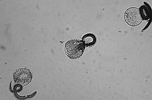
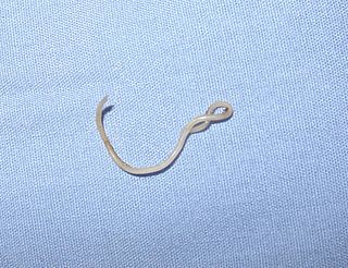
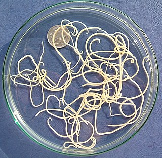
Ascaris lumbricoides is a large parasitic worm that causes ascariasis in humans. A roundworm of genus Ascaris, it is the most common parasitic worm in humans. An estimated 807 million–1.2 billion people are infected with A. lumbricoides worldwide. People living in tropical and subtropical countries are at greater risk of infection.

<span class="mw-page-title-main">Trichuriasis</span> Infection by Trichuris trichiura (whipworm)

Trichuriasis, also known as whipworm infection, is an infection by the parasitic worm Trichuris trichiura (whipworm). If infection is only with a few worms, there are often no symptoms. In those who are infected with many worms, there may be abdominal pain, fatigue and diarrhea. The diarrhea sometimes contains blood. Infections in children may cause poor intellectual and physical development. Low red blood cell levels may occur due to loss of blood.

<span class="mw-page-title-main">Ascariasis</span> Disease caused by the parasitic roundworm Ascaris lumbricoides

Ascariasis is a disease caused by the parasitic roundworm Ascaris lumbricoides. Infections have no symptoms in more than 85% of cases, especially if the number of worms is small. Symptoms increase with the number of worms present and may include shortness of breath and fever in the beginning of the disease. These may be followed by symptoms of abdominal swelling, abdominal pain, and diarrhea. Children are most commonly affected, and in this age group the infection may also cause poor weight gain, malnutrition, and learning problems.

Toxocariasis is an illness of humans caused by the dog roundworm and, less frequently, the cat roundworm. These are the most common intestinal roundworms of dogs, coyotes, wolves and foxes and domestic cats, respectively. Humans are among the many "accidental" or paratenic hosts of these roundworms.

Gnathostomiasis, also known as larva migrans profundus, is the human infection caused by the nematode Gnathostoma spinigerum and/or Gnathostoma hispidum, which infects vertebrates.

<i>Baylisascaris</i> Genus of worms

Baylisascaris is a genus of roundworms that infect more than fifty animal species.

Spirometra is a genus of pseudophyllid cestodes that reproduce in canines and felines, but can also cause pathology in humans if infected. As an adult, this tapeworm lives in the small intestine of its definitive host and produces eggs that pass with the animal's feces. When the eggs reach water, the eggs hatch into coracidia which are eaten by copepods. The copepods are eaten by a second intermediate host to continue the life cycle. Humans can become infected if they accidentally eat frog legs or fish with the plerocercoid stage encysted in the muscle. In humans, an infection of Spirometra is termed sparganosis.

<i>Echinococcus multilocularis</i> Species of flatworm

Echinococcus multilocularis, the fox tapeworm, is a small cyclophyllid tapeworm found extensively in the northern hemisphere. E. multilocularis, along with other members of the Echinococcus genus, produce diseases known as echinococcosis. Unlike E. granulosus,E. multilocularis produces many small cysts that spread throughout the internal organs of the infected animal. The resultant disease is called Alveolar echinococcosis, and is caused by ingesting the eggs of E. multilocularis.

<i>Ascaris suum</i> Species of roundworm

Ascaris suum, also known as the large roundworm of pig, is a parasitic nematode that causes ascariasis in pigs. While roundworms in pigs and humans are today considered as two species with different hosts, cross-infection between humans and pigs is possible; some researchers have thus argued they are the same species. Ascariasis is associated with contact to pigs and pig manure in Denmark.

<span class="mw-page-title-main">Paragonimiasis</span> Medical condition

Paragonimiasis is a food-borne parasitic disease caused by several species of lung flukes belonging to genus Paragonimus. Infection is acquired by eating crustaceans such as crabs and crayfishes which host the infective forms called metacercariae, or by eating raw or undercooked meat of mammals harboring the metacercariae from crustaceans.

Spirometra erinaceieuropaei is a parasitic tapeworm that infects domestic animals and humans. The medical term for this infection in humans and other animals is sparganosis. Morphologically, these worms are similar to other worms in the genus Spirometra. They have a long body consisting of three sections: the scolex, the neck, and the strobilia. They have a complex life cycle that consists of three hosts, and can live in varying environments and bodily tissues. Humans can contract this parasite in three main ways. Historically, humans are considered a paratenic host; however, the first case of an adult S. erinaceieuropaei infection in humans was reported in 2017. Spirometra tapeworms exist worldwide and infection is common in animals, but S. erinaceieuropaei infections are rare in humans. Treatment for infection typically includes surgical removal and anti-worm medication.

<i>Toxocara canis</i> Species of roundworm

Toxocara canis is a worldwide-distributed helminth parasite that primarily infects dogs and other canids, but can also infect other animals including humans. The name is derived from the Greek word "toxon," meaning bow or quiver, and the Latin word "caro," meaning flesh. T. canis live in the small intestine of the definitive host. This parasite is very common in puppies and somewhat less common in adult dogs. In adult dogs, infection is usually asymptomatic but may be characterized by diarrhea. By contrast, untreated infection with Toxocara canis can be fatal in puppies, causing diarrhea, vomiting, pneumonia, enlarged abdomen, flatulence, poor growth rate, and other complications.

Angiostrongyliasis is an infection by a roundworm of the Angiostrongylus type. Symptoms may vary from none, to mild, to meningitis.

<i>Toxascaris leonina</i> Species of roundworm

Toxascaris leonina is a common parasitic roundworm found in dogs, cats, foxes, and related host species. T. leonina is an ascarid nematode, a worldwide distributed helminth parasite which is in a division of eukaryotic parasites that, unlike external parasites such as lice and fleas, live inside their host. The definitive hosts of T. leonina include canids and felines (cats), while the intermediate hosts are usually rodents, such as mice or rats. Infection occurs in the definitive host when the animal eats an infected rodent. While T. leonina can occur in either dogs or cats, it is far more frequent in cats.

Gnathostoma hispidum is a nematode (roundworm) that infects many vertebrate animals including humans. Infection of Gnathostoma hispidum, like many species of Gnathostoma causes the disease gnathostomiasis due to the migration of immature worms in the tissues.

<i>Alaria</i> (flatworm) Genus of flukes

Alaria is a genus of flatworms, or trematodes, in the family Diplostomidae.

Eustrongylidosis is a parasitic disease that mainly affects wading birds worldwide; however, the parasite's complex, indirect lifecycle involves other species, such as aquatic worms and fish. Moreover, this disease is zoonotic, which means the parasite can transmit disease from animals to humans. Eustrongylidosis is named after the causative agent Eustrongylides, and typically occurs in eutrophicated waters where concentrations of nutrients and minerals are high enough to provide ideal conditions for the parasite to thrive and persist. Because eutrophication has become a common issue due to agricultural runoff and urban development, cases of eustrongylidosis are becoming prevalent and hard to control. Eustrongylidosis can be diagnosed before or after death by observing behavior and clinical signs, and performing fecal flotations and necropsies. Methods to control it include preventing eutrophication and providing hosts with uninfected food sources in aquaculture farms. Parasites are known to be indicators of environmental health and stability, so should be studied further to better understand the parasite's lifecycle and how it affects predator-prey interactions and improve conservation efforts.

<i>Anisakis simplex</i> Species of parasitic nematode

Anisakis simplex, known as the herring worm, is a species of nematode in the genus Anisakis. Like other nematodes, it infects and settles in the organs of marine animals, such as salmon, mackerels and squids. It is commonly found in cold marine waters, such as the Pacific Ocean and Atlantic Ocean.

<span class="mw-page-title-main">Cat worm infections</span> Worm infections in cats

Cat worm infections, the infection of cats (Felidae) with parasitic worms, occur frequently. Most worm species occur worldwide in both domestic and other cats, but there are regional, species and lifestyle differences in the frequency of infestation. According to the classification of the corresponding parasites in the zoological system, infections can be divided into those caused by nematode and flatworms - in the case of the latter, mainly cestoda and trematoda - while other strains are of no veterinary significance. While threadworms usually do not require an intermediate host for their reproduction, the development cycle of flatworms always proceeds via alternate hosts.

<span class="mw-page-title-main">Nematode infection in dogs</span> Threadworm infections of dogs are frequent

Nematode infection in dogs - the infection of dogs with parasitic nemamotodes - are, along with tapeworm infections and infections with protozoa, frequent parasitoses in veterinary practice. Nematodes, as so-called endoparasites, colonize various internal organs - most of them the digestive tract - and the skin. To date, about 30 different species of nematode have been identified in domestic dogs; they are essentially also found in wild dog species. However, the majority of them often cause no or only minor symptoms of disease in adult animals. The infection therefore does not necessarily have to manifest itself in a worm disease (helminthosis). For most nematodes, an infection can be detected by examining the feces for eggs or larvae. Roundworm infection in dogs and the hookworm in dogs is of particular health significance in Central Europe, as they can also be transmitted to humans (zoonosis). Regular deworming can significantly reduce the frequency of infection and thus the risk of infection for humans and dogs.

References

  1. "Baylisascaris procyonis (Stefanski & Zarnowski, 1951)". Global Biodiversity Information Facility . Retrieved 24 February 2024.
  2. Carol Snarey (Nov 2010). "Etymologia: Baylisascaris". Emerg Infect Dis. 16 (11): 1819. doi:10.3201/eid1611.ET1611. PMC   3294543 .
  3. 1 2 3 Drisdelle R (2010). Parasites. Tales of Humanity's Most Unwelcome Guests. Univ. of California Publishers, 2010. p. 189f. ISBN   978-0-520-25938-6.
  4. 1 2 3 4 5 6 7 8 9 Gavin, P. J.; Kazacos, K. R.; Shulman, S. T. (2005). "Baylisascariasis". Clinical Microbiology Reviews. 18 (4): 703–18. doi:10.1128/CMR.18.4.703-718.2005. PMC   1265913 . PMID   16223954.
  5. 1 2 Sircar, Anita D.; Abanyie, Francisca; Blumberg, Dean; Chin-Hong, Peter; Coulter, Katrina S.; Cunningham, Dennis; Huskins, W. Charles; Langelier, Charles; Reid, Michael; Scott, Brian J.; Shirley, Debbie-Ann; Babik, Jennifer M.; Belova, Aleksandra; Sapp, Sarah G. H.; McAuliffe, Isabel; Rivera, Hilda N.; Yabsley, Michael J.; Montgomery, Susan P. (2016). "Raccoon Roundworm Infection Associated with Central Nervous System Disease and Ocular Disease — Six States, 2013–2015". MMWR. Morbidity and Mortality Weekly Report. 65 (35): 930–933. doi: 10.15585/mmwr.mm6535a2 . PMID   27608169.
  6. 1 2 Sapp, Sarah G.H.; Rascoe, Lisa N.; Wilkins, Patricia P.; Handali, Sukwan; Gray, Elizabeth B.; Eberhard, Mark; Woodhall, Dana M.; Montgomery, Susan P.; Bailey, Karen L.; Lankau, Emily W.; Yabsley, Michael J. (2016). "Baylisascaris procyonis Roundworm Seroprevalence among Wildlife Rehabilitators, United States and Canada, 2012–2015". Emerging Infectious Diseases. 22 (12): 2128–2131. doi:10.3201/eid2212.160467. PMC   5189140 . PMID   27869612.
  7. 1 2 3 4 5 6 Sorvillo, F.; Ash, L. R.; Berlin, O. G.; Morse, S. A. (2002). "Baylisascaris procyonis: An Emerging Helminthic Zoonosis". Emerging Infectious Diseases. 8 (4): 355–359. doi:10.3201/eid0804.010273. PMC   2730233 . PMID   11971766.
  8. Weinstein, Sara B.; Lake, Camille M.; Chastain, Holly M.; Fisk, David; Handali, Sukwan; Kahn, Philip L.; Montgomery, Susan P.; Wilkins, Patricia P.; Kuris, Armand M.; Lafferty, Kevin D. (2017). "Seroprevalence of Baylisascaris procyonis Infection among Humans, Santa Barbara County, California, USA, 2014–2016". Emerging Infectious Diseases. 23 (8): 1397–1399. doi: 10.3201/eid2308.170222 . PMC   5547801 . PMID   28726612.
  9. Beasley, JC et al., (2013) "Baylisascarisprocyonis Infections in White- Footed Mice: Predicting patterns of Infection from landscape Habitat Attributes," Journal of Parasitology, 99(5). , p. 743.
  10. Beasley, JC et al., (2013). "Baylisascarisprocyonis Infections in White- Footed Mice: Predicting patterns of Infection from landscape Habitat Attributes," Journal of Parasitology, 99(5), p. 745.
  11. "Allegheny Woodrat - NYS Dept. Of Environmental Conservation".
  12. "Allegheny Woodrat in Alabama". Archived from the original on 2007-12-03. Retrieved 2008-01-14.
  13. Sorvillo, Frank (2002). "Baylisascaris procyonis: An Emerging Helminthic Zoonosis". Emerging Infectious Diseases. 8 (4): 355–359. doi:10.3201/eid0804.010273. PMC   2730233 . PMID   11971766.