Cranial nerve examination

Last updated
Cranial nerve examination
Eye dilate.gif
One component of the examination (III) uses the pupillary light reflex to assess the status of the oculomotor nerve (CNIII).
Purposepart of the neurological examination

The cranial nerve exam is a type of neurological examination. It is used to identify problems with the cranial nerves by physical examination. It has nine components. Each test is designed to assess the status of one or more of the twelve cranial nerves (I-XII). These components correspond to testing the sense of smell (I), visual fields and acuity (II), eye movements (III, IV, VI) and pupils (III, sympathetic and parasympathetic), sensory function of face (V), strength of facial (VII) and shoulder girdle muscles (XI), hearing and balance (VII, VIII), taste (VII, IX, X), pharyngeal movement and reflex (IX, X), tongue movements (XII).

Contents

Components

CN I

The first test is for the olfactory nerve. Smell is tested in each nostril separately by placing stimuli under one nostril and occluding the opposing nostril. The stimuli used should be non-irritating and identifiable. Some example stimuli include cinnamon, cloves, and toothpaste. Loss of the sense of smell is called anosmia and can be either unilaateral or bilateral. Bilateral loss can occur with rhinitis, smoking, or aging. Unilateral loss indicates a possible nerve lesion or deviated septum. This test is usually skipped on a cranial nerve exam. [1]

The short axons of the first cranial nerve regenerate on a regular basis. The neurons in the olfactory epithelium have a limited life span, and new cells grow to replace the ones that die off. The axons from these neurons grow back into the CNS by following the existing axons—representing one of the few examples of such growth in the mature nervous system. If all of the fibers are sheared when the brain moves within the cranium, such as in a motor vehicle accident, then no axons can find their way back to the olfactory bulb to re-establish connections. If the nerve is not completely severed, the anosmia may be temporary as new neurons can eventually reconnect. [2]

CN II

Vision via the optic nerve is examined both in fields of vision, and in clarity of vision.

CN III, CN IV, CN VI

The three nerves that control the extraocular muscles are the oculomotor nerve (CN III), the trochlear nerve (CN IV), and the abducens nerve (CN VI). As the name suggests, the abducens nerve is responsible for abducting the eye, which it controls through contraction of the lateral rectus muscle. The trochlear nerve controls the superior oblique muscle to rotate the eye along its axis in the orbit medially, which is called intorsion, and is a component of focusing the eyes on an object close to the face. The oculomotor nerve controls all the other extraocular muscles, as well as a muscle of the upper eyelid. Movements of the two eyes need to be coordinated to locate and track visual stimuli accurately. When moving the eyes to locate an object in the horizontal plane, or to track movement horizontally in the visual field, the lateral rectus muscle of one eye and medial rectus muscle of the other eye are both active. The lateral rectus is controlled by neurons of the abducens nucleus in the superior medulla, whereas the medial rectus is controlled by neurons in the oculomotor nucleus of the midbrain. [2]

CN V

Testing the trigeminal nerve involves testing its three branches.

CN VII

The facial nerve is tested by inspecting for facial asymmetry and involuntary movements. The individual is asked to:

  1. Raise both eyebrows
  2. Frown
  3. Close both eyes tightly so that you can not open them. Test muscular strength by trying to open them
  4. Show both upper and lower teeth
  5. Smile
  6. Puff out both cheeks

The sensory component is tested for taste. Testing this is as simple as introducing salty, sour, bitter, or sweet stimuli to either side of the tongue. The patient should respond to the taste stimulus before retracting the tongue into the mouth. Stimuli applied to specific locations on the tongue will dissolve into the saliva and may stimulate taste buds connected to either the left or right of the nerves, masking any lateral deficits. [2]

CN VIII

The vestibulocochlear nerve is tested for hearing and balance.

More sensitive hearing tests are Rinne test and Weber test. The Rinne test involves using a tuning fork to distinguish between conductive hearing and sensorineural hearing. Conductive hearing relies on vibrations being conducted through the ossicles of the middle ear. Sensorineural hearing is the transmission of sound stimuli through the neural components of the inner ear and cranial nerve. A vibrating tuning fork is placed on the mastoid process and the patient indicates when the sound produced from this is no longer present. Then the fork is immediately moved to just next to the ear canal so the sound travels through the air. If the sound is not heard through the ear, meaning the sound is conducted better through the temporal bone than through the ossicles, a conductive hearing deficit is present. [2]

The Weber test also uses a tuning fork to differentiate between conductive versus sensorineural hearing loss. In this test, the tuning fork is placed at the top of the skull, and the sound of the tuning fork reaches both inner ears by travelling through bone. In a healthy patient, the sound would appear equally loud in both ears. With unilateral conductive hearing loss, however, the tuning fork sounds louder in the ear with hearing loss. This is because the sound of the tuning fork has to compete with background noise coming from the outer ear, but in conductive hearing loss, the background noise is blocked in the damaged ear, allowing the tuning fork to sound relatively louder in that ear. With unilateral sensorineural hearing loss, however, damage to the cochlea or associated nervous tissue means that the tuning fork sounds quieter in that ear. [2]

CN IX, CN X

The glossopharyngeal nerve (CN IX) and vagus nerve (CN X) are tested for:

CN XI

The accessory nerve is tested for:

CN XII

The hypoglossal nerve has a sole motor function for most of the muscles of the tongue:

See also

Related Research Articles

<span class="mw-page-title-main">Cranial nerves</span> Nerves that emerge directly from the brain and the brainstem

Cranial nerves are the nerves that emerge directly from the brain, of which there are conventionally considered twelve pairs. Cranial nerves relay information between the brain and parts of the body, primarily to and from regions of the head and neck, including the special senses of vision, taste, smell, and hearing.

<span class="mw-page-title-main">Optic chiasm</span> Part of the brain where the optic nerves cross

In neuroanatomy, the optic chiasm, or optic chiasma, is the part of the brain where the optic nerves cross. It is located at the bottom of the brain immediately inferior to the hypothalamus. The optic chiasm is found in all vertebrates, although in cyclostomes, it is located within the brain.

<span class="mw-page-title-main">Optic nerve</span> Second cranial nerve, which connects the eyes to the brain

In neuroanatomy, the optic nerve, also known as the second cranial nerve, cranial nerve II, or simply CN II, is a paired cranial nerve that transmits visual information from the retina to the brain. In humans, the optic nerve is derived from optic stalks during the seventh week of development and is composed of retinal ganglion cell axons and glial cells; it extends from the optic disc to the optic chiasma and continues as the optic tract to the lateral geniculate nucleus, pretectal nuclei, and superior colliculus.

<span class="mw-page-title-main">Abducens nerve</span> Cranial nerve VI, for eye movements

The abducens nerve or abducent nerve, also known as the sixth cranial nerve, cranial nerve VI, or simply CN VI, is a cranial nerve in humans and various other animals that controls the movement of the lateral rectus muscle, one of the extraocular muscles responsible for outward gaze. It is a somatic efferent nerve.

<span class="mw-page-title-main">Facial nerve</span> Cranial nerve VII, for the face and tasting

The facial nerve, also known as the seventh cranial nerve, cranial nerve VII, or simply CN VII, is a cranial nerve that emerges from the pons of the brainstem, controls the muscles of facial expression, and functions in the conveyance of taste sensations from the anterior two-thirds of the tongue. The nerve typically travels from the pons through the facial canal in the temporal bone and exits the skull at the stylomastoid foramen. It arises from the brainstem from an area posterior to the cranial nerve VI and anterior to cranial nerve VIII.

Articles related to anatomy include:

<span class="mw-page-title-main">Oculomotor nerve</span> Cranial nerve III, for eye movements

The oculomotor nerve, also known as the third cranial nerve, cranial nerve III, or simply CN III, is a cranial nerve that enters the orbit through the superior orbital fissure and innervates extraocular muscles that enable most movements of the eye and that raise the eyelid. The nerve also contains fibers that innervate the intrinsic eye muscles that enable pupillary constriction and accommodation. The oculomotor nerve is derived from the basal plate of the embryonic midbrain. Cranial nerves IV and VI also participate in control of eye movement.

<span class="mw-page-title-main">Vestibulocochlear nerve</span> Cranial nerve VIII, for hearing and balance

The vestibulocochlear nerve or auditory vestibular nerve, also known as the eighth cranial nerve, cranial nerve VIII, or simply CN VIII, is a cranial nerve that transmits sound and equilibrium (balance) information from the inner ear to the brain. Through olivocochlear fibers, it also transmits motor and modulatory information from the superior olivary complex in the brainstem to the cochlea.

<span class="mw-page-title-main">Vestibulo–ocular reflex</span> Reflex where rotation of the head causes eye movement to stabilize vision

The vestibulo-ocular reflex (VOR) is a reflex acting to stabilize gaze during head movement, with eye movement due to activation of the vestibular system. The reflex acts to stabilize images on the retinas of the eye during head movement. Gaze is held steadily on a location by producing eye movements in the direction opposite that of head movement. For example, when the head moves to the right, the eyes move to the left, meaning the image a person sees stays the same even though the head has turned. Since slight head movement is present all the time, VOR is necessary for stabilizing vision: people with an impaired reflex find it difficult to read using print, because the eyes do not stabilise during small head tremors, and also because damage to reflex can cause nystagmus.

<span class="mw-page-title-main">Hearing test</span> Evaluation of the sensitivity of a persons sense of hearing

A hearing test provides an evaluation of the sensitivity of a person's sense of hearing and is most often performed by an audiologist using an audiometer. An audiometer is used to determine a person's hearing sensitivity at different frequencies. There are other hearing tests as well, e.g., Weber test and Rinne test.

<span class="mw-page-title-main">Eye examination</span> Series of tests assessing vision and pertaining to the eyes

An eye examination is a series of tests performed to assess vision and ability to focus on and discern objects. It also includes other tests and examinations pertaining to the eyes. Eye examinations are primarily performed by an optometrist, ophthalmologist, or an orthoptist. Health care professionals often recommend that all people should have periodic and thorough eye examinations as part of routine primary care, especially since many eye diseases are asymptomatic.

<span class="mw-page-title-main">Accommodation reflex</span> Reflex action of the human eye

The accommodation reflex is a reflex action of the eye, in response to focusing on a near object, then looking at a distant object, comprising coordinated changes in vergence, lens shape (accommodation) and pupil size. It is dependent on cranial nerve II, superior centers (interneuron) and cranial nerve III. The change in the shape of the lens is controlled by ciliary muscles inside the eye. Changes in contraction of the ciliary muscles alter the focal distance of the eye, causing nearer or farther images to come into focus on the retina; this process is known as accommodation. The reflex, controlled by the parasympathetic nervous system, involves three responses: pupil constriction, lens accommodation, and convergence.

<span class="mw-page-title-main">Weber test</span> Screening test for hearing

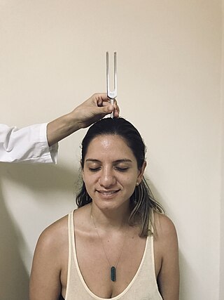
The Weber test is a screening test for hearing performed with a tuning fork. It can detect unilateral (one-sided) conductive hearing loss and unilateral sensorineural hearing loss. The test is named after Ernst Heinrich Weber (1795–1878). Conductive hearing ability is mediated by the middle ear composed of the ossicles: the malleus, the incus, and the stapes. Sensorineural hearing ability is mediated by the inner ear composed of the cochlea with its internal basilar membrane and attached cochlear nerve. The outer ear consisting of the pinna, ear canal, and ear drum or tympanic membrane transmits sounds to the middle ear but does not contribute to the conduction or sensorineural hearing ability save for hearing transmissions limited by cerumen impaction.

<span class="mw-page-title-main">Audiometry</span> Branch of audiology measuring hearing sensitivity

Audiometry is a branch of audiology and the science of measuring hearing acuity for variations in sound intensity and pitch and for tonal purity, involving thresholds and differing frequencies. Typically, audiometric tests determine a subject's hearing levels with the help of an audiometer, but may also measure ability to discriminate between different sound intensities, recognize pitch, or distinguish speech from background noise. Acoustic reflex and otoacoustic emissions may also be measured. Results of audiometric tests are used to diagnose hearing loss or diseases of the ear, and often make use of an audiogram.

<span class="mw-page-title-main">Eye movement</span> Movement of the eyes

Eye movement includes the voluntary or involuntary movement of the eyes. Eye movements are used by a number of organisms to fixate, inspect and track visual objects of interests. A special type of eye movement, rapid eye movement, occurs during REM sleep.

<span class="mw-page-title-main">Lateral rectus muscle</span> Muscle on lateral side of the eye

The lateral rectus muscle is a muscle on the lateral side of the eye in the orbit. It is one of six extraocular muscles that control the movements of the eye. The lateral rectus muscle is responsible for lateral movement of the eyeball, specifically abduction. Abduction describes the movement of the eye away from the midline, allowing the eyeball to move horizontally in the lateral direction, bringing the pupil away from the midline of the body.

<span class="mw-page-title-main">Extraocular muscles</span> Seven extrinsic muscles of the human eye

The extraocular muscles, or extrinsic ocular muscles, are the seven extrinsic muscles of the human eye. Six of the extraocular muscles, the four recti muscles, and the superior and inferior oblique muscles, control movement of the eye and the other muscle, the levator palpebrae superioris, controls eyelid elevation. The actions of the six muscles responsible for eye movement depend on the position of the eye at the time of muscle contraction.

Many types of sense loss occur due to a dysfunctional sensation process, whether it be ineffective receptors, nerve damage, or cerebral impairment. Unlike agnosia, these impairments are due to damages prior to the perception process.

<span class="mw-page-title-main">Chiasm (anatomy)</span> Nerve crossings outside the central nervous system

In anatomy a chiasm is the spot where two structures cross, forming an X-shape. Examples of chiasms are:

<span class="mw-page-title-main">Visual pathway lesions</span> Overview about the lesions of visual pathways

The visual pathway consists of structures that carry visual information from the retina to the brain. Lesions in that pathway cause a variety of visual field defects. In the visual system of human eye, the visual information processed by retinal photoreceptor cells travel in the following way:
Retina→Optic nerve→Optic chiasma →Optic tract→Lateral geniculate body→Optic radiation→Primary visual cortex

References

  1. Jon Brillman; Scott Kahan (1 March 2005). In A Page Neurology. Lippincott Williams & Wilkins. pp. 4–. ISBN   978-1-4051-0432-6 . Retrieved 27 June 2011.
  2. 1 2 3 4 5 6 7 Creative Commons by small.svg  This article incorporates text available under the CC BY 4.0 license.Ju, William (November 11, 2023). Neuroscience. Toronto: University of Toronto. 1.6 The Neurological Exam.
  3. "Examination of the Cranial Nerves". 22 April 2016.
  4. "Examination of the Cranial Nerves". Archived from the original on 2021-03-09. Retrieved 2014-06-26.