Gastrointestinal cancer

Last updated
Gastrointestinal cancer
Specialty Gastroenterology oncology
Symptoms upper Hematemesis Melena lower Coffee ground vomiting Hematochezia

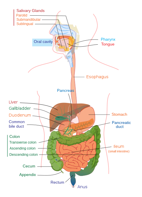
Gastrointestinal cancer refers to malignant conditions of the gastrointestinal tract (GI tract) and accessory organs of digestion, including the esophagus, stomach, biliary system, pancreas, small intestine, large intestine, rectum and anus. The symptoms relate to the organ affected and can include obstruction (leading to difficulty swallowing or defecating), abnormal bleeding or other associated problems. The diagnosis often requires endoscopy, followed by biopsy of suspicious tissue. The treatment depends on the location of the tumor, as well as the type of cancer cell and whether it has invaded other tissues or spread elsewhere. These factors also determine the prognosis.

Contents

Overall, the GI tract and the accessory organs of digestion (pancreas, liver, gall bladder) are responsible for more cancers and more deaths from cancer than any other system in the body. [1] [2] There is significant geographic variation in the rates of different gastrointestinal cancers. [1]

Upper digestive tract

Esophageal cancer

Age-standardised death rates from esophageal cancer, as reported by the WHO in 2004. Oesophagus cancer world map - Death - WHO2004.svg
Age-standardised death rates from esophageal cancer, as reported by the WHO in 2004.

Esophageal cancer is the sixth-most-common cancer in the world, and its incidence is increasing. [4] Some three to five males are affected for each female. [4] An "esophageal cancer belt", in which the incidence of esophageal squamous cell carcinoma (SCC) is more than a hundred times that of adjacent areas, extends from northeastern China through central Asia to northern Iran. [1] Ethiopia also has a notably high incidence. [4] There are two main types of esophageal cancer—adenocarcinoma and squamous cell carcinoma. Worldwide, the incidence of each type is about the same, but in developed countries like North America and Europe adenocarcinoma is the more common. [4]

Cancer of the esophagus is often detected late inasmuch as there are typically no early symptoms. Nevertheless, if the cancer is caught soon enough, patients can have a five-year survival rate of 90% or above. By the time esophageal cancer is usually detected, though, it might have spread beyond the esophageal wall, and the survival rate drops significantly. In China, the overall five-year survival rate for advanced esophageal cancer is about 20%, and in the United States it is about 15%. [4]

Stomach cancer

Cancer of the stomach, also called gastric cancer, is the fourth-most-common type of cancer and the second-highest cause of cancer death globally. [2] Eastern Asia (China, Japan, Korea, Mongolia) is a high-risk area for gastric cancer, and North America, Australia, New Zealand and western and northern Africa are areas with low risk. [5] The most common type of gastric cancer is adenocarcinoma, which causes about 750,000 deaths each year. [6] Important factors that may contribute to the development of gastric cancer include diet, smoking and alcohol consumption, genetic aspects (including a number of heritable syndromes) and infections (for example, Helicobacter pylori or Epstein-Barr virus) and pernicious anemia. [1] [6] Chemotherapy improves survival compared to best supportive care, however the optimal regimen is unclear. [7]

Pancreatic cancer

Upper and lower human gastrointestinal tract Digestive system diagram edit.svgpharynxoral cavity
Upper and lower human gastrointestinal tract

Pancreatic cancer is the fifth most-common cause of cancer-related deaths in the United States, [8] and the seventh most-common in Europe. [9] In 2008, globally there were 280,000 new cases of pancreatic cancer reported and 265,000 deaths. [10] These cancers are classified as endocrine or nonendocrine tumors. The most common is ductal adenocarcinoma. [1] The most significant risk factors for pancreatic cancer are advanced age (over 60) and smoking. [8] Chronic pancreatitis, diabetes or other conditions may also be involved in their development. [2] Early pancreatic cancer does not tend to result in any symptoms, but when a tumor is advanced, a patient may experience severe pain in the upper abdomen, possibly radiating to the back. [8] Another symptom might be jaundice, a yellowing of the skin and eyes. [9]

Pancreatic cancer has a poor prognosis, [2] with a five-year survival rate of less than 5%. By the time the cancer is diagnosed, it is usually at an advanced, inoperable stage. [9] Only one in about fifteen to twenty patients is curative surgery attempted. [11] Pancreatic cancer tends to be aggressive, and it resists radiotherapy and chemotherapy. [10]

Liver cancer

People get liver cancer (also called hepatocellular carcinoma, HCC or hepatoma) typically from a prolonged Hepatitis B or C infection or as a result of cirrhosis from chronic alcoholism. Liver cancer may bring about yellowing of the skin and eyes (jaundice), itching (pruritus), or cause a buildup of fluid in the abdomen (ascites). A person may feel an enlarging mass, or the cancer might be revealed by abnormal liver function tests. [12] :664–666

An attending practitioner might order a biopsy, an MRI or a CT scan, and a patient might be monitored through blood tests (including alpha-fetoprotein, liver-function tests or ultrasound. These cancers are typically treated according to their TNM stage and whether or not cirrhosis is present. Options include surgical resection, embolisation, ablation or a liver transplant. [13] :969–970

Gallbladder cancer

Cancers of the gallbladder are typically adenocarcinomas, and are common in elderly women. Gallbladder cancer is strongly associated with gallstones, a porcelain gallbladder appearance on ultrasound, and the presence of polyps within the gallbladder. Gallbladder cancer may manifest with weight loss, jaundice, and pain in the upper right of. It is typically diagnosed with ultrasound and staged with CT. The prognosis for gallbladder cancer is poor. [13] :981

Other

Lower digestive tract

Colorectal cancer

Example of an invasive colorectal cancer. Colon cancer 2.jpg
Example of an invasive colorectal cancer.

Colorectal cancer is a disease of old age. It typically originates in the secretory cells lining the gut, and risk factors include diets low in vegetable fibre and high in fat. If a younger person gets such a cancer, it is often associated with hereditary syndromes like Peutz-Jegher's, hereditary nonpolyposis colorectal cancer, or familial adenomatous polyposis. [12] :619–620 Colorectal cancer can be detected through the bleeding of a polyp, colicky bowel pain, a bowel obstruction or the biopsy of a polyp at a screening colonoscopy. A constant feeling of having to go to the toilet or anemia might also point to this kind of cancer. [13] :911

Use of a colonoscope can find these cancers, and a biopsy can reveal the extent of the involvement of the bowel wall. Removal of a section of the colon is necessary for treatment, with or without chemotherapy. Colorectal cancer has a comparatively good prognosis when detected early. [13] :911–912

Anal cancer

An important anatomic landmark in anal cancer is the pectinate line (dentate line), which is located about 1–2 cm from the anal verge (where the anal mucosa of the anal canal becomes skin). [14] Anal cancers located above this line (towards the head) are more likely to be carcinomas, whilst those located below (towards the feet) are more likely to be squamous cell carcinomas that may ulcerate. Anal cancer is strongly associated with ulcerative colitis and the sexually transmissible infections HPV and HIV. Anal cancer may be a cause of constipation or tenesmus, or may be felt as a palpable mass, although it may occasionally present as an ulcerative form. [15] :580

Anal cancer is investigated by biopsy and may be treated by excision and radiotherapy, or with external beam radiotherapy and adjunctive chemotherapy. The five-year survival rate with the latter procedure is above 70%. [15] :580

Gastrointestinal carcinoid tumor

A gastrointestinal carcinoid tumor is a rare, slow-growing form of cancer that affects certain cells in the lining of the stomach and intestines. The cells it affects make hormones that regulate the production of digestive juices and muscles that move food through the stomach and intestines. This kind of cancer usually occurs in the appendix, small intestine, or rectum. Its presence is associated with an increased risk of cancers affecting the other parts of the digestive system. It is usually treated with surgery. [16]

Field defects

A "field defect" or "field cancerization" is a region of tissue that precedes and predisposes to the development of cancer. Field defects occur in progression to gastrointestinal tract cancers. [17] These field defects may contain visible gross manifestations, epigenetic alterations and/or mutations.

Esophagus

Adenocarcinomas of the esophagus tend to arise in a field defect called Barrett's esophagus, a red patch of tissue in the generally pink lower esophagus. A diagnosis of Barrett’s esophagus is confirmed by a metaplastic change of the esophageal mucosa from squamous to columnar mucosa with intestinal metaplasia. Barrett's esophagus is the dominant pre-malignant lesion of esophageal adenocarcinoma, [18] and has prevalent epigenetic alterations. [19]

Esophageal squamous-cell carcinomas may occur as second primary tumors associated with head and neck cancer, due to field cancerization (i.e. a regional reaction to long-term carcinogenic exposure). [20] [21] A field defect associated with progression towards squamous cell carcinoma can be identified with epigenetic markers. [22]

Stomach

Gastric cancer develops within areas (field defects) of the stomach with atrophic gastritis and intestinal metaplasia: these lesions represent the cancerization field in which (intestinal-type) gastric cancers develop. [23] In one study, the field defect was clearly demonstrated in gastric carcinogenesis using miRNA high throughput data from normal gastric mucosa (from patients who had never had a gastric malignant neoplasm), non-tumor tissue adjacent to a gastric cancer, and gastric cancer tissue. Greater than five-fold reductions were found in four miRNAs in tumor-adjacent tissues and gastric cancers as compared to those miRNA levels in normal gastric tissues. [24]

Large intestine

Longitudinally opened freshly resected colon segment showing a cancer and four polyps. Plus a schematic diagram indicating a likely field defect (a region of tissue that precedes and predisposes to the development of cancer) in this colon segment. The diagram indicates sub-clones and sub-sub-clones that were precursors to the tumors. Image of resected colon segment with cancer & 4 nearby polyps plus schematic of field defects with sub-clones.jpg
Longitudinally opened freshly resected colon segment showing a cancer and four polyps. Plus a schematic diagram indicating a likely field defect (a region of tissue that precedes and predisposes to the development of cancer) in this colon segment. The diagram indicates sub-clones and sub-sub-clones that were precursors to the tumors.

When a segment of the large intestine, containing a cancer, is removed, the area adjacent to the cancer (and removed with it) may show additional neoplasia in the form of polyps (see image). This is visual evidence of a field defect. Some of these polyps may be premalignant neoplastic tumors. As shown by Hofstad et al., [25] when polyps are allowed to remain in the colon and are observed for three years, about 40% of polyps are seen to grow larger, likely progressing towards cancer. Luo et al. [26] summarized the substantial body of evidence that field cancerization occurs in the colon, often due to aberrant DNA methylation.

Etiology

Bile acids are synthesized in the liver to facilitate digestion of dietary fats. High exposure of the gastrointestinal tract to bile acids can occur in several different settings, but most significantly is prevalent among individuals who have a high dietary fat intake. High bile acid exposure has been implicated in several cancers of both the upper and lower digestive tract. [27] The deleterious effects on cells of elevated bile acid exposure include induction of reactive oxygen species, induction of DNA damage leading to mutation, and induction of apoptosis in the short term and selection for apoptosis resistance over the long term. [27] High levels of bile acids also alter the microbiome and act as signaling molecules, altering the microenvironment of the colon. [28]

Related Research Articles

<span class="mw-page-title-main">Gastroenterology</span> Branch of medicine focused on the digestive system and its disorders

Gastroenterology is the branch of medicine focused on the digestive system and its disorders. The digestive system consists of the gastrointestinal tract, sometimes referred to as the GI tract, which includes the esophagus, stomach, small intestine and large intestine as well as the accessory organs of digestion which include the pancreas, gallbladder, and liver. The digestive system functions to move material through the GI tract via peristalsis, break down that material via digestion, absorb nutrients for use throughout the body, and remove waste from the body via defecation. Physicians who specialize in the medical specialty of gastroenterology are called gastroenterologists or sometimes GI doctors. Some of the most common conditions managed by gastroenterologists include gastroesophageal reflux disease, gastrointestinal bleeding, irritable bowel syndrome, inflammatory bowel disease (IBD) which includes Crohn's disease and ulcerative colitis, peptic ulcer disease, gallbladder and biliary tract disease, hepatitis, pancreatitis, colitis, colon polyps and cancer, nutritional problems, and many more.

<span class="mw-page-title-main">Barrett's esophagus</span> Medical condition

Barrett's esophagus is a condition in which there is an abnormal (metaplastic) change in the mucosal cells lining the lower portion of the esophagus, from stratified squamous epithelium to simple columnar epithelium with interspersed goblet cells that are normally present only in the small intestine and large intestine. This change is considered to be a premalignant condition because it is associated with a high incidence of further transition to esophageal adenocarcinoma, an often-deadly cancer.

<span class="mw-page-title-main">Adenocarcinoma</span> Medical condition

Adenocarcinoma is a type of cancerous tumor that can occur in several parts of the body. It is defined as neoplasia of epithelial tissue that has glandular origin, glandular characteristics, or both. Adenocarcinomas are part of the larger grouping of carcinomas, but are also sometimes called by more precise terms omitting the word, where these exist. Thus invasive ductal carcinoma, the most common form of breast cancer, is adenocarcinoma but does not use the term in its name—however, esophageal adenocarcinoma does to distinguish it from the other common type of esophageal cancer, esophageal squamous cell carcinoma. Several of the most common forms of cancer are adenocarcinomas, and the various sorts of adenocarcinoma vary greatly in all their aspects, so that few useful generalizations can be made about them.

<span class="mw-page-title-main">Esophageal cancer</span> Gastrointestinal system cancer that is located in the esophagus

Esophageal cancer is cancer arising from the esophagus—the food pipe that runs between the throat and the stomach. Symptoms often include difficulty in swallowing and weight loss. Other symptoms may include pain when swallowing, a hoarse voice, enlarged lymph nodes ("glands") around the collarbone, a dry cough, and possibly coughing up or vomiting blood.

<span class="mw-page-title-main">Stomach cancer</span> Cancerous tumor originating in the stomach lining

Stomach cancer, also known as gastric cancer, is a cancer that develops from the lining of the stomach. Most cases of stomach cancers are gastric carcinomas, which can be divided into a number of subtypes, including gastric adenocarcinomas. Lymphomas and mesenchymal tumors may also develop in the stomach. Early symptoms may include heartburn, upper abdominal pain, nausea, and loss of appetite. Later signs and symptoms may include weight loss, yellowing of the skin and whites of the eyes, vomiting, difficulty swallowing, and blood in the stool, among others. The cancer may spread from the stomach to other parts of the body, particularly the liver, lungs, bones, lining of the abdomen, and lymph nodes.

<span class="mw-page-title-main">Gastrointestinal disease</span> Medical condition

Gastrointestinal diseases refer to diseases involving the gastrointestinal tract, namely the esophagus, stomach, small intestine, large intestine and rectum, and the accessory organs of digestion, the liver, gallbladder, and pancreas.

<span class="mw-page-title-main">Neoplasm</span> Abnormal mass of tissue as a result of abnormal growth or division of cells

A neoplasm is a type of abnormal and excessive growth of tissue. The process that occurs to form or produce a neoplasm is called neoplasia. The growth of a neoplasm is uncoordinated with that of the normal surrounding tissue, and persists in growing abnormally, even if the original trigger is removed. This abnormal growth usually forms a mass, when it may be called a tumour or tumor.

<span class="mw-page-title-main">Precancerous condition</span> Medical condition

A precancerous condition is a condition, tumor or lesion involving abnormal cells which are associated with an increased risk of developing into cancer. Clinically, precancerous conditions encompass a variety of abnormal tissues with an increased risk of developing into cancer. Some of the most common precancerous conditions include certain colon polyps, which can progress into colon cancer, monoclonal gammopathy of undetermined significance, which can progress into multiple myeloma or myelodysplastic syndrome. and cervical dysplasia, which can progress into cervical cancer. Bronchial premalignant lesions can progress to squamous cell carcinoma of the lung.

<span class="mw-page-title-main">Endoscopic ultrasound</span> Medical imaging procedure

Endoscopic ultrasound (EUS) or echo-endoscopy is a medical procedure in which endoscopy is combined with ultrasound to obtain images of the internal organs in the chest, abdomen and colon. It can be used to visualize the walls of these organs, or to look at adjacent structures. Combined with Doppler imaging, nearby blood vessels can also be evaluated.

The International Cancer Genome Consortium (ICGC) is a voluntary scientific organization that provides a forum for collaboration among the world's leading cancer and genomic researchers. The ICGC was launched in 2008 to coordinate large-scale cancer genome studies in tumours from 50 cancer types and/or subtypes that are of main importance across the globe.

<span class="mw-page-title-main">Chromoendoscopy</span>

Chromoendoscopy is a medical procedure wherein dyes are instilled into the gastrointestinal tract at the time of visualization with fibre-optic endoscopy. The purposes of chromoendoscopy is chiefly enhance the characterization of tissues, although dyes may be used for other functional purposes. The detail achieved with chromoendoscopy can often allow for identification of the tissue type or pathology based upon the pattern uncovered.

Endoscopic mucosal resection is a technique used to remove cancerous or other abnormal lesions found in the digestive tract. It is one method of performing a mucosectomy.

<span class="mw-page-title-main">Field cancerization</span> Biological process

Field cancerization or field effect is a biological process in which large areas of cells at a tissue surface or within an organ are affected by carcinogenic alterations. The process arises from exposure to an injurious environment, often over a lengthy period.

<span class="mw-page-title-main">Pancreatic tumor</span> Medical condition

A pancreatic tumor is an abnormal growth in the pancreas. In adults, almost 90% are pancreatic cancer and a few are benign. Pancreatic tumors are rare in children.

<span class="mw-page-title-main">Oesophagogastric junctional adenocarcinoma</span>

Oesophagogastric junctional adenocarcinoma is a cancer of the lower part of the oesophagus with a rising incidence in Western countries. This disease is often linked to Barrett's oesophagus.

<span class="mw-page-title-main">Human digestive system</span> Digestive system in humans

The human digestive system consists of the gastrointestinal tract plus the accessory organs of digestion. Digestion involves the breakdown of food into smaller and smaller components, until they can be absorbed and assimilated into the body. The process of digestion has three stages: the cephalic phase, the gastric phase, and the intestinal phase.

Gastrointestinal intraepithelial neoplasia is also known as gastrointestinal dysplasia. Gastrointestinal dysplasia refers to abnormal growth of the epithelial tissue lining the gastrointestinal tract including the esophagus, stomach, and colon. Pancreatic, biliary, and rectal Intraepithelial Neoplasia are discussed separately. The regions of abnormal growth are confined by the basement membrane adjacent to the epithelial tissue and are thought to represent pre-cancerous lesions. 

<span class="mw-page-title-main">Squamous-cell carcinoma</span> Carcinoma that derives from squamous epithelial cells

Squamous-cell carcinoma (SCC), also known as epidermoid carcinoma, comprises a number of different types of cancer that begin in squamous cells. These cells form on the surface of the skin, on the lining of hollow organs in the body, and on the lining of the respiratory and digestive tracts.

<span class="mw-page-title-main">CMTM3</span> Protein-coding gene in the species Homo sapiens

CKLF-like MARVEL transmembrane domain-containing protein 3, also termed chemokine-like factor superfamily 3, is a member of the CKLF-like MARVEL transmembrane domain-containing family of proteins. In humans, CMTM2 protein is encoded by the CMTM3 gene located in band 22.1 on the long arm of chromosome 16. This protein is expressed in a wide range of tissues, including fetal tissues. It is highly expressed in the male reproductive system, particularly testicular tissues and may play a role in the development of this tissue. It is also highly expressed in the immune system including circulating blood cells, i.e. B lymphocytes, CD4+ T lymphocytes, and monocytes. However, CMTM3 protein is weakly expressed or unexpressed in the malignant tissues of several types of cancers. In many but not all of theses cancers, this decreased or lack of expression appears due to methylation of the GpC islands in the promoter region, and thereby the silencing, of the CMTM3 gene.

References

  1. 1 2 3 4 5 Yamada T, Alpers DH, et al. (2009). Textbook of gastroenterology (5th ed.). Chichester, West Sussex: Blackwell Pub. pp. 603, 1028. ISBN   978-1-4051-6911-0. OCLC   404100761.
  2. 1 2 3 4 Bjelakovic, G; Nikolova, D; Simonetti, RG; Gluud, C (Jul 16, 2008). "Antioxidant supplements for preventing gastrointestinal cancers". The Cochrane Database of Systematic Reviews (3): CD004183. doi:10.1002/14651858.CD004183.pub3. PMID   18677777.
  3. "Death and DALY estimates for 2004 by cause for WHO Member States (Persons, all ages)". WHO. Retrieved 12 October 2013.
  4. 1 2 3 4 5 Yang, S; Wu, S; Huang, Y; Shao, Y; Chen, XY; Xian, L; Zheng, J; Wen, Y; Chen, X; Li, H; Yang, C (Dec 12, 2012). "Screening for oesophageal cancer". The Cochrane Database of Systematic Reviews. 12: CD007883. doi:10.1002/14651858.CD007883.pub2. PMID   23235651.
  5. Bennett, C; Wang, Y; Pan, T (Oct 7, 2009). "Endoscopic mucosal resection for early gastric cancer". The Cochrane Database of Systematic Reviews. 2009 (4): CD004276. doi:10.1002/14651858.CD004276.pub3. PMC   7199372 . PMID   19821324.
  6. 1 2 O'Connor, A; McNamara, D; O'Moráin, CA (Sep 23, 2013). "Surveillance of gastric intestinal metaplasia for the prevention of gastric cancer". The Cochrane Database of Systematic Reviews. 9 (9): CD009322. doi:10.1002/14651858.CD009322.pub2. PMID   24062262.
  7. Wagner, Anna Dorothea; Syn, Nicholas LX; Moehler, Markus; Grothe, Wilfried; Yong, Wei Peng; Tai, Bee-Choo; Ho, Jingshan; Unverzagt, Susanne (2017-08-29). "Chemotherapy for advanced gastric cancer" (PDF). Cochrane Database of Systematic Reviews. 2017 (8): CD004064. doi:10.1002/14651858.cd004064.pub4. PMC   6483552 . PMID   28850174.
  8. 1 2 3 Arcidiacono, PG; Calori, G; Carrara, S; McNicol, ED; Testoni, PA (Mar 16, 2011). "Celiac plexus block for pancreatic cancer pain in adults". The Cochrane Database of Systematic Reviews. 2019 (3): CD007519. doi:10.1002/14651858.CD007519.pub2. PMC   6464722 . PMID   21412903.
  9. 1 2 3 Moss, AC; Morris, E; Mac Mathuna, P (Apr 19, 2006). MacMathuna, Padraic (ed.). "Palliative biliary stents for obstructing pancreatic carcinoma". The Cochrane Database of Systematic Reviews. 2006 (2): CD004200. doi:10.1002/14651858.CD004200.pub4. PMC   6769023 . PMID   16625598.
  10. 1 2 Gurusamy, Kurinchi Selvan; Kumar, Senthil; Davidson, Brian R; Fusai, Giuseppe; Gurusamy, Kurinchi Selvan (2012). "Resection versus other treatments for locally advanced pancreatic cancer". In Gurusamy, Kurinchi Selvan (ed.). Cochrane Database of Systematic Reviews. doi:10.1002/14651858.CD010244.
  11. Gurusamy, Kurinchi Selvan; Allen, Victoria B; Kalia, Amun; Davidson, Brian R; Gurusamy, Kurinchi Selvan (2011). "Diagnostic accuracy of laparoscopy for assessing the resectability in pancreatic and periampullary cancer". In Gurusamy, Kurinchi Selvan (ed.). Cochrane Database of Systematic Reviews. doi:10.1002/14651858.CD009323.
  12. 1 2 Vinay Kumar; et al., eds. (2007). Robbins basic pathology (8th ed.). Philadelphia: Saunders/Elsevier. ISBN   978-1-4160-2973-1. OCLC   804094752.
  13. 1 2 3 4 Nicki R. Colledge; Brian R. Walker; Stuart H. Ralston, eds. (2010). Davidson's principles and practice of medicine. Illustrated by Robert Britton (21st ed.). Edinburgh: Churchill Livingstone/Elsevier. ISBN   978-0-7020-3085-7. OCLC   455157186.
  14. Bruce G. Wolff; et al., eds. (2007). The ASCRS textbook of colon and rectal surgery. New York: Springer. p. 1. ISBN   978-0-387-24846-2.
  15. 1 2 Fauci AS, Braunwald E, Kasper D, Hauser S, Longo D, Jameson J, Loscalzo J (2008). Harrison's principles of internal medicine (17th ed.). New York: McGraw-Hill Medical. ISBN   978-0-07-147692-8. OCLC   237889182.
  16. "Gastrointestinal Carcinoid Tumor". National Cancer Institute at the National Institutes of Health. 1980-01-01. Retrieved 15 October 2013.
  17. Bernstein C, Bernstein H, Payne CM, Dvorak K, Garewal H (2008). "Field defects in progression to gastrointestinal tract cancers". Cancer Lett. 260 (1–2): 1–10. doi:10.1016/j.canlet.2007.11.027. PMC   2744582 . PMID   18164807.
  18. Halland M, Katzka D, Iyer PG (2015). "Recent developments in pathogenesis, diagnosis and therapy of Barrett's esophagus". World J. Gastroenterol. 21 (21): 6479–90. doi: 10.3748/wjg.v21.i21.6479 . PMC   4458759 . PMID   26074687.
  19. Agarwal A, Polineni R, Hussein Z, Vigoda I, Bhagat TD, Bhattacharyya S, Maitra A, Verma A (2012). "Role of epigenetic alterations in the pathogenesis of Barrett's esophagus and esophageal adenocarcinoma". Int J Clin Exp Pathol. 5 (5): 382–96. PMC   3396065 . PMID   22808291.
  20. Priante AV, Castilho EC, Kowalski LP (April 2011). "Second primary tumors in patients with head and neck cancer". Current Oncology Reports. 13 (2): 132–7. doi:10.1007/s11912-010-0147-7. PMID   21234721. S2CID   207335139.
  21. Scherübl H, Steinberg J, Schwertner C, Mir-Salim P, Stölzel U, de Villiers EM (June 2008). "'Field cancerization' im oberen Aerodigestivtrakt" [Coincidental squamous cell cancers of the esophagus, head, and neck: risk and screening]. HNO (in German). 56 (6): 603–8. doi:10.1007/s00106-007-1616-7. PMID   17928979.
  22. Lee YC, Wang HP, Wang CP, Ko JY, Lee JM, Chiu HM, Lin JT, Yamashita S, Oka D, Watanabe N, Matsuda Y, Ushijima T, Wu MS (2011). "Revisit of field cancerization in squamous cell carcinoma of upper aerodigestive tract: better risk assessment with epigenetic markers". Cancer Prev Res (Phila). 4 (12): 1982–92. doi: 10.1158/1940-6207.CAPR-11-0096 . PMID   21952583.
  23. Rugge M, Capelle LG, Cappellesso R, Nitti D, Kuipers EJ (2013). "Precancerous lesions in the stomach: from biology to clinical patient management". Best Pract Res Clin Gastroenterol. 27 (2): 205–23. doi:10.1016/j.bpg.2012.12.007. PMID   23809241.
  24. Assumpção MB, Moreira FC, Hamoy IG, Magalhães L, Vidal A, Pereira A, Burbano R, Khayat A, Silva A, Santos S, Demachki S, Ribeiro-Dos-Santos Â, Assumpção P (2015). "High-Throughput miRNA Sequencing Reveals a Field Effect in Gastric Cancer and Suggests an Epigenetic Network Mechanism". Bioinform Biol Insights. 9: 111–7. doi:10.4137/BBI.S24066. PMC   4496000 . PMID   26244015.
  25. Hofstad B, Vatn MH, Andersen SN, Huitfeldt HS, Rognum T, Larsen S, Osnes M (1996). "Growth of colorectal polyps: redetection and evaluation of unresected polyps for a period of three years". Gut. 39 (3): 449–56. doi:10.1136/gut.39.3.449. PMC   1383355 . PMID   8949653.
  26. Luo Y, Yu M, Grady WM (2014). "Field cancerization in the colon: a role for aberrant DNA methylation?". Gastroenterol Rep (Oxf). 2 (1): 16–20. doi:10.1093/gastro/got039. PMC   3920999 . PMID   24760232.
  27. 1 2 Bernstein H, Bernstein C, Payne CM, Dvorak K. Bile acids as endogenous etiologic agents in gastrointestinal cancer. World J Gastroenterol. 2009 Jul 21;15(27):3329-40. doi: 10.3748/wjg.15.3329. PMID: 19610133; PMCID: PMC2712893
  28. Bernstein H, Bernstein C (January 2023). "Bile acids as carcinogens in the colon and at other sites in the gastrointestinal system". Exp Biol Med (Maywood). 248 (1): 79–89. doi:10.1177/15353702221131858. PMC   9989147 . PMID   36408538.