Intraretinal microvascular abnormalities

Last updated
Intraretinal microvascular abnormalities
Human eye cross-sectional view grayscale.png
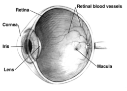
Human eye cross-sectional/ retina view
Specialty Ophthalmology

Intraretinal microvascular abnormalities (IRMA) are abnormalities of the blood vessels that supply the retina of the eye, a sign of diabetic retinopathy. [1] IRMA can be difficult to distinguish from and is likely a precursor to retinal neovascularization. One way to distinguish IRMA from retinal neovascularization is to perform fluorescein angiography. Since IRMA blood vessels are patent, unlike neovascular vessels, they do not leak, and therefore exhibit hyperfluorescence on fluorescein angiography.

Contents

IRMA is deeper in the retina than neovascularization, has blurrier edges, is more of a burgundy than a red, does not appear on the optic disc, and is usually seen after a shorter period of poorly controlled diabetes than neovascularization.

Related Research Articles

<span class="mw-page-title-main">Retinopathy</span> Medical condition

Retinopathy is any damage to the retina of the eyes, which may cause vision impairment. Retinopathy often refers to retinal vascular disease, or damage to the retina caused by abnormal blood flow. Age-related macular degeneration is technically included under the umbrella term retinopathy but is often discussed as a separate entity. Retinopathy, or retinal vascular disease, can be broadly categorized into proliferative and non-proliferative types. Frequently, retinopathy is an ocular manifestation of systemic disease as seen in diabetes or hypertension. Diabetes is the most common cause of retinopathy in the U.S. as of 2008. Diabetic retinopathy is the leading cause of blindness in working-aged people. It accounts for about 5% of blindness worldwide and is designated a priority eye disease by the World Health Organization.

<span class="mw-page-title-main">Diabetic retinopathy</span> Medical condition

Diabetic retinopathy, is a medical condition in which damage occurs to the retina due to diabetes mellitus. It is a leading cause of blindness in developed countries.

<span class="mw-page-title-main">Microangiopathy</span> Medical condition

Microangiopathy is a disease of the microvessels, small blood vessels in the microcirculation. It can be contrasted to macroangiopathies such as atherosclerosis, where large and medium-sized arteries are primarily affected.

<span class="mw-page-title-main">Retinal detachment</span> Medical condition of the eye

Retinal detachment is a disorder of the eye in which the retina peels away from its underlying layer of support tissue. Initial detachment may be localized, but without rapid treatment the entire retina may detach, leading to vision loss and blindness. It is a surgical emergency.

<span class="mw-page-title-main">Fluorescein angiography</span> Technique for examining the circulation of the retina and choroid of the eye

Fluorescein angiography (FA), fluorescent angiography (FAG), or fundus fluorescein angiography (FFA) is a technique for examining the circulation of the retina and choroid using a fluorescent dye and a specialized camera. Sodium fluorescein is added into the systemic circulation, the retina is illuminated with blue light at a wavelength of 490 nanometers, and an angiogram is obtained by photographing the fluorescent green light that is emitted by the dye. The fluorescein is administered intravenously in intravenous fluorescein angiography (IVFA) and orally in oral fluorescein angiography (OFA). The test is a dye tracing method.

<span class="mw-page-title-main">Electroretinography</span>

Electroretinography measures the electrical responses of various cell types in the retina, including the photoreceptors, inner retinal cells, and the ganglion cells. Electrodes are placed on the surface of the cornea or on the skin beneath the eye to measure retinal responses. Retinal pigment epithelium (RPE) responses are measured with an EOG test with skin-contact electrodes placed near the canthi. During a recording, the patient's eyes are exposed to standardized stimuli and the resulting signal is displayed showing the time course of the signal's amplitude (voltage). Signals are very small, and typically are measured in microvolts or nanovolts. The ERG is composed of electrical potentials contributed by different cell types within the retina, and the stimulus conditions can elicit stronger response from certain components.

<span class="mw-page-title-main">Central serous chorioretinopathy</span> Eye disease characterized by leakage of fluid under the retina

Central serous chorioretinopathy, also known as central serous retinopathy (CSR), is an eye disease that causes visual impairment, often temporary, usually in one eye. When the disorder is active it is characterized by leakage of fluid under the retina that has a propensity to accumulate under the central macula. This results in blurred or distorted vision (metamorphopsia). A blurred or gray spot in the central visual field is common when the retina is detached. Reduced visual acuity may persist after the fluid has disappeared.

Neovascularization is the natural formation of new blood vessels, usually in the form of functional microvascular networks, capable of perfusion by red blood cells, that form to serve as collateral circulation in response to local poor perfusion or ischemia.

Rubeosis iridis is a medical condition of the iris of the eye in which new abnormal blood vessels are found on the surface of the iris.

Charcot–Bouchard aneurysms are aneurysms of the brain vasculature which occur in small blood vessels. Charcot–Bouchard aneurysms are most often located in the lenticulostriate vessels of the basal ganglia and are associated with chronic hypertension. Charcot–Bouchard aneurysms are a common cause of cerebral hemorrhage.

<span class="mw-page-title-main">Angioid streaks</span> Medical condition

Angioid streaks, also called Knapp streaks or Knapp striae, are small breaks in Bruch's membrane, an elastic tissue containing membrane of the retina that may become calcified and crack. Up to 50% of angioid streak cases are idiopathic. It may occur secondary to blunt trauma, or it may be associated with many systemic diseases. The condition is usually asymptomatic, but decrease in vision may occur due to choroidal neovascularization.

The PASCAL Photocoagulator is an integrated semi-automatic pattern scan laser photocoagulation system designed to treat ocular diseases using a single shot or predetermined pattern array. The device is for ophthalmologists, particularly those that focus in vitreo-retinal surgery, a type of eye surgery. It was developed by OptiMedica, an ophthalmic medical device company located in Silicon Valley.

<span class="mw-page-title-main">Fundus photography</span> Medical imaging of the eyes

Fundus photography involves photographing the rear of an eye, also known as the fundus. Specialized fundus cameras consisting of an intricate microscope attached to a flash enabled camera are used in fundus photography. The main structures that can be visualized on a fundus photo are the central and peripheral retina, optic disc and macula. Fundus photography can be performed with colored filters, or with specialized dyes including fluorescein and indocyanine green.

<span class="mw-page-title-main">Macular telangiectasia</span> Disease of the retina affecting central vision

Macular telangiectasia is a condition of the retina, the light-sensing tissue at the back of the eye that causes gradual deterioration of central vision, interfering with tasks such as reading and driving.

<span class="mw-page-title-main">Familial exudative vitreoretinopathy</span> Retinal vascular disease

Familial exudative vitreoretinopathy is a genetic disorder affecting the growth and development of blood vessels in the retina of the eye. This disease can lead to visual impairment and sometimes complete blindness in one or both eyes. FEVR is characterized by incomplete vascularization of the peripheral retina. This can lead to the growth of new blood vessels which are prone to leakage and hemorrhage and can cause retinal folds, tears, and detachments. Treatment involves laser photocoagulation of the avascular portions of the retina to reduce new blood vessel growth and risk of complications including leakage of retinal blood vessels and retinal detachments.

<span class="mw-page-title-main">Branch retinal vein occlusion</span> Medical condition

Branch retinal vein occlusion is a common retinal vascular disease of the elderly. It is caused by the occlusion of one of the branches of central retinal vein.

<span class="mw-page-title-main">Radiation retinopathy</span> Medical condition

Radiation retinopathy is damage to retina due to exposure to ionizing radiation. Radiation retinopathy has a delayed onset, typically after months or years of radiation, and is slowly progressive. In general, radiation retinopathy is seen around 18 months after treatment with external-beam radiation and with brachytherapy. The time of onset of radiation retinopathy is between 6 months to 3 years.

Optical coherence tomography angiography (OCTA) is a non-invasive imaging technique based on optical coherence tomography (OCT) developed to visualize vascular networks in the human retina, choroid, skin and various animal models. OCTA may make use of speckle variance optical coherence tomography.

<span class="mw-page-title-main">Indocyanine green angiography</span> Diagnostic procedure

Indocyanine green angiography (ICGA) is a diagnostic procedure used to examine choroidal blood flow and associated pathology. Indocyanine green (ICG) is a water soluble cyanine dye which shows fluorescence in near-infrared (790–805 nm) range, with peak spectral absorption of 800-810 nm in blood. The near infrared light used in ICGA penetrates ocular pigments such as melanin and xanthophyll, as well as exudates and thin layers of sub-retinal vessels. Age-related macular degeneration is the third main cause of blindness worldwide, and it is the leading cause of blindness in industrialized countries. Indocyanine green angiography is widely used to study choroidal neovascularization in patients with exudative age-related macular degeneration. In nonexudative AMD, ICGA is used in classification of drusen and associated subretinal deposits.

Sickle cell retinopathy can be defined as retinal changes due to blood vessel damage in the eye of a person with a background of sickle cell disease. It can likely progress to loss of vision in late stages due to vitreous hemorrhage or retinal detachment. Sickle cell disease is a structural red blood cell disorder leading to consequences in multiple systems. It is characterized by chronic red blood cell destruction, vascular injury, and tissue ischemia causing damage to the brain, eyes, heart, lungs, kidneys, spleen, and musculoskeletal system.

References

  1. Shimouchi, Akito; Ishibazawa, Akihiro; Ishiko, Satoshi; Omae, Tsuneaki; Ro-Mase, Tomoko; Yanagi, Yasuo; Yoshida, Akitoshi (9 March 2020). "A Proposed Classification of Intraretinal Microvascular Abnormalities in Diabetic Retinopathy Following Panretinal Photocoagulation". Investigative Ophthalmology & Visual Science. 61 (3): 34. doi:10.1167/iovs.61.3.34. ISSN   1552-5783. PMC   7401423 . PMID   32191287 . Retrieved 12 September 2022.

Further reading