Mercury(II) reductase

Last updated
mercury (II) reductase
Mercuric reductase protein structure.jpg
Identifiers
EC no. 1.16.1.1
CAS no. 67880-93-7
Databases
IntEnz IntEnz view
BRENDA BRENDA entry
ExPASy NiceZyme view
KEGG KEGG entry
MetaCyc metabolic pathway
PRIAM profile
PDB structures RCSB PDB PDBe PDBsum
Gene Ontology AmiGO / QuickGO
Search
PMC articles
PubMed articles
NCBI proteins

Mercury(II) reductase (EC 1.16.1.1), commonly known as MerA, is an oxidoreductase enzyme and flavoprotein that catalyzes the reduction of Hg 2+ to Hg0. Mercury(II) reductase is found in the cytoplasm of many eubacteria [1] in both aerobic and anaerobic environments [2] and serves to convert toxic mercury ions into relatively inert elemental mercury.

Contents

Gene

Mercury(II) reductase, commonly known as MerA, is encoded in a structural gene found on the mer loci or as transposon 501 (Tn501). [3] It shares the same promoter region as mercury transport class proteins, such as MerP and MerT, and regulatory factor MerD. [1] MerA transcription is regulated by both MerR and MerD. [1]

Function

Free mercury ions can bind to metalloproteins, particularly those with cysteine residues, and can cause incorrect conformations resulting in function loss. This can cause death in many bacteria, as can many other heavy metals, and thus, needs to be removed from the cell or transformed into a chemically inert form. Mercury(II) reductase takes Hg2+ and catalyzes its reduction into Hg0 which is then released from the cell as a vapour. Mercury in its elemental form does not have the ability to form stable complexes with amino acid residues in proteins so is less dangerous than its ionic form.

Mechanism

Hg2+ + NADPH → Hg0 + H+ + NADP+

Active site mechanism of mercuric reductase Active site mechanism of mercuric reductase.png
Active site mechanism of mercuric reductase

1. Hg2+ + 2Cys-S → Cys-S-Hg-S-Cys

2. FAD + NADPH → FADH + NADP+

3. Cys-S-Hg-S-Cys + FADH → H+ + Hg0 + FAD + 2Cys-S

The substrates used in mercuric(II) reductase, as shown above, are Hg2+ and NADPH. In the catalytic active site of the enzyme, Hg2+ is held as a complex with two cysteine thiolates in a linear geometry. [4] NADPH from the cytoplasm of the cell undergo a hydride transfer with an embedded FAD forming FADH. [4] The resulting FADH then reduces Hg2+ into Hg0, in turn being oxidized back into FAD. [4] After reduction, the mercury is then released from the enzyme as a volatile vapour.

Mercury(II) reductase cannot completely reduce organomercury compounds such as methyl mercury. Thus, MerB cleaves the carbon-mercury bonds via protonolysis and forms a mercury dithiolate complex, upon which MerB transports the mercury directly to MerA for reduction. [5]

Structure

The active form of mercury(II) reductase is found as a homodimer. [4] It has a quaternary conformation and the monomer is composed of two domains. [4]

NmerA

One of the domains of mercuric reductase, NmerA, has a structural fold of βαββαβ. [6] It is attached to the active site through linkers made of around 30 amino acids. [6] NmerA contains two cysteine residues which function in the acquisition of Hg2+ from other proteins or inorganic ligands such as MerT and direct transport to the catalytic active site of MerA. [7] Very few mercuric(II) reductases have been found to lack the NmerA domain. [6]

Active site

The active site of MerA consists of four cysteine residues, a FAD, and a tyrosine residue. When bound to a Hg2+, a complex is formed with at least two cysteine thiolates at any time until release. [4] Two cysteine residues (Cys-136 and Cys-141) are buried within the protein and the other two cysteine residues (Cys-558' and Cys-559') are found near the surface near the C terminus. [4] The buried cysteine residues function as the site of catalysis whereas the surface cysteine residues function as transport to the site of catalysis. [4]

During Hg2+ transfer to the catalytic active site from the C terminus cysteine residues, a trigonal planar intermediate is formed stabilized by hydrogen bonding of a water molecule to the thiolates. [4] The water molecule is held in place by hydrogen bonding from the hydroxyl group of a nearby tyrosine residue (Tyr-194). [4]

Mercury transport

Various proteins assist in transporting mercury to mercury(II) reductase. MerP, a periplasmic mercury transport protein found in gram negative bacteria, transports mercury through the outer membrane into the inner membrane where it holds the mercury for another protein to bind to it and transport it to mercury(II) reductase. [1] MerT, a membrane bound protein found in both gram negative and gram positive bacteria, binds to free floating mercury. [1] Mercury(II) reductase can directly take mercury from MerT and MerP. [1]

When mercury enters the cell and is not bound to a membrane protein, mercury(II) reductase can transport it to its active site on its own depending on the size of its ligands. [8] If the ligands attached to mercury are large, mercury(II) reductase uses the C-terminus cysteine residues to transport the mercury to its active site. [8] If the ligands are small, mercury can go directly the active site for reduction. [8] The ligands can be removed by the NmerA domain. [8]

In the case of organomercury compounds, MerB breaks the Hg-C bonds and transports the Hg to mercuric(II) reductase. [5]

Regulation

When not bound to Hg2+, mercury(II) reductase acts as an oxidase creating toxic hydrogen peroxide. [1] Thus, excess of the enzyme can result in bacterial death. Bacteria developed two regulatory proteins, MerR and MerD, for mercury(II) reductase. [1] There are two promoter regions on the mer loci: The first region encodes regulator protein MerR, and the second region encodes the structural mer genes and the gene for the regulatory protein MerD. Both promoter regions overlap. [1]

MerR binds to an operator in the structural mer gene promoter called MerO. [1] This binding causes the DNA of the mer loci to bend to where RNA polymerase can not read the region. However, Hg2+ can bind to MerR and allosterically change the shape of the DNA, so that RNA polymerase can read the promoter region of the structural genes. [1] Since both promoter regions overlap when apoMerR is bound to MerO, the change in DNA conformation causes neither the structural genes nor the regulatory genes to be read. This makes MerR a negative autoregulator. [1]

MerR forms a stable trigonal planar complex with Hg2+, which causes it to be released much later than when mercury(II) reductase has reduced all free Hg2+ in the cytoplasm. [1] Thus, it causes an excess in production of mercuric(II) reductase. To circumvent this problem, MerD also binds to MerO in order to act antagonistically to Hg2+ bound MerR. [1] MerD is produced when MerR is active with Hg2+ since MerD is encoded in the structural mer genes.

Applications and uses

In waste-water treatment procedures, mercury is sometimes removed from the water by making the water flow through a biofilm rich with mercury(II) reductase-containing bacteria. [1]

Related Research Articles

<span class="mw-page-title-main">Metalloprotein</span> Protein that contains a metal ion cofactor

Metalloprotein is a generic term for a protein that contains a metal ion cofactor. A large proportion of all proteins are part of this category. For instance, at least 1000 human proteins contain zinc-binding protein domains although there may be up to 3000 human zinc metalloproteins.

<span class="mw-page-title-main">Active site</span> Active region of an enzyme

In biology and biochemistry, the active site is the region of an enzyme where substrate molecules bind and undergo a chemical reaction. The active site consists of amino acid residues that form temporary bonds with the substrate, the binding site, and residues that catalyse a reaction of that substrate, the catalytic site. Although the active site occupies only ~10–20% of the volume of an enzyme, it is the most important part as it directly catalyzes the chemical reaction. It usually consists of three to four amino acids, while other amino acids within the protein are required to maintain the tertiary structure of the enzymes.

<span class="mw-page-title-main">Metallothionein</span> Family of proteins

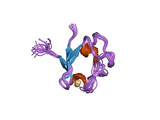
Metallothionein (MT) is a family of cysteine-rich, low molecular weight proteins. They are localized to the membrane of the Golgi apparatus. MTs have the capacity to bind both physiological and xenobiotic heavy metals through the thiol group of its cysteine residues, which represent nearly 30% of its constituent amino acid residues.

Thioredoxin reductases are enzymes that reduce thioredoxin (Trx). Two classes of thioredoxin reductase have been identified: one class in bacteria and some eukaryotes and one in animals. In bacteria TrxR also catalyzes the reduction of glutaredoxin like proteins known as NrdH. Both classes are flavoproteins which function as homodimers. Each monomer contains a FAD prosthetic group, a NADPH binding domain, and an active site containing a redox-active disulfide bond.

Ferredoxins are iron–sulfur proteins that mediate electron transfer in a range of metabolic reactions. The term "ferredoxin" was coined by D.C. Wharton of the DuPont Co. and applied to the "iron protein" first purified in 1962 by Mortenson, Valentine, and Carnahan from the anaerobic bacterium Clostridium pasteurianum.

<span class="mw-page-title-main">Flavin adenine dinucleotide</span> Redox-active coenzyme

In biochemistry, flavin adenine dinucleotide (FAD) is a redox-active coenzyme associated with various proteins, which is involved with several enzymatic reactions in metabolism. A flavoprotein is a protein that contains a flavin group, which may be in the form of FAD or flavin mononucleotide (FMN). Many flavoproteins are known: components of the succinate dehydrogenase complex, α-ketoglutarate dehydrogenase, and a component of the pyruvate dehydrogenase complex.

<span class="mw-page-title-main">Flavoprotein</span> Protein family

Flavoproteins are proteins that contain a nucleic acid derivative of riboflavin. These proteins are involved in a wide array of biological processes, including removal of radicals contributing to oxidative stress, photosynthesis, and DNA repair. The flavoproteins are some of the most-studied families of enzymes.

<span class="mw-page-title-main">Rubredoxin</span>

Rubredoxins are a class of low-molecular-weight iron-containing proteins found in sulfur-metabolizing bacteria and archaea. Sometimes rubredoxins are classified as iron-sulfur proteins; however, in contrast to iron-sulfur proteins, rubredoxins do not contain inorganic sulfide. Like cytochromes, ferredoxins and Rieske proteins, rubredoxins are thought to participate in electron transfer in biological systems. Recent work in bacteria and algae have led to the hypothesis that some rubredoxins may instead have a role in delivering iron to metalloproteins.

Copper proteins are proteins that contain one or more copper ions as prosthetic groups. Copper proteins are found in all forms of air-breathing life. These proteins are usually associated with electron-transfer with or without the involvement of oxygen (O2). Some organisms even use copper proteins to carry oxygen instead of iron proteins. A prominent copper proteins in humans is in cytochrome c oxidase (cco). The enzyme cco mediates the controlled combustion that produces ATP.

Nitrite reductase refers to any of several classes of enzymes that catalyze the reduction of nitrite. There are two classes of NIR's. A multi haem enzyme reduces NO2 to a variety of products. Copper containing enzymes carry out a single electron transfer to produce nitric oxide.

Oxidative protein folding is a process that is responsible for the formation of disulfide bonds between cysteine residues in proteins. The driving force behind this process is a redox reaction, in which electrons pass between several proteins and finally to a terminal electron acceptor.

<span class="mw-page-title-main">Nitrate reductase</span> Class of enzymes

Nitrate reductases are molybdoenzymes that reduce nitrate to nitrite. This reaction is critical for the production of protein in most crop plants, as nitrate is the predominant source of nitrogen in fertilized soils.

Nitrile hydratases are mononuclear iron or non-corrinoid cobalt enzymes that catalyse the hydration of diverse nitriles to their corresponding amides

<span class="mw-page-title-main">MMP7</span> Protein-coding gene in humans

Matrilysin also known as matrix metalloproteinase-7 (MMP-7), pump-1 protease (PUMP-1), or uterine metalloproteinase is an enzyme in humans that is encoded by the MMP7 gene. The enzyme has also been known as matrin, putative metalloproteinase-1, matrix metalloproteinase pump 1, PUMP-1 proteinase, PUMP, metalloproteinase pump-1, putative metalloproteinase, MMP). Human MMP-7 has a molecular weight around 30 kDa.

In enzymology, a ferredoxin-NADP+ reductase (EC 1.18.1.2) abbreviated FNR, is an enzyme that catalyzes the chemical reaction

<span class="mw-page-title-main">Thiosulfate dehydrogenase</span>

Thiosulfate dehydrogenase is an enzyme that catalyzes the chemical reaction:

<span class="mw-page-title-main">Ars operon</span>

In molecular biology, the ars operon is an operon found in several bacterial taxon. It is required for the detoxification of arsenate, arsenite, and antimonite. This system transports arsenite and antimonite out of the cell. The pump is composed of two polypeptides, the products of the arsA and arsB genes. This two-subunit enzyme produces resistance to arsenite and antimonite. Arsenate, however, must first be reduced to arsenite before it is extruded. A third gene, arsC, expands the substrate specificity to allow for arsenate pumping and resistance. ArsC is an approximately 150-residue arsenate reductase that uses reduced glutathione (GSH) to convert arsenate to arsenite with a redox active cysteine residue in the active site. ArsC forms an active quaternary complex with GSH, arsenate, and glutaredoxin 1 (Grx1). The three ligands must be present simultaneously for reduction to occur.

<span class="mw-page-title-main">Mercury transporter</span>

The mercury transporter superfamily is a family of transmembrane bacterial transporters of mercury ions. The common origin of all Mer superfamily members has been established. The common elements between family members are included in TMSs 1-2. A representative list of the subfamilies and proteins that belong to those subfamilies is available in the Transporter Classification Database.

<span class="mw-page-title-main">Nickel superoxide dismutase</span>

Nickel superoxide dismutase (Ni-SOD) is a metalloenzyme that, like the other superoxide dismutases, protects cells from oxidative damage by catalyzing the disproportionation of the cytotoxic superoxide radical to hydrogen peroxide and molecular oxygen. Superoxide is a reactive oxygen species that is produced in large amounts during photosynthesis and aerobic cellular respiration. The equation for the disproportionation of superoxide is shown below:

<span class="mw-page-title-main">L-ornithine N5 monooxygenase</span> Enzyme

L-ornithine N5 monooxygenase (EC 1.14.13.195 or EC 1.14.13.196) is an enzyme which catalyzes one of the following chemical reactions:

L-ornithine + NADPH + O2 N(5)-hydroxy-L-ornithine + NADP+ + H2O L-ornithine + NAD(P)H + O2 N(5)-hydroxy-L-ornithine + NAD(P)+ + H2O

References

  1. 1 2 3 4 5 6 7 8 9 10 11 12 13 14 15 Barkay T, Miller SM, Summers AO (June 2003). "Bacterial mercury resistance from atoms to ecosystems". FEMS Microbiology Reviews. 27 (2–3): 355–84. doi: 10.1016/s0168-6445(03)00046-9 . PMID   12829275.
  2. Ní Chadhain SM, Schaefer JK, Crane S, Zylstra GJ, Barkay T (October 2006). "Analysis of mercuric reductase (merA) gene diversity in an anaerobic mercury-contaminated sediment enrichment". Environmental Microbiology. 8 (10): 1746–52. doi:10.1111/j.1462-2920.2006.01114.x. PMID   16958755.
  3. Moore MJ, Walsh CT (February 1989). "Mutagenesis of the N- and C-terminal cysteine pairs of Tn501 mercuric ion reductase: consequences for bacterial detoxification of mercurials". Biochemistry. 28 (3): 1183–94. doi:10.1021/bi00429a036. PMID   2540817.
  4. 1 2 3 4 5 6 7 8 9 10 Lian P, Guo HB, Riccardi D, Dong A, Parks JM, Xu Q, Pai EF, Miller SM, Wei DQ, Smith JC, Guo H (November 2014). "X-ray structure of a Hg2+ complex of mercuric reductase (MerA) and quantum mechanical/molecular mechanical study of Hg2+ transfer between the C-terminal and buried catalytic site cysteine pairs". Biochemistry. 53 (46): 7211–22. doi:10.1021/bi500608u. PMC   4245977 . PMID   25343681.
  5. 1 2 Lafrance-Vanasse J, Lefebvre M, Di Lello P, Sygusch J, Omichinski JG (January 2009). "Crystal structures of the organomercurial lyase MerB in its free and mercury-bound forms: insights into the mechanism of methylmercury degradation". The Journal of Biological Chemistry. 284 (2): 938–44. doi: 10.1074/jbc.m807143200 . PMID   19004822.
  6. 1 2 3 Hong L, Sharp MA, Poblete S, Biehl R, Zamponi M, Szekely N, Appavou MS, Winkler RG, Nauss RE, Johs A, Parks JM, Yi Z, Cheng X, Liang L, Ohl M, Miller SM, Richter D, Gompper G, Smith JC (July 2014). "Structure and dynamics of a compact state of a multidomain protein, the mercuric ion reductase". Biophysical Journal. 107 (2): 393–400. Bibcode:2014BpJ...107..393H. doi:10.1016/j.bpj.2014.06.013. PMC   4104034 . PMID   25028881.
  7. Ledwidge R, Patel B, Dong A, Fiedler D, Falkowski M, Zelikova J, Summers AO, Pai EF, Miller SM (August 2005). "NmerA, the metal binding domain of mercuric ion reductase, removes Hg2+ from proteins, delivers it to the catalytic core, and protects cells under glutathione-depleted conditions". Biochemistry. 44 (34): 11402–16. doi:10.1021/bi050519d. PMID   16114877.
  8. 1 2 3 4 Engst S, Miller SM (March 1999). "Alternative routes for entry of HgX2 into the active site of mercuric ion reductase depend on the nature of the X ligands". Biochemistry. 38 (12): 3519–29. doi:10.1021/bi982680c. PMID   10090738.