
The Apicomplexa are a large phylum of parasitic alveolates. Most of them possess a unique form of organelle that comprises a type of non-photosynthetic plastid called an apicoplast, and an apical complex structure. The organelle is an adaptation that the apicomplexan applies in penetration of a host cell.

Plasmodium is a genus of unicellular eukaryotes that are obligate parasites of vertebrates and insects. The life cycles of Plasmodium species involve development in a blood-feeding insect host which then injects parasites into a vertebrate host during a blood meal. Parasites grow within a vertebrate body tissue before entering the bloodstream to infect red blood cells. The ensuing destruction of host red blood cells can result in malaria. During this infection, some parasites are picked up by a blood-feeding insect, continuing the life cycle.

Plasmodium falciparum is a unicellular protozoan parasite of humans, and the deadliest species of Plasmodium that causes malaria in humans. The parasite is transmitted through the bite of a female Anopheles mosquito and causes the disease's most dangerous form, falciparum malaria. It is responsible for around 50% of all malaria cases. P. falciparum is therefore regarded as the deadliest parasite in humans. It is also associated with the development of blood cancer and is classified as a Group 2A (probable) carcinogen.

CD36, also known as platelet glycoprotein 4, fatty acid translocase (FAT), scavenger receptor class B member 3 (SCARB3), and glycoproteins 88 (GP88), IIIb (GPIIIB), or IV (GPIV) is a protein that in humans is encoded by the CD36 gene. The CD36 antigen is an integral membrane protein found on the surface of many cell types in vertebrate animals. It imports fatty acids inside cells and is a member of the class B scavenger receptor family of cell surface proteins. CD36 binds many ligands including collagen, thrombospondin, erythrocytes parasitized with Plasmodium falciparum, oxidized low density lipoprotein, native lipoproteins, oxidized phospholipids, and long-chain fatty acids.

The Aconoidasida are a class of apicomplexan parasites created by Mehlhorn et al in 1980.
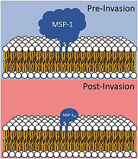
Merozoitesurface proteins are both integral and peripheral membrane proteins found on the surface of a merozoite, an early life cycle stage of a protozoan. Merozoite surface proteins, or MSPs, are important in understanding malaria, a disease caused by protozoans of the genus Plasmodium. During the asexual blood stage of its life cycle, the malaria parasite enters red blood cells to replicate itself, causing the classic symptoms of malaria. These surface protein complexes are involved in many interactions of the parasite with red blood cells and are therefore an important topic of study for scientists aiming to combat malaria.

A rhoptry is a specialized secretory organelle. They are club-shaped organelles connected by thin necks to the extreme apical pole of the parasite. These organelles, like micronemes, are characteristic of the motile stages of Apicomplexa protozoans. They can vary in number and shape and contain numerous enzymes that are released during the penetration process. The proteins they contain are important in the interaction between the host and the parasite, including the formation of the parasitophorous vacuole.

Dense granules are specialized secretory organelles. Dense granules are found only in platelets and are smaller than alpha granules. The origin of these dense granules is still unknown, however, it is thought that may come from the mechanism involving the endocytotic pathway. Dense granules are a sub group of lysosome-related organelles (LRO). There are about three to eight of these in a normal human platelet.
An apicoplast is a derived non-photosynthetic plastid found in most Apicomplexa, including Toxoplasma gondii, Plasmodium falciparum and other Plasmodium spp., but not in others such as Cryptosporidium. It originated from algae through secondary endosymbiosis. The apicoplast is surrounded by four membranes within the outermost part of the endomembrane system. The apicoplast hosts important metabolic pathways like fatty acid synthesis, isoprenoid precursor synthesis and parts of the heme biosynthetic pathway
Acidocalcisomes are rounded electron-dense acidic organelles, rich in calcium and polyphosphate and between 100 nm and 200 nm in diameter.
Acroeimeria is a genus of parasites that contains those species which initially develop immediately beneath the brush-border of the intestinal epithelium, but the meronts and gamonts of which are early on extruded to form a layer on the surface of the gut mucosa. Morphologically they are similar to the Eimeria to which they are closely related. The genus was described in 1989 by Paperna and Landsberg.
The genus Polychromophilus consists of obligate intracellular eukaryotic parasites that infect bats from every continent except Antarctica. They are transmitted by bat flies, which act as an insect vector as well as the parasite’s site of sporogeny. Polychromophilus follows a fairly typical Haemospororidian lifecycle, with gametocytes and gametes restricted to the bloodstream of the host and meronts infecting organs – most notably the lungs and the liver. The type species is Polychromophilus melanipherus, and was described by Dionisi in 1898.

Immunity Related Guanosine Triphosphatases or IRGs are proteins activated as part of an early immune response. IRGs have been described in various mammals but are most well characterized in mice. IRG activation in most cases is induced by an immune response and leads to clearance of certain pathogens.

In molecular biology, apical membrane antigen 1 is a novel antigen of Plasmodium falciparum which has been cloned. It contains a hydrophobic domain typical of an integral membrane protein. The antigen is designated apical membrane antigen 1 (AMA-1) by virtue of appearing to be located in the apical complex. AMA-1 appears to be transported to the merozoite surface close to the time of schizont rupture.

In molecular biology, Duffy binding proteins are found in Plasmodium. Plasmodium vivax and Plasmodium knowlesi merozoites invade Homo sapiens erythrocytes that express Duffy blood group surface determinants. The Duffy receptor family is localised in micronemes, an organelle found in all organisms of the phylum Apicomplexa.
The genus Schellackia comprises obligate unicellular eukaryotic parasites within the phylum Apicomplexa, and infects numerous species of lizards and amphibians worldwide. Schellackia is transmitted via insect vectors, primarily mites and mosquitoes, which take up the parasite in blood meals. These vectors then subsequently infect reptilian and amphibian which consume the infected insects. The parasites deform erythrocytes of the host into crescents, and can be visualised using a blood smear.
The parasitophorous vacuole (PV) is a structure produced by apicomplexan parasites in the cells of its host. The PV allows the parasite to develop while protected from the phagolysosomes of the host cell.
Plasmodium falciparum erythrocyte membrane protein 1 (PfEMP1) is a family of proteins present on the membrane surface of red blood cells that are infected by the malarial parasite Plasmodium falciparum. PfEMP1 is synthesized during the parasite's blood stage inside the RBC, during which the clinical symptoms of falciparum malaria are manifested. Acting as both an antigen and adhesion protein, it is thought to play a key role in the high level of virulence associated with P. falciparum. It was discovered in 1984 when it was reported that infected RBCs had unusually large-sized cell membrane proteins, and these proteins had antibody-binding (antigenic) properties. An elusive protein, its chemical structure and molecular properties were revealed only after a decade, in 1995. It is now established that there is not one but a large family of PfEMP1 proteins, genetically regulated (encoded) by a group of about 60 genes called var. Each P. falciparum is able to switch on and off specific var genes to produce a functionally different protein, thereby evading the host's immune system. RBCs carrying PfEMP1 on their surface stick to endothelial cells, which facilitates further binding with uninfected RBCs, ultimately helping the parasite to both spread to other RBCs as well as bringing about the fatal symptoms of P. falciparum malaria.
Maurer's clefts are membranous structures seen in the red blood cell during infection with Plasmodium falciparum. The function and contents of Maurer's clefts are not completely known; however, they appear to play a role in trafficking of Plasmodium falciparum erythrocyte membrane protein 1 (PfEMP1) and other adhesins to the red blood cell surface.

A symbiosome is a specialised compartment in a host cell that houses an endosymbiont in a symbiotic relationship.
