
A gland is a cell or an organ in an animal's body that produces and secretes different substances that the organism needs, either into the bloodstream or into a body cavity or outer surface. A gland may also function to remove unwanted substances such as urine from the body.

Brown algae are a large group of multicellular algae comprising the class Phaeophyceae. They include many seaweeds located in colder waters of the Northern Hemisphere. Brown algae are the major seaweeds of the temperate and polar regions. Many brown algae, such as members of the order Fucales, commonly grow along rocky seashores. Most brown algae live in marine environments, where they play an important role both as food and as a potential habitat. For instance, Macrocystis, a kelp of the order Laminariales, may reach 60 m (200 ft) in length and forms prominent underwater kelp forests that contain a high level of biodiversity. Another example is Sargassum, which creates unique floating mats of seaweed in the tropical waters of the Sargasso Sea that serve as the habitats for many species. Some members of the class, such as kelps, are used by humans as food.

The lamina propria is a thin layer of connective tissue that forms part of the moist linings known as mucous membranes or mucosae, which line various tubes in the body, such as the respiratory tract, the gastrointestinal tract, and the urogenital tract.

In botany, a stipe is a stalk that supports some other structure. The precise meaning is different depending on which taxonomic group is being described.

Wakame(Undaria pinnatifida) is a species of kelp native to cold, temperate coasts of the northwest Pacific Ocean. As an edible seaweed, it has a subtly sweet, but distinctive and strong flavour and satiny texture. It is most often served in soups and salads.

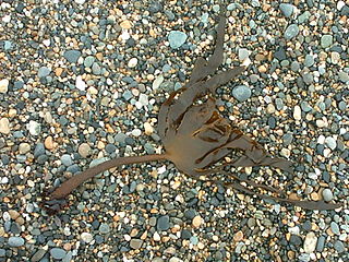
Alaria esculenta is an edible seaweed, also known as dabberlocks or badderlocks, or winged kelp, and occasionally as Atlantic Wakame. It is a traditional food along the coasts of the far north Atlantic Ocean. It may be eaten fresh or cooked in Greenland, Iceland, Scotland and Ireland. It is the only one of twelve species of Alaria to occur in both Ireland and in Great Britain.
The vascular organ of lamina terminalis (VOLT), organum vasculosum of the lamina terminalis(OVLT), or supraoptic crest is a sensory organ, one of the circumventricular organs of the third ventricle within the lamina terminalis. It is covered with pia mater, and lined with ependyma. It overlies the paraventricular nucleus of hypothalamus, and is involved in the secretion of vasopressin. The VOLT monitors the presence of peptides and macromolecules in the bloodstream, and conveys the information to the hypothalamus.

Circumventricular organs (CVOs) are structures in the brain characterized by their extensive and highly permeable capillaries, unlike those in the rest of the brain where there exists a blood–brain barrier (BBB) at the capillary level. Although the term "circumventricular organs" was originally proposed in 1958 by Austrian anatomist Helmut O. Hofer concerning structures around the brain ventricular system, the penetration of blood-borne dyes into small specific CVO regions was discovered in the early 20th century. The permeable CVOs enabling rapid neurohumoral exchange include the subfornical organ (SFO), the area postrema (AP), the vascular organ of lamina terminalis, the median eminence, the pituitary neural lobe, and the pineal gland.

The lamina terminalis is a thin layer that forms the median portion of the wall of the forebrain. It stretches from the interventricular foramen to the recess at the base of the optic stalk and contains the vascular organ of the lamina terminalis, which regulates the osmotic concentration of the blood. The lamina terminalis is immediately anterior to the tuber cinereum; together they form the pituitary stalk.

The apex of the posterior grey column, one of the three grey columns of the spinal cord, is capped by a V-shaped or crescentic mass of translucent, gelatinous neuroglia, termed the substantia gelatinosa of Rolando, which contains both neuroglia cells, and small neurons. The gelatinous appearance is due to an abundance of neuropil with a very low concentration of myelinated fibers. It extends the entire length of the spinal cord and into the medulla oblongata where it becomes the spinal trigeminal nucleus.

The Fucales (fucoids) are an order in the brown algae. The list of families in the Fucales, as well as additional taxonomic information on algae, is publicly accessible at Algaebase.
The history of phycology is the history of the scientific study of algae. Human interest in plants as food goes back into the origins of the species, and knowledge of algae can be traced back more than two thousand years. However, only in the last three hundred years has that knowledge evolved into a rapidly developing science.

Conceptacles are specialized cavities of marine and freshwater algae that contain the reproductive organs. They are situated in the receptacle and open by a small ostiole. Conceptacles are present in Corallinaceae, and Hildenbrandiales, as well as the brown Fucales. In the Fucales there is no haploid phase in the reproductive cycle and therefore no alternation of generations. The thallus is a sporophyte. The diploid plants produce male (antheridia) and female (oogonia) gametangia by meiosis. The gametes are released into the surrounding water; after fusion, the zygote settles and begins growth.

Seaweed, or macroalgae, refers to thousands of species of macroscopic, multicellular, marine algae. The term includes some types of Rhodophyta (red), Phaeophyta (brown) and Chlorophyta (green) macroalgae. Seaweed species such as kelps provide essential nursery habitat for fisheries and other marine species and thus protect food sources; other species, such as planktonic algae, play a vital role in capturing carbon and producing at least 50% of Earth's oxygen.

Red algae, or Rhodophyta, make up one of the oldest groups of eukaryotic algae. The Rhodophyta comprises one of the largest phyla of algae, containing over 7,000 recognized species within over 900 genera amidst ongoing taxonomic revisions. The majority of species (6,793) are Florideophyceae, and mostly consist of multicellular, marine algae, including many notable seaweeds. Red algae are abundant in marine habitats. Approximately 5% of red algae species occur in freshwater environments, with greater concentrations in warmer areas. Except for two coastal cave dwelling species in the asexual class Cyanidiophyceae, no terrestrial species exist, which may be due to an evolutionary bottleneck in which the last common ancestor lost about 25% of its core genes and much of its evolutionary plasticity.
Ecklonia cava, also called paddle weed, kajime, noro-kajime, or gamtae (Korean: 감태), is an edible marine brown alga species found in the ocean off Japan and Korea.
Tegument is a term in helminthology for the outer body covering of members of the phylum Platyhelminthes. The name is derived from a Latin word tegumentum or tegere, meaning "to cover". It is characteristic of flatworms including the broad groups of tapeworms and flukes. Once considered to be a non-living component, it is now known to be a dynamic cellular structure. In fact it is a living structure consisting of proteins, lipids, carbohydrates and RNA. It forms the protective layer and the host-parasite interface of the worms, serving both secretory and absorptive functions.

The reticular membrane is a thin, stiff lamina that extends from the outer hair cells to the Hensen's cells. The RM is composed of "minute-fiddle-shaped cuticular structures" called the phalangeal extensions of the outer hair cells, interspaced with extensions coming from the outer phalangeal cells. The RM separates endolymph in the cochlear duct from underlying corticolymph and perilymph of the scala tympani.
Odonata are insects with an incomplete metamorphosis (hemimetabolous). The aquatic larva or nymph hatches from an egg, and develops through eight to seventeen instars before leaving the water and emerging as the winged adult or imago.
