Statistical physics is a branch of physics that evolved from a foundation of statistical mechanics, which uses methods of probability theory and statistics, and particularly the mathematical tools for dealing with large populations and approximations, in solving physical problems. It can describe a wide variety of fields with an inherently stochastic nature. Its applications include many problems in the fields of physics, biology, chemistry, neuroscience. Its main purpose is to clarify the properties of matter in aggregate, in terms of physical laws governing atomic motion.
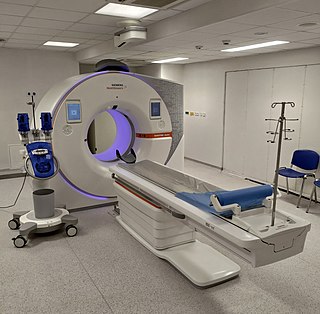
A CT scan or computed tomography scan is a medical imaging technique used in radiology (x-ray) to obtain detailed internal images of the body noninvasively for diagnostic purposes. The personnel that perform CT scans are called radiographers or radiology technologists.
Medical physics deals with the application of the concepts and methods of physics to the prevention, diagnosis and treatment of human diseases with a specific goal of improving human health and well-being. Since 2008, medical physics has been included as a health profession according to International Standard Classification of Occupation of the International Labour Organization.

External beam radiotherapy (EBRT) is the most common form of radiotherapy. The patient sits or lies on a couch and an external source of ionizing radiation is pointed at a particular part of the body. In contrast to brachytherapy and unsealed source radiotherapy, in which the radiation source is inside the body, external beam radiotherapy directs the radiation at the tumour from outside the body. Orthovoltage ("superficial") X-rays are used for treating skin cancer and superficial structures. Megavoltage X-rays are used to treat deep-seated tumours, whereas megavoltage electron beams are typically used to treat superficial lesions extending to a depth of approximately 5 cm. X-rays and electron beams are by far the most widely used sources for external beam radiotherapy. A small number of centers operate experimental and pilot programs employing beams of heavier particles, particularly protons, owing to the rapid dropoff in absorbed dose beneath the depth of the target.
Superficial X-rays are low-energy X-rays that do not penetrate very far before they are absorbed. They are produced by X-ray tubes operating at voltages in the 10–100 kV range, and therefore have peak energies in the 10–100 keV range. Precise naming and definitions of energy ranges may vary, and X-rays at the lower end of this range may also be known as Grenz rays. They are useful in radiation therapy for the treatment of various benign or malignant skin problems, including skin cancer and severe eczema. They have a useful depth of up to 5 mm. In some locations, orthovoltage treatment is being replaced by electron therapy or brachytherapy.
The Hounsfield scale, named after Sir Godfrey Hounsfield, is a quantitative scale for describing radiodensity. It is frequently used in CT scans, where its value is also termed CT number.

In the field of medical treatment, proton therapy, or proton radiotherapy, is a type of particle therapy that uses a beam of protons to irradiate diseased tissue, most often to treat cancer. The chief advantage of proton therapy over other types of external beam radiotherapy is that the dose of protons is deposited over a narrow range of depth, which results in minimal entry, exit, or scattered radiation dose to healthy nearby tissues.

The Bragg peak is a pronounced peak on the Bragg curve which plots the energy loss of ionizing radiation during its travel through matter. For protons, α-rays, and other ion rays, the peak occurs immediately before the particles come to rest. It is named after William Henry Bragg, who discovered it in 1903.

In radiotherapy, radiation treatment planning (RTP) is the process in which a team consisting of radiation oncologists, radiation therapist, medical physicists and medical dosimetrists plan the appropriate external beam radiotherapy or internal brachytherapy treatment technique for a patient with cancer.

Tomotherapy is a radiation therapy modality, in which the patient is scanned across a modulated strip-beam, so that only one “slice” of the target is exposed at any one time by the linear accelerator (linac) beam. The three components distinctive to this modality are: (1) a collimator pair that defines the length of the strip, (2) a binary multileaf collimator whose leaves open and close during treatment to modulate the strip’s intensity, and (3) a couch that scans the patient across the beam at a fixed speed during the treatment delivery.
The computed tomography dose index (CTDI) is a commonly used radiation exposure index in X-ray computed tomography (CT), first defined in 1981. The unit of CTDI is the gray (Gy) and it can be used in conjunction with patient size to estimate the absorbed dose. The CTDI and absorbed dose may differ by more than a factor of two for small patients such as children.

Positron emission tomography–magnetic resonance imaging (PET–MRI) is a hybrid imaging technology that incorporates magnetic resonance imaging (MRI) soft tissue morphological imaging and positron emission tomography (PET) functional imaging.

Cone beam computed tomography is a medical imaging technique consisting of X-ray computed tomography where the X-rays are divergent, forming a cone.
Computational human phantoms are models of the human body used in computerized analysis. Since the 1960s, the radiological science community has developed and applied these models for ionizing radiation dosimetry studies. These models have become increasingly accurate with respect to the internal structure of the human body.
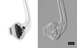
Phase-contrast X-ray imaging or phase-sensitive X-ray imaging is a general term for different technical methods that use information concerning changes in the phase of an X-ray beam that passes through an object in order to create its images. Standard X-ray imaging techniques like radiography or computed tomography (CT) rely on a decrease of the X-ray beam's intensity (attenuation) when traversing the sample, which can be measured directly with the assistance of an X-ray detector. However, in phase contrast X-ray imaging, the beam's phase shift caused by the sample is not measured directly, but is transformed into variations in intensity, which then can be recorded by the detector.
Xie George Xu is currently the Edward E. Hood Chair Professor of Engineering at Rensselaer Polytechnic Institute (RPI), Troy, New York, United States.
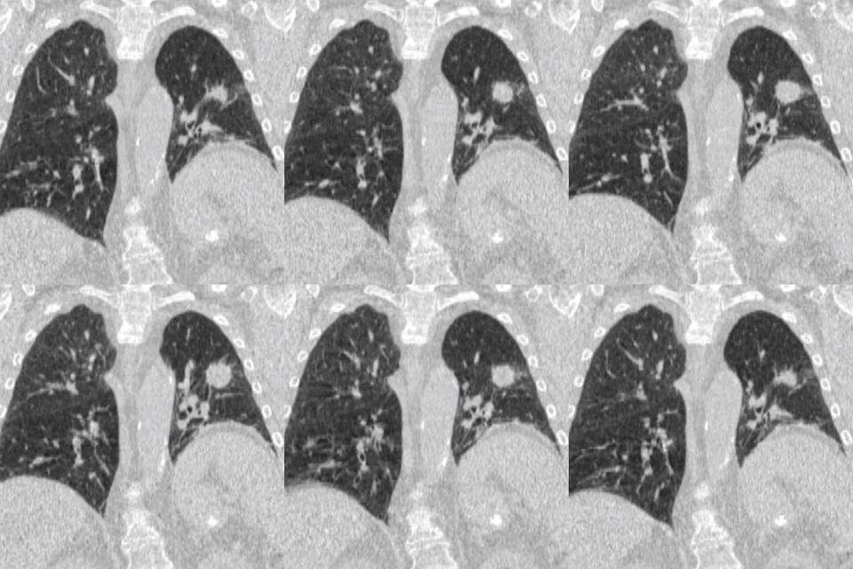
Four-dimensional computed tomography (4DCT) is a type of CT scanning which records multiple images over time. It allows playback of the scan as a video, so that physiological processes can be observed and internal movement can be tracked. The name is derived from the addition of time to traditional 3D computed tomography. Alternatively, the phase of a particular process, such as respiration, may be considered the fourth dimension.
Jeffrey Harold Siewerdsen is an American physicist and biomedical engineer who is a Professor of Biomedical Engineering, Computer Science, Radiology, and Neurosurgery at Johns Hopkins University. He is Co-Director of the Carnegie Center for Surgical Innovation at Johns Hopkins School of Medicine and is a member of the Malone Center for Engineering in Healthcare. He is among the original inventors of cone-beam CT-guided radiotherapy as well as weight-bearing cone-beam CT for musculoskeletal radiology and orthopedic surgery. His work also includes the early development of flat-panel detectors on mobile C-arms for intraoperative cone-beam CT in image-guided surgery. He developed early models for the signal and noise performance of flat-panel detectors and later extended such analysis to dual-energy imaging and 3D imaging performance in cone-beam CT. His core laboratory at Johns Hopkins University is the ISTAR Lab in the Department of Biomedical Engineering at the Johns Hopkins Hospital.
Photon-counting computed tomography (PCCT) is a form of X-ray computed tomography (CT) in which X-rays are detected using a photon-counting detector (PCD). The first clinically-approved PCCT system was cleared by the Food and Drug Administration (FDA) in September 2021. Photon-counting CT offers several significant advantages over other existing CT imaging techniques and continues to inspire and make possible completely novel applications. A photon-counting CT system employs a photon-counting detector (PCD) which registers the interactions of individual photons. By keeping track of the deposited energy in each interaction, the detector pixels of a PCD each record an approximate energy spectrum, making it a spectral or energy-resolved CT technique. In contrast, more conventional CT scanners use energy-integrating detectors (EIDs), where the total energy deposited in a pixel during a fixed period of time is registered. These EIDs thus register only photon intensity, comparable to black-and-white photography, whereas PCDs register also spectral information, similar to color photography.
Spectral imaging is an umbrella term for energy-resolved X-ray imaging in medicine. The technique makes use of the energy dependence of X-ray attenuation to either increase the contrast-to-noise ratio, or to provide quantitative image data and reduce image artefacts by so-called material decomposition. Dual-energy imaging, i.e. imaging at two energy levels, is a special case of spectral imaging and is still the most widely used terminology, but the terms "spectral imaging" and "spectral CT" have been coined to acknowledge the fact that photon-counting detectors have the potential for measurements at a larger number of energy levels.


