Purpureocillium lilacinum

Last updated

Purpureocillium lilacinum
Paecilomyces lilacinus.jpg
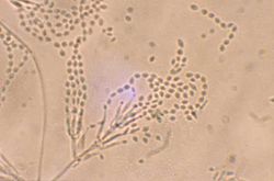
Divergent phialides and long, tangled chains of elliptical conidia borne from more complex fruiting structures characteristic of Purpureocillium lilacinum; magnification 460X.
Scientific classification OOjs UI icon edit-ltr.svg
Domain: Eukaryota
Kingdom: Fungi
Division: Ascomycota
Class: Sordariomycetes
Order: Hypocreales
Family: Ophiocordycipitaceae
Genus: Purpureocillium
Species:
P. lilacinum
Binomial name
Purpureocillium lilacinum
(Thom) Luangsa-ard, Hou- braken, Hywel-Jones & Samson (2011)
Synonyms [1]

PaecilliumLuangsa-ard, Hywel-Jones & Samson nom. prov. (2007) [2]
Penicillium lilacinumThom (1910)
Penicillium amethystinumWehmer (1923)
Spicaria rubidopurpureaAoki (1941)Paecilomyces lilacinus(Thom) Samson (1974)

Contents

Purpureocillium lilacinum is a species of filamentous fungus in the family Ophiocordycipitaceae. [3] It has been isolated from a wide range of habitats, including cultivated and uncultivated soils, forests, grassland, deserts, estuarine sediments and sewage sludge, and insects. It has also been found in nematode eggs, and occasionally from females of root-knot and cyst nematodes. In addition, it has frequently been detected in the rhizosphere of many crops. The species can grow at a wide range of temperatures – from 8 to 38 °C (46 to 100 °F) for a few isolates, with optimal growth in the range 26 to 30 °C (79 to 86 °F). It also has a wide pH tolerance and can grow on a variety of substrates. [4] [5] P. lilacinum has shown promising results for use as a biocontrol agent to control the growth of destructive root-knot nematodes.

Taxonomy and phylogeny

The species was originally described by American mycologist Charles Thom in 1910, under than name Penicillium lilacinum. [6] Taxonomic synonyms include Penicillium amethystinum Wehmer and Spicaria rubidopurpurea Aoki. [1] In 1974, Robert A. Samson transferred the species to Paecilomyces . [4] Publications in the 2000s (decade) indicated that the genus Paecilomyces was not monophyletic, [7] and that close relatives were Paecilomyces nostocoides, Isaria takamizusanensis and Nomuraea atypicola. [8] The new genus Purpureocillium was created to hold the taxon. The generic name refers to the purple conidia produced by the fungus. [9]

Description

Purpureocillium lilacinum forms a dense mycelium which gives rise to conidiophores. These bear phialides from the ends of which spores are formed in long chains. Spores germinate when suitable moisture and nutrients are available. Colonies on malt agar grow rather fast, attaining a diameter of 5–7 cm within 14 days at 25 °C (77 °F), consisting of a basal felt with a floccose overgrowth of aerial mycelium; at first white, but when sporulating changing to various shades of vinaceous. The reverse side is sometimes uncolored but usually in vinaceous shades. The vegetative hyphae are smooth-walled, hyaline, and 2.5–4.0  µm wide. Conidiophores arising from submerged hyphae, 400–600 µm in length, or arising from aerial hyphae and half as long. Phialides consisting of a swollen basal part, tapering into a thin distinct neck. Conidia are in divergent chains, ellipsoid to fusiform in shape, and smooth walled to slightly roughened. Chlamydospores are absent. [4]

Life cycles

Purpureocillium lilacinum is highly adaptable in its life strategy: depending on the availability of nutrients in the surrounding microenvironments it may be entomopathogenic, [10] [11] [12] mycoparasitic, [13] saprophytic, [14] as well as nematophagous.

Human pathogenicity

Purpureocillium lilacinum is an infrequent cause of human disease. [15] [16] Most reported cases involve patients with compromised immune systems, indwelling foreign devices, or intraocular lens implants. [17] [18] Research of the last decade suggests it may be an emerging pathogen of both immunocompromised [19] as well as immunocompetent adults. [20] It is one of the most common species causing hyalohyphomycosis along with Paecilomyces variotii . [9]

Biocontrol agent

P. lilacinum is used as a biocontrol agent for several nematode species, such as Meloidogyne incognita, shown here. A juvenile root-knot nematode (Meloidogyne incognita) penetrates a tomato root - USDA-ARS.jpg
P. lilacinum is used as a biocontrol agent for several nematode species, such as Meloidogyne incognita , shown here.

Plant-parasitic nematodes cause significant economic losses to a wide variety of crops. Chemical control is a widely used option for plant-parasitic nematode management. However, chemical nematicides are now being reappraised in respect of environmental hazard, high costs, limited availability in many developing countries or their diminished effectiveness following repeated applications.

Control of plant-parasitic nematodes

Purpureocillium lilacinum was first observed in association with nematode eggs in 1966 [21] and the fungus was subsequently found parasitising the eggs of Meloidogyne incognita in Peru. [22] It has now been isolated from many cyst and root-knot nematodes and from soil in many locations. [23] [24] Several successful field trials using P. lilacinum against pest nematodes were conducted in Peru. [22] The Peruvian isolate was then sent to nematologists in 46 countries for testing, as part of the International Meloidogyne project, resulting in many more field trials on a range of crops in many soil types and climates. [25] Field trials, glasshouse trials and in vitro testing of P. lilacinum continues and more isolates have been collected from soil, nematodes and occasionally from insects. Isolates vary in their pathogenicity to plant-parasitic nematodes. Some isolates are aggressive parasites while others, though morphologically indistinguishable, are less or non-pathogenic. Sometimes isolates that looked promising in vitro or in glasshouse trials have failed to provide control in the field. [26]

Enzymes

Many enzymes produced by P. lilacinum have been studied. A basic serine protease with biological activity against Meloidogyne hapla eggs has been identified. [27] One strain of P. lilacinum has been shown to produce proteases and a chitinase, enzymes that could weaken a nematode egg shell so as to enable a narrow infection peg to push through. [28]

Egg infection

Before infecting a nematode egg, P. lilacinum flattens against the egg surface and becomes closely appressed to it. P. lilacinum produces simple appressoria anywhere on the nematode egg shell either after a few hyphae grow along the egg surface, or after a network of hyphae form on the egg. The presence of appressoria appears to indicate that the egg is, or is about to be, infected. In either case, the appressorium appears the same, as a simple swelling at the end of a hypha, closely appressed to the eggshell. Adhesion between the appressorium and nematode egg surface must be strong enough to withstand the opposing force produced by the extending tip of a penetration hypha. [29] When the hypha has penetrated the egg, it rapidly destroys the juvenile within, before growing out of the now empty egg shell to produce conidiophores and to grow towards adjacent eggs.

Mycotoxins

Paecilotoxin is a mycotoxin isolated from the fungus. [30] Its significance is unknown. Khan et al. (2003) tested one strain of P. lilacinum for the production of paecilotoxin and were unable to show toxin production in that strain, suggesting that toxin synthesis may vary among isolates. [31] [32]

Related Research Articles

<span class="mw-page-title-main">Hypha</span> Long, filamentous structure in fungi and Actinobacteria

A hypha is a long, branching, filamentous structure of a fungus, oomycete, or actinobacterium. In most fungi, hyphae are the main mode of vegetative growth, and are collectively called a mycelium.

<span class="mw-page-title-main">Root-knot nematode</span> Genus of parasitic worms

Root-knot nematodes are plant-parasitic nematodes from the genus Meloidogyne. They exist in soil in areas with hot climates or short winters. About 2000 plants worldwide are susceptible to infection by root-knot nematodes and they cause approximately 5% of global crop loss. Root-knot nematode larvae infect plant roots, causing the development of root-knot galls that drain the plant's photosynthate and nutrients. Infection of young plants may be lethal, while infection of mature plants causes decreased yield.

<i>Meloidogyne incognita</i> Nematode worm, plant disease, many hosts

Meloidogyne incognita, also known as the southern root-nematode or cotton root-knot nematode is a plant-parasitic roundworm in the family Heteroderidae. This nematode is one of the four most common species worldwide and has numerous hosts. It typically incites large, usually irregular galls on roots as a result of parasitism.

<span class="mw-page-title-main">Entomopathogenic fungus</span> Fungus that can act as a parasite of insects

An entomopathogenic fungus is a fungus that can kill or seriously disable insects.

<i>Metarhizium robertsii</i> Species of fungus

Metarhizium robertsii – formerly known as M. anisopliae, and even earlier as Entomophthora anisopliae (basionym) – is a fungus that grows naturally in soils throughout the world and causes disease in various insects by acting as a parasitoid. Ilya I. Mechnikov named it after the insect species from which it was originally isolated – the beetle Anisoplia austriaca. It is a mitosporic fungus with asexual reproduction, which was formerly classified in the form class Hyphomycetes of the phylum Deuteromycota.

<i>Paecilomyces</i> Genus of fungi

Paecilomyces is a genus of fungi. A number of species in this genus are plant pathogens.

Hyphomycetes are a form classification of fungi, part of what has often been referred to as fungi imperfecti, Deuteromycota, or anamorphic fungi. Hyphomycetes lack closed fruit bodies, and are often referred to as moulds. Most hyphomycetes are now assigned to the Ascomycota, on the basis of genetic connections made by life-cycle studies or by phylogenetic analysis of DNA sequences; many remain unassigned phylogenetically.

Clonostachys rosea f. rosea, also known as Gliocladium roseum, is a species of fungus in the family Bionectriaceae. It colonizes living plants as an endophyte, digests material in soil as a saprophyte and is also known as a parasite of other fungi and of nematodes. It produces a wide range of volatile organic compounds which are toxic to organisms including other fungi, bacteria, and insects, and is of interest as a biological pest control agent.

Acremonium strictum is an environmentally widespread saprotroph species found in soil, plant debris, and rotting mushrooms. Isolates have been collected in North and Central America, Asia, Europe and Egypt. A. strictum is an agent of hyalohyphomycosis and has been identified as an increasingly frequent human pathogen in immunosuppressed individuals, causing localized, disseminated and invasive infections. Although extremely rare, A. strictum can infect immunocompetent individuals, as well as neonates. Due to the growing number of infections caused by A. strictum in the past few years, the need for new medical techniques in the identification of the fungus as well as for the treatment of human infections has risen considerably.

<span class="mw-page-title-main">Nematophagous fungus</span> Carnivorous fungi specialized in trapping and digesting nematodes

Nematophagous fungi are carnivorous fungi specialized in trapping and digesting nematodes. Around 160 species are known. Species exist that live inside the nematodes from the beginning and others that catch them, mostly with glue traps or in rings, some of which constrict on contact. Some species possess both types of traps. Another technique is to stun the nematodes using toxins, a method employed by Coprinus comatus, Stropharia rugosoannulata, and the family Pleurotaceae. The habit of feeding on nematodes has arisen many times among fungi, as is demonstrated by the fact that nematophagous species are found in all major fungal groups. Nematophagous fungi can be useful in controlling those nematodes that eat crops. Purpureocillium, for example, can be used as a bio-nematicide.

<span class="mw-page-title-main">Nematode</span> Phylum of worms with tubular digestive systems with openings at both ends

The nematodes, roundworms or eelworms constitute the phylum Nematoda. They are a diverse animal phylum inhabiting a broad range of environments. Most species are free-living, feeding on microorganisms, but there are many that are parasitic. The parasitic worms (helminths) are the cause of soil-transmitted helminthiases.

<i>Purpureocillium</i> Genus of fungi

Purpureocillium is a fungal genus in the Ophiocordycipitaceae family. The genus now contains at least 5 species with the type species Purpureocillium lilacinum, a common soil mold. It has been isolated from a wide range of habitats, including cultivated and uncultivated soils, forests, grassland, deserts, estuarine sediments and sewage sludge, and insects. It has also been found in nematode eggs, and occasionally from females of root-knot and cyst nematodes. In addition, it has frequently been detected in the rhizosphere of many crops. The species can grow at a wide range of temperatures – from 8 to 38 °C for a few isolates, with optimal growth in the range 26 to 30 °C. It also has a wide pH tolerance and can grow on a variety of substrates. P. lilacinum has shown promising results for use as a biocontrol agent to control the growth of destructive root-knot nematodes.

<i>Rhopalomyces elegans</i> Species of fungus

Rhopalomyces elegans is a common species of zygomycete fungus, and the type species of the genus Rhopalomyces. Widely distributed, it is found in soil, rotting plant material, and animal dung. It is a facultative parasite of nematode eggs.

Pasteuria is a genus of mycelial and endospore-forming, nonmotile gram-positive bacteria that are obligate parasites of some nematodes and crustaceans. The genus of Pasteuria was previously classified within the family Alicyclobacillaceae, but has since been moved to the family Pasteuriaceae.

<i>Harposporium anguillulae</i> Species of fungus

Harposporium anguillulae is a member of the genus Harposporium. It is an endoparasitic nematophagous fungus that attacks nematodes and eelworms and is isolated commonly from field and agricultural soils as well as used as an experimental organism in the laboratory.

<i>Arthrobotrys oligospora</i> Species of fungus

Arthrobotrys oligospora was discovered in Europe in 1850 by Georg Fresenius. A. oligospora is the model organism for interactions between fungi and nematodes. It is the most common nematode-capturing fungus, and most widespread nematode-trapping fungus in nature. It was the first species of fungi documented to actively capture nematodes.

Dactylellina haptotyla is a common soil-living fungus that develops structures to capture nematodes as nutrient source. In the presence of nematodes, spores can germinate into sticky knobs or non-constricting loops. The fungus traps nematodes with sticky knobs and non-constricting loops, then breakdown the cuticle, and penetrates the body of nematodes to obtain nutrients. For its predatory nature, Dactylellina haptotyla is also considered as nematode-trapping fungus or carnivorous fungus.

Paecilomyces marquandii is a soil-borne filamentous fungus distributed throughout temperate to tropical latitudes worldwide including forest, grassland, sewage sludge and strongly metal polluted area characterized by high tolerance in heavy metals. Simultaneous toxic action of zinc and alachlor result an increase in uptake of metal in this fungus but disrupts the cell membrane. Paecilomyces marquandii is known to parasitize the mushroom, Cuphophyllus virgineus, in the family, Hygrophoraceae. Paecilomyces marquandii is categorised as a biosafety risk group 1 in Canada and is not thought to be a significant pathogen of humans or animals.

Arthrobotrys dactyloides is a species of fungus in the family Orbiliaceae. It is nematophagous, forming loops of hypha to trap nematodes, on which it then feeds.

Arthrobotrys musiformis is a species of nematode catching fungi, genus Arthrobotrys. This, like other Arthrobotrys species, captures and feeds on nematodes. It is widespread, with its initial discovery being in Norfolk, Virginia. This species demonstrates promising anti-helminth potential, and is hypothesized to reduce the number of parasitic nematodes in plants and livestock as either a biocontrol or through isolating metabolites.

References

  1. 1 2 "Paecilomyces lilacinus (Thom) Samson 1974". MycoBank. International Mycological Association. Retrieved 2011-07-17.
  2. Domsch KH, Gams W, Anderson TH, eds. (2007). Compendium of Soil Fungi (2nd ed.). Lubrecht & Cramer Ltd. p. 322. ISBN   978-3-9803083-8-0.
  3. Spatafora; et al. (2015). "New 1F1N Species Combinations in Ophiocordycipitaceae (Hypocreales)". IMA Fungus. 6 (2): 357–362. doi:10.5598/imafungus.2015.06.02.07. PMC   4681259 . PMID   26734546.
  4. 1 2 3 Samson RA. (1974). "Paecilomyces and some allied hyphomycetes". Studies in Mycology. 6: 58.
  5. Anderson TH, Domsch KH, Gams W (1995). Compendium of Soil Fungi. Lubrecht & Cramer Ltd. ISBN   978-3-9803083-8-0.
  6. Thom C. (1910). Bulletin of the Bureau of Animal Industry US Department of Agriculture. 118: 73.{{cite journal}}: Missing or empty |title= (help)
  7. Inglis PW, Tigano MS (2006). "Identification and taxonomy of some entomopathogenic Paecilomyces spp. (Ascomycota) isolates using rDNA-ITS Sequences" (PDF). Genetics and Molecular Biology. 29 (1): 132–6. doi: 10.1590/s1415-47572006000100025 .
  8. Sung GH, Hywel-Jones NL, Sung JM, Luangsa-ard JJ, Shrestha B, Spatafora JW (2007). "Phylogenetic classification of Cordyceps and the clavicipitaceous fungi". Studies in Mycology. 57 (1): 5–59. doi:10.3114/sim.2007.57.01. PMC   2104736 . PMID   18490993.
  9. 1 2 Luangsa-Ard J, Houbraken J, van Doorn T, Hong SB, Borman AM, Hywel-Jones NL, Samson RA (2011). "Purpureocillium, a new genus for the medically important Paecilomyces lilacinus". FEMS Microbiology Letters. 321 (2): 141–9. doi: 10.1111/j.1574-6968.2011.02322.x . PMID   21631575.
  10. Rombach MC, Aguda RM, Shepard BM, Roberts DW (1986). "Infection of rice brown planthopper, Nilaparvata lugens (Homoptera: Delphacidae), by field application of entomopathogenic hyphomycetes (Deuteromycotina)". Environmental Entomology. 15 (5): 1070–3. doi:10.1093/ee/15.5.1070. ISSN   0046-225X.
  11. Marti GA, Lastra CC, Pelizza SA, García JJ (2006). "Isolation of Paecilomyces lilacinus (Thom) Samson (Ascomycota: Hypocreales) from the Chagas disease vector, Triatoma infestans Klug (Hemiptera: Reduviidae) in an endemic area in Argentina". Mycopathologia. 162 (5): 369–72. doi:10.1007/s11046-006-0072-3. hdl: 11336/81093 . PMID   17123036. S2CID   25132425.
  12. Fiedler Ż, Sosnowska D (2007). "Nematophagous fungus Paecilomyces lilacinus (Thom) Samson is also a biological agent for control of greenhouse insects and mite pests". BioControl. 52 (4): 547–8. doi:10.1007/s10526-006-9052-2. S2CID   39116161.
  13. Gupta SC, Leathers TD, Wicklow DT (1993). "Hydrolytic enzymes secreted by Paecilomyces lilacinus cultured on sclerotia of Aspergillus flavus" (PDF). Applied Microbiology and Biotechnology. 39 (1): 99–103. doi:10.1007/bf00166856. hdl:10113/25102. S2CID   20548355. Archived from the original (PDF) on 2012-04-25. Retrieved 2018-07-23.
  14. Tigrano-Milani MS, Carneiro RG, de Faria MR, Frazão HS, McCoy CW (1995). "Isozyme characterization and pathogenicity of Paecilomyces fumosoroseus and P. lilacinus to Diabrotica speciosa (Coleoptera: Chrysomelidae) and Meloidogyne javanica (Nematoda: Tylenchidae)". Biological Control. 5 (3): 378–82. doi:10.1006/bcon.1995.1044.
  15. Saberhagen C, Klotz SA, Bartholomew W, Drews D, Dixon A (1997). "Infection due to Paecilomyces lilacinus: a challenging clinical identification". Clinical Infectious Diseases. 25 (6): 1411–3. doi: 10.1086/516136 . PMID   9431388.
  16. Westenfeld F, Alston WK, Winn WC (1996). "Complicated soft tissue infection with prepatellar bursitis caused by Paecilomyces lilacinus in an immunocompetent host: case report and review". Journal of Clinical Microbiology. 34 (6): 1559–62. doi:10.1128/JCM.34.6.1559-1562.1996. PMC   229063 . PMID   8735119.
  17. O'Day DM (1977). "Fungal endophthalmitis caused by Paecilomyces lilacinus after intraocular lens implantation". American Journal of Ophthalmology. 83 (1): 130–1. doi:10.1016/0002-9394(77)90206-9. PMID   299984.
  18. Pettit TH, Olson RJ, Foos RY, Martin WJ (1980). "Fungal endophthalmitis following intraocular lens implantation. A surgical epidemic". Archives of Ophthalmology. 98 (6): 1025–39. doi:10.1001/archopht.1980.01020031015002. PMID   7190003.
  19. Safdar A. (2002). "Progressive cutaneous hyalohyphomycosis due to Paecilomyces lilacinus: rapid response to treatment with caspofungin and itraconazole". Clinical Infectious Diseases. 34 (10): 1415–7. doi: 10.1086/340260 . PMID   11981740.
  20. Carey J, D’Amico R, Sutton DA, Rinaldi MG. Paecilomyces lilacinus vaginitis in an immunocompetent patient. Emerg Infect Dis [serial online] 2003 Sep
  21. Lysek H. (1996). "Study of biology of geohelminths. II. The importance of some soil microorganisms for the viability of geohelminth eggs in the soil". Acta Universitatis Palackianae Olomucensis. 40: 83–90.
  22. 1 2 Jatala P, Kaltenbach R, Bocangel M (1979). "Biological control of Meloidogyne incognita acrita and Globodera pallida on potatoes". Journal of Nematology. 11: 303.
  23. Stirling, GR (1991). Biological Control of Plant Parasitic Nematodes. UK: CABI Publishing. p. 282.
  24. Stirling GR, West LM (1991). "Fungal parasites of root-knot nematode eggs from tropical and sub-tropical regions of Australia". Australasian Plant Pathology. 20 (4): 149–54. doi:10.1071/APP9910149. S2CID   34778287.
  25. Jatala P. (1986). "Biological control of plant-parasitic nematodes". Annual Review of Phytopathology. 24: 453–89. doi:10.1146/annurev.py.24.090186.002321.
  26. Gomes Carniero RMD, Cayrol J (1991). "Relationship between inoculum density of the nematophagous fungus Paecilomyces lilacinus and control of Meloidogyne arenaria on tomato". Revue de Nématologie. 14 (4): 629–34.
  27. Bonants PJM; Fitters PFL; Thijs H; den Belder E; Waalwijk C; Henfling JWDM. (1995). "A basic serine protease from Paecilomyces lilacinus with biological activity against Meloidogyne hapla eggs". Microbiology. 141 (Pt 4): 775–84. doi: 10.1099/13500872-141-4-775 . PMID   7773385.
  28. Khan A, Williams KL, Nevalainen HK (2004). "Effects of Paecilomyces lilacinus protease and chitinase on the eggshell structures and hatching of Meloidogyne javanica juveniles". Biological Control. 31 (3): 346–52. doi:10.1016/j.biocontrol.2004.07.011.
  29. Money NP. (1998). "Mechanics of invasive fungal growth and the significance of turgor in plant infection". Molecular genetics of host-specific toxins in plant disease. Netherlands: Kluwer Academic Publishers. pp. 261–71.
  30. Mikami Y, Yazawa K, Fukushima K, Arai T, Udagawa S, Samson RA (December 1989). "Paecilotoxin production in clinical or terrestrial isolates of Paecilomyces lilacinus strains". Mycopathologia. 108 (3): 195–9. doi:10.1007/BF00436225. PMID   2615806. S2CID   681219.
  31. Khan A, Williams K, Nevalainen H (2003). "Testing the nematophagous bioloigcal control strain Paecilomyces lilacinus 251 for paecilotoxin production". FEMS Microbiology Letters. 227 (1): 107–11. doi: 10.1016/S0378-1097(03)00654-2 . PMID   14568155.
  32. Park JO, Hargreaves JR, McConville EJ, Stirling GR, Ghisalberti EL (2004). "Production of leucinostatins and nematicidal activity of Australian isolates of Paecilomyces lilacinus (Thom) Samson". Letters in Applied Microbiology. 38 (4): 271–6. doi:10.1111/j.1472-765X.2004.01488.x. PMID   15214724. S2CID   38406684.