SR protein

Last updated
Solution structure of the RRM domain of the mouse SR protein Sfrs9 based on 1wg4 . Protein SFRS9 PDB 1wg4.png
Solution structure of the RRM domain of the mouse SR protein Sfrs9 based on 1wg4 .

SR proteins are a conserved family of proteins involved in RNA splicing. SR proteins are named because they contain a protein domain with long repeats of serine and arginine amino acid residues, whose standard abbreviations are "S" and "R" respectively. SR proteins are ~200-600 amino acids in length and composed of two domains, the RNA recognition motif (RRM) region and the RS domain. [1] SR proteins are more commonly found in the nucleus than the cytoplasm, but several SR proteins are known to shuttle between the nucleus and the cytoplasm.

Contents

SR proteins were discovered in the 1990s in Northern Ireland, Belfast and in amphibian oocytes, and later in humans. In general, metazoans appear to have SR proteins and unicellular organisms lack SR proteins.

SR proteins are important in constitutive and alternative pre-mRNA splicing, mRNA export, genome stabilization, nonsense-mediated decay, and translation. SR proteins alternatively splice pre-mRNA by preferentially selecting different splice sites on the pre-mRNA strands to create multiple mRNA transcripts from one pre-mRNA transcript. Once splicing is complete the SR protein may or may not remain attached to help shuttle the mRNA strand out of the nucleus. As RNA Polymerase II is transcribing DNA into RNA, SR proteins attach to newly made pre-mRNA to prevent the pre-mRNA from binding to the coding DNA strand to increase genome stabilization. Topoisomerase I and SR proteins also interact to increase genome stabilization. SR proteins can control the concentrations of specific mRNA that is successfully translated into protein by selecting for nonsense-mediated decay codons during alternative splicing. SR proteins can alternatively splice NMD codons into its own mRNA transcript to auto-regulate the concentration of SR proteins. Through the mTOR pathway and interactions with polyribosomes, SR proteins can increase translation of mRNA.

Ataxia telangiectasia, neurofibromatosis type 1, several cancers, HIV-1, and spinal muscular atrophy have all been linked to alternative splicing by SR proteins.

History

SR proteins were discovered independently through the use of two different monoclonal antibodies. The first antibody, mAb104 found SR proteins in the nucleus of amphibian oocytes. The mAb104 antibody binds to a phosphoepitope on the C-terminal domain of SR proteins. mAb104 also binds to active sites of RNA polymerase II transcription. [2] This antibody allowed identification of four SR proteins (SRp20, SRp40, SRp55 and SRp75) and demonstrated their conservation among vertebrates and invertebrates. [1] The second antibody, B52 was used in Drosophila. B52 is closely related to the splicing factor SF2/ASF and bound to both RNA and DNA in Drosophila. The discovery of SR proteins in Drosophila revealed three SR proteins, SWAP (suppressor-of-white-apricot), Tra and Tra-2 (transformer and transformer-2 respectively). [3] [4] [5]

Examples of genes

The following is a list of 14 human genes encoding SR proteins involved in splicing:

GeneAliasesProtein Locus
SRSF1 SFRS1; ASF; SF2; SF2p33; SFRS1; SRp30aSerine/arginine-rich splicing factor 117q22
SRSF2 SFRS2; PR264; SC-35; SC35; SFRS2; SFRS2A; SRp30bSerine/arginine-rich splicing factor 217q25
SRSF3 SFRS3; SRp20Serine/arginine-rich splicing factor 36p21
SRSF4 SFRS4; SRP75Serine/arginine-rich splicing factor 41p35
SRSF5 HRS; SFRS5; SRP40Serine/arginine-rich splicing factor 514q24
SRSF6 B52; SFRS6; SRP55Serine/arginine-rich splicing factor 620q13
SRSF7 9G8; AAG3; SFRS7Serine/arginine-rich splicing factor 72p22
SRSF8 SFRS2B; SRp46 (human only)Serine/arginine-rich splicing factor 811q21
SRSF9 SFRS9; SRp30cSerine/arginine-rich splicing factor 912q24
SRSF10 TASR1; SRp38; SRrp40; SFRS13ASerine/arginine-rich splicing factor 101p36.11
SRSF11 NET2; SFRS11; dJ677H15.2; p54Serine/arginine-rich splicing factor 111p31
SRSF12 SRrp35; SFRS13BSerine/arginine-rich splicing factor 126q15
TRA2A AWMS1; HSU53209Transformer 2 Alpha Homolog7p15.3
TRA2B PPP1R156; SFRS10; SRFS10; TRAN2BTransformer 2 Beta Homolog3q27.2

[6]

Structure

SR proteins are characterized by an RS domain and at least one RNA recognition motif (RRM). The RRM is typically located near the N-terminus. The RS domain is located near the C-terminal end of an SR protein. RS domains regulate protein-protein interactions of SR proteins. Based on sequence analysis, SR proteins are suspected to be intrinsically disordered proteins resulting in an unstructured RS domain. Eight unphosphorylated repeats of arginine and serine in the RS domain take a helical form with arginine on the outside to reduce charge and in a phosphorylated state, the eight repeats of arginine and serine form a 'claw' shape. [1] [7] [8]

SR proteins can have more than one RRM domain. The second RRM domain is called the RNA recognition motif homolog (RRMH). RRM domains are located near the N-terminus end of SR proteins. The RRM domain mediates the RNA interactions of the SR proteins by binding to exon splicing enhancer sequences. The RRMH usually has weaker interactions with RNA compared to the RRM domain. From NMR, the RRM domain of SRSF1, an SR protein, has a RNA binding fold structure. The RRM domain may also protect the phosphorylated RS domain, which suggests that the RS domain fits into the RRM domain. [3] [7] [9]

SR proteins translocating out of the nucleus with TAP SR proteins translocation into and out of the nucleus.png
SR proteins translocating out of the nucleus with TAP

Location and translocation

SR proteins can be found in both the cytosol and in nuclear speckles in the nucleus. SR proteins are mostly found in the nucleus. Localization depends on the phosphorylation of the RS domain of the SR protein. Phosphorylation of the RS domain causes the SR proteins to enter and remain in the nucleus. Partial dephosphorylation of the RS domain causes the SR proteins to leave the nucleus and SR proteins with unphosphorylated RS domains are found in the cytosol. [10] [11] [12]

SR proteins are located in two different types of nuclear speckles, interchromatin granule clusters and perichromatin fibrils. Interchromatin granule clusters are for the storage and reassembly of pre-mRNA splicing proteins. Perichromatin fibrils are areas of gene transcription and where SR proteins associate with RNA polymerase II for co-transcriptional splicing. [1] [12]

Two protein kinases are thought to play a role in the localization of SR proteins in the nucleus. SR protein kinase 1 (SRPK1) binds to and phosphorylates 10-12 serine residues on the N-terminal portion of the RS domain of SR proteins located in the cytosol. SR proteins can translocate into the nucleus after the serines are phosphorylated. The phosphorylated SR protein moves into the nucleus and relocates to a nuclear speckle. The second protein kinase, CLK1, then phosphorylates the remaining serines on the RS domain of the SR protein causing it to translocate out of the nuclear speckle and become associated with RNA polymerase II for co-transcriptional splicing of RNA. [3] [7]

Movement of SR proteins out of the nucleus is controlled by a different mechanism. SR proteins that do not leave the nucleus are called nonshuttling SR proteins and those that do leave the nucleus are called shuttling SR proteins. SRp20 (SFRS3) and 9G8 (SFRS7) are two examples of mammalian shuttling SR proteins. Both recognize and bind poly-A RNA to transport RNA. Most SR proteins that do not shuttle out of the nucleus with an RNA transcript have nuclear retention signals. Shuttling SR proteins associate with the nuclear export factor TAP for export out of the nucleus. Methylation of arginine residues in the RRM may also contribute to the export of SR proteins out of the nucleus. [9] [11]

Function

SR proteins have been shown to have roles in alternative and constitutive splicing resulting in differential gene expression and also play a part in mRNA export, genome stabilization, non-sense mediated decay, and translation. [1] [2]

Splicing

The first step for SR proteins to begin alternative splicing of an RNA transcript is for SR proteins to bind on to the carboxyl-terminal domain (CTD) of the largest subunit of RNA polymerase II. The CTD is made of the conserved repeating heptapeptide sequence YSPTSPS. Different steps of transcription have different levels of phosphorylation of the CTD of RNA polymerase II. Before initiation of transcription, the CTD has low levels of phosphorylation, but it is subsequently hyperphosphorylated during initiation and elongation. The RS domain of SR proteins interacts with the hyperphosphorylated CTD during elongation of transcription. [2] [12]

RNA polymerase II moves from initiation to elongation once P-TEFb kinase phosphorylates Ser5 and Ser2 on RNA polymerase II. SR proteins interact with CDK9, the kinase component of P-TEFb leading to the phosphorylation of Ser2. SR proteins bind to the phosphorylated Ser2 on the CTD. The positioning of SR proteins on the RNA polymerase II allows the SR proteins to "see" the new RNA transcript first. SR proteins then moves from the RNA polymerase II to the pre-mRNA transcript. [1] [2]

Once on the new RNA transcript, SR proteins can then stimulate the formation of the spliceosome. SR proteins promote the binding of U1 snRNP and U2AF snRNP to the new RNA transcript to begin the formation of the spliceosome. SR proteins also help U2 recognize and bind to the branch site of the intron that is to be excised. Later in spliceosome formation, SR proteins help recruit U4/U6 and U5 snRNPs. [8] [12]

SR proteins are important for selecting splice sites for alternative splicing. SR proteins recognize intron and exon enhancers and silencers. SR proteins combine with SR-like proteins to select exon splicing enhancers on RNA transcripts causing U2 snRNP to bind to the upstream, adjacent branch site causing spliceosome assembly at the specific 3' site selected by the SR proteins. [12] [13]

SR proteins' alternative splicing promoting activities are in contrast to those of hnRNPs. hnRNPs bind to exon splicing silencers, ESS, and inhibit the inclusion of exons, thus hnRNPs are splicing repressors. SR proteins and hnRNPs compete for binding to ESEs and ESSs sequences in exons. Binding is based on concentrations of SR proteins and hnRNPs in cells. If the cell has a high concentration of SR proteins then SR proteins are more likely to bind to ESEs compared to hnRNPs binding to ESS. If the cell has a high concentration of hnRNPs then hnRNPs can outcompete SR proteins for ESSs compared to ESEs. [14] [15]

SR proteins may work in an antagonistic fashion, competing with each other to bind to exonic splicing enhancers. Some evidence suggests that selection of the mRNA splicing variant depends upon the relative ratios of SR proteins. SR proteins appear to be redundant. Experiments have shown that knocking down SR proteins with RNAi shows no detectable phenotype in C. elegans . After knocking down one specific SR protein another different SR protein can make up for the lost function of the SR protein that was knocked down. Specific SR proteins' activities are important for specific tissues and developmental stages. [13] [16]

Exon dependent roles

SR proteins select alternative upstream 3' splice sites by recruiting U2AF35 and U2AF65 to specific ESE pyrimidine sequences in the exon of the pre-mRNA transcript. [8] [17]

SR proteins can also alternatively select different downstream 5' splice sites by binding to ESE upstream of the splice site. The suspected mechanism is that alternative 5' splice sites are chosen when SR proteins bind to upstream ESE and interacts with U1-70K and together recruit U1 to the 5' splice site. [8] [17]

In constitutive splicing SR proteins bind to U2AF and U1-70K to bridge the gap between the two components of the spliceosome to mark the 3' and 5' splice sites. Constitutively spliced exons have many different SR protein binding sequences that act as constitutive splicing enhancers. The difference between alternative and constitutive splicing is that during alternative splicing the splice site choice is regulated. [8] [17]

Exon independent roles

Exon independent roles of SR proteins are called exon independent because it is not known if SR proteins must bind to exons in order for them to perform exon independent activities. SR proteins can bind to U1 and U2AF while they are bound to the 3' and 5' splice sites at the same time without binding to the pre-mRNA transcript. The SR protein thus creates a bridge across the intron in what is called a cross-intron interaction. SR proteins also recruit the tri-snRNP molecule U4/U6·U5 to the maturing spliceosome complex by interacting with RS domains in the tri-snRNP. SR proteins might be able to bind directly to the 5' splice site and recruit the U1 complex of the spliceosome. [8] [17]

mRNA export

SR proteins can be either shuttling SR proteins or nonshuttling SR proteins. Some SR proteins associate with RNA export factor TAP, a nuclear export factor, to shuttle RNA out of the nucleus. The shuttling property of the SR protein is determined by the phosphorylation status of the RS domain. When hyperphosphorylated, SR proteins bind to pre-mRNA transcripts, but SR proteins become partially dephosphorylated during transcription allowing them to interact with NXF1. Thus the phosphorylation of the RS domain determines if the SR proteins stays with the RNA transcript after co-transcription splicing and while the mRNP matures. If the RS domain remains phosphorylated, then the SR protein will not shuttle from the nucleus to the cytosol. The phosphorylated SR protein will be sorted away from the mRNA transcript further preventing shuttling of the phosphorylated SR proteins. If the RS domain becomes partially dephosphorylated then the SR protein will shuttle out of the nucleus into the cytosol. The methylation and charge of arginine residues in the RRM domain also contributes to the export of SR proteins associated with mRNA. [9] [10] [11]

Genomic stabilization

SR proteins can increase genome stability by preventing the formation of R loops in the DNA strand that is actively being transcribed during transcription. SR protein SC35 has the ability to bind to the largest subunit of RNA polymerase II at the phosphorylated C-terminal domain. Once RNA polymerase II begins making the new RNA strand, SR proteins move from the C-terminal domain of the RNA polymerase II to the new RNA strand. The movement of SR proteins from the RNA polymerase II to the new RNA strand prevents the new RNA strand, which is complementary to the template DNA strand, from binding to the template DNA strand thus preventing R loops. [2] [11]

SR proteins can also stabilize DNA during transcription through an interaction with Topoisomerase I. When Topoisomerase I, Topo I, reduces supercoiling caused by transcription when it is bound to DNA. When Topo I is not bound to DNA it can phosphorylate the SR protein SF2/ASF. Topo I and SF2/ASF interact when SF2/ASF is hypophosphorylated during transcription elongation. SR proteins can become hypophosphorylated during elongation decreasing their affinity for RNA polymerase II causing SR proteins to move to Topo I. When Topo I complexes with SF2/ASF, it can no longer undo the supercoiling of DNA causing elongation to pause. Topo I phosphorylates S2F/ASF increasing the SR proteins affinity for RNA poly II moving S2F/ASF from the Topo I back to RNA poly II allowing elongation to continue. [2]

Nonsense-mediated decay

SR proteins can alternatively splice pre-mRNA transcripts to include nonsense-mediated decay (NMD) codons in the mRNA. The most common method of an NMD response in cells is alternative splicing. If a pre-mRNA transcript has a duplicated 5' splice site and SR proteins are over expressed then NMD can be upregulated. The splice variant with the NMD codon is chosen more often during splicing and the cell is more sensitive to NMD further down stream during translation. It is estimated that close to 30% of alternatively spliced mRNA are degraded by NMD. SR protein concentrations in cells can be auto-regulated by NMD codons in SR proteins pre-mRNA. For example, SC35 SR protein can alternatively splice a SC35 pre-mRNA to include a NMD codon in the mRNA. The location of SR protein binding on a pre-mRNA strand and which SR proteins are binding determine the NMD activity of a cell. [9] [18]

Translation

SR proteins can indirectly and directly influence translation. SR proteins SF2/ASF alternatively splices the transcript of MNK2. MNK2 is a kinase that initiates translation. High levels of SF2/ASF produce an isoform of MNK2 that increases cap-dependent translation by promoting phosphorylation of MAPK-independent eIF4E. SF2/ASF recruits components of the mTOR pathway, specifically S6K1. SF2/ASF creates an oncogenic form of S6K1 to increase the prevalence of cap-dependent translation. SF2/ASF can also interact with polyribosomes to directly influence translation of mRNA into protein by recruiting component of the mTOR pathway. SF2/ASF increases the phosphorylation of rpS6 and eIF4B by S6K1. 9G8 increases the translation of unspliced mRNA with a constitutive transport sequence. [1] [3]

Diseases

Genetic diversity is increased by the alternative splicing activities of SR proteins, but splicing can also result in mutations in mRNA strands. Mutations in pre-mRNA can affect the correct splice site selection for SR proteins. [1] Mutations in mRNA, because of nonsense-associated altered splicing by SR proteins, have been linked to ataxia telangiectasia, neurofibromatosis type 1, several cancers, HIV-1, and spinal muscular atrophy.

Cancer

Several SR proteins have been implicated in cancer. Elevated levels of SF2/ASF, SC35, and SRp20 have all been associated with breast and ovarian cancer development. [1] SF2/ASF is also upregulated in lung, kidney, and liver tumors. SFRS1, the gene that codes for SF2/ASF, is a known proto-oncogene. Mutations in the ESE sequence of BRCA1 have been linked to irregular exon skipping because SF2/ASF cannot recognize the ESE. [8]

HIV

Three SR proteins have been implicated in HIV-1, SRp75, SF2/ASF, and SRp40. [1] All three SR proteins are important for alternatively splicing the viral pre-mRNA. HIV can also change the concentrations of specific SR proteins in the cell. New drug treatments for HIV infections are looking to target specific SR proteins to prevent the virus from replicating in cells. One treatment works by blocking SR proteins from selecting 3' splice sites for an important HIV-1 regulatory protein.

Spinal muscular atrophy

Spina muscular atrophy is caused by a transition from cytosine to thymine. The transition mutation results in exon 7 being skipped during splicing. The exon could be skipped for two reasons. The first is that the mutation prevents SF2/ASF from recognizing the correct ESE. The second is that the mutation creates an ESS for an hnRNP to bind and block splicing of the exon. [1]

See also

Related Research Articles

<span class="mw-page-title-main">RNA splicing</span> Process in molecular biology

RNA splicing is a process in molecular biology where a newly-made precursor messenger RNA (pre-mRNA) transcript is transformed into a mature messenger RNA (mRNA). It works by removing all the introns and splicing back together exons. For nuclear-encoded genes, splicing occurs in the nucleus either during or immediately after transcription. For those eukaryotic genes that contain introns, splicing is usually needed to create an mRNA molecule that can be translated into protein. For many eukaryotic introns, splicing occurs in a series of reactions which are catalyzed by the spliceosome, a complex of small nuclear ribonucleoproteins (snRNPs). There exist self-splicing introns, that is, ribozymes that can catalyze their own excision from their parent RNA molecule. The process of transcription, splicing and translation is called gene expression, the central dogma of molecular biology.

<span class="mw-page-title-main">Alternative splicing</span> Process by which a gene can code for multiple proteins

Alternative splicing, or alternative RNA splicing, or differential splicing, is an alternative splicing process during gene expression that allows a single gene to code for multiple proteins. In this process, particular exons of a gene may be included within or excluded from the final, processed messenger RNA (mRNA) produced from that gene. This means the exons are joined in different combinations, leading to different (alternative) mRNA strands. Consequently, the proteins translated from alternatively spliced mRNAs will contain differences in their amino acid sequence and, often, in their biological functions.

<span class="mw-page-title-main">Spliceosome</span> Molecular machine that removes intron RNA from the primary transcript

A spliceosome is a large ribonucleoprotein (RNP) complex found primarily within the nucleus of eukaryotic cells. The spliceosome is assembled from small nuclear RNAs (snRNA) and numerous proteins. Small nuclear RNA (snRNA) molecules bind to specific proteins to form a small nuclear ribonucleoprotein complex, which in turn combines with other snRNPs to form a large ribonucleoprotein complex called a spliceosome. The spliceosome removes introns from a transcribed pre-mRNA, a type of primary transcript. This process is generally referred to as splicing. An analogy is a film editor, who selectively cuts out irrelevant or incorrect material from the initial film and sends the cleaned-up version to the director for the final cut.

<span class="mw-page-title-main">Primary transcript</span> RNA produced by transcription

A primary transcript is the single-stranded ribonucleic acid (RNA) product synthesized by transcription of DNA, and processed to yield various mature RNA products such as mRNAs, tRNAs, and rRNAs. The primary transcripts designated to be mRNAs are modified in preparation for translation. For example, a precursor mRNA (pre-mRNA) is a type of primary transcript that becomes a messenger RNA (mRNA) after processing.

RNA-binding proteins are proteins that bind to the double or single stranded RNA in cells and participate in forming ribonucleoprotein complexes. RBPs contain various structural motifs, such as RNA recognition motif (RRM), dsRNA binding domain, zinc finger and others. They are cytoplasmic and nuclear proteins. However, since most mature RNA is exported from the nucleus relatively quickly, most RBPs in the nucleus exist as complexes of protein and pre-mRNA called heterogeneous ribonucleoprotein particles (hnRNPs). RBPs have crucial roles in various cellular processes such as: cellular function, transport and localization. They especially play a major role in post-transcriptional control of RNAs, such as: splicing, polyadenylation, mRNA stabilization, mRNA localization and translation. Eukaryotic cells express diverse RBPs with unique RNA-binding activity and protein–protein interaction. According to the Eukaryotic RBP Database (EuRBPDB), there are 2961 genes encoding RBPs in humans. During evolution, the diversity of RBPs greatly increased with the increase in the number of introns. Diversity enabled eukaryotic cells to utilize RNA exons in various arrangements, giving rise to a unique RNP (ribonucleoprotein) for each RNA. Although RBPs have a crucial role in post-transcriptional regulation in gene expression, relatively few RBPs have been studied systematically.It has now become clear that RNA–RBP interactions play important roles in many biological processes among organisms.

Small nuclear RNA (snRNA) is a class of small RNA molecules that are found within the splicing speckles and Cajal bodies of the cell nucleus in eukaryotic cells. The length of an average snRNA is approximately 150 nucleotides. They are transcribed by either RNA polymerase II or RNA polymerase III. Their primary function is in the processing of pre-messenger RNA (hnRNA) in the nucleus. They have also been shown to aid in the regulation of transcription factors or RNA polymerase II, and maintaining the telomeres.

Heterogeneous nuclear ribonucleoproteins (hnRNPs) are complexes of RNA and protein present in the cell nucleus during gene transcription and subsequent post-transcriptional modification of the newly synthesized RNA (pre-mRNA). The presence of the proteins bound to a pre-mRNA molecule serves as a signal that the pre-mRNA is not yet fully processed and therefore not ready for export to the cytoplasm. Since most mature RNA is exported from the nucleus relatively quickly, most RNA-binding protein in the nucleus exist as heterogeneous ribonucleoprotein particles. After splicing has occurred, the proteins remain bound to spliced introns and target them for degradation.

<span class="mw-page-title-main">HNRNPA1</span> Protein-coding gene in the species Homo sapiens

Heterogeneous nuclear ribonucleoprotein A1 is a protein that in humans is encoded by the HNRNPA1 gene. Mutations in hnRNP A1 are causative of amyotrophic lateral sclerosis and the syndrome multisystem proteinopathy.

<span class="mw-page-title-main">U2AF2</span> Protein-coding gene in the species Homo sapiens

Splicing factor U2AF 65 kDa subunit is a protein that in humans is encoded by the U2AF2 gene.

<span class="mw-page-title-main">HNRNPA2B1</span>

Heterogeneous nuclear ribonucleoproteins A2/B1 is a protein that in humans is encoded by the HNRNPA2B1 gene.

snRNP70 Protein-coding gene in the species Homo sapiens

snRNP70 also known as U1 small nuclear ribonucleoprotein 70 kDa is a protein that in humans is encoded by the SNRNP70 gene. snRNP70 is a small nuclear ribonucleoprotein that associates with U1 spliceosomal RNA, forming the U1snRNP a core component of the spliceosome. The U1-70K protein and other components of the spliceosome complex form detergent-insoluble aggregates in both sporadic and familial human cases of Alzheimer's disease. U1-70K co-localizes with Tau in neurofibrillary tangles in Alzheimer's disease.

<span class="mw-page-title-main">HNRPF</span> Protein-coding gene in the species Homo sapiens

Heterogeneous nuclear ribonucleoprotein F is a protein that in humans is encoded by the HNRNPF gene.

<span class="mw-page-title-main">HNRPH1</span> Protein-coding gene in the species Homo sapiens

Heterogeneous nuclear ribonucleoprotein H is a protein that in humans is encoded by the HNRNPH1 gene.

<span class="mw-page-title-main">SFRS7</span>

Serine/arginine-rich splicing factor 7 (SRSF7) also known as splicing factor, arginine/serine-rich 7 (SFRS7) or splicing factor 9G8 is a protein that in humans is encoded by the SRSF7 gene.

<span class="mw-page-title-main">HNRNPAB</span> Protein-coding gene in humans

Heterogeneous nuclear ribonucleoprotein A/B, also known as HNRNPAB, is a protein which in humans is encoded by the HNRNPAB gene. Although this gene is named HNRNPAB in reference to its first cloning as an RNA binding protein with similarity to HNRNP A and HNRNP B, it is not a member of the HNRNP A/B subfamily of HNRNPs, but groups together closely with HNRNPD/AUF1 and HNRNPDL.

<span class="mw-page-title-main">PTBP1</span> Protein-coding gene in the species Homo sapiens

Polypyrimidine tract-binding protein 1 is a protein that in humans is encoded by the PTBP1 gene.

<span class="mw-page-title-main">Serine/arginine-rich splicing factor 1</span> Protein-coding gene in the species Homo sapiens

Serine/arginine-rich splicing factor 1 (SRSF1) also known as alternative splicing factor 1 (ASF1), pre-mRNA-splicing factor SF2 (SF2) or ASF1/SF2 is a protein that in humans is encoded by the SRSF1 gene. ASF/SF2 is an essential sequence specific splicing factor involved in pre-mRNA splicing. SRSF1 is the gene that codes for ASF/SF2 and is found on chromosome 17. The resulting splicing factor is a protein of approximately 33 kDa. ASF/SF2 is necessary for all splicing reactions to occur, and influences splice site selection in a concentration-dependent manner, resulting in alternative splicing. In addition to being involved in the splicing process, ASF/SF2 also mediates post-splicing activities, such as mRNA nuclear export and translation.

<span class="mw-page-title-main">Exon junction complex</span> Protein complex assembled on mRNA

An exon junction complex (EJC) is a protein complex which forms on a pre-messenger RNA strand at the junction of two exons which have been joined together during RNA splicing. The EJC has major influences on translation, surveillance and localization of the spliced mRNA. It is first deposited onto mRNA during splicing and is then transported into the cytoplasm. There it plays a major role in post-transcriptional regulation of mRNA. It is believed that exon junction complexes provide a position-specific memory of the splicing event. The EJC consists of a stable heterotetramer core, which serves as a binding platform for other factors necessary for the mRNA pathway. The core of the EJC contains the protein eukaryotic initiation factor 4A-III bound to an adenosine triphosphate (ATP) analog, as well as the additional proteins Magoh and Y14. The binding of these proteins to nuclear speckled domains has been measured recently and it may be regulated by PI3K/AKT/mTOR signaling pathways. In order for the binding of the complex to the mRNA to occur, the eIF4AIII factor is inhibited, stopping the hydrolysis of ATP. This recognizes EJC as an ATP dependent complex. EJC also interacts with a large number of additional proteins; most notably SR proteins. These interactions are suggested to be important for mRNA compaction. The role of EJC in mRNA export is controversial.

<span class="mw-page-title-main">RNA recognition motif</span>

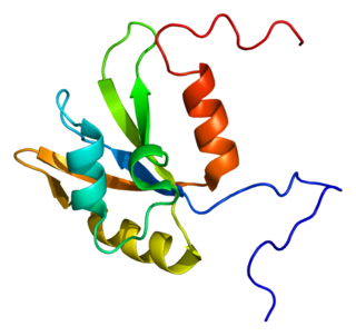
RNA recognition motif, RNP-1 is a putative RNA-binding domain of about 90 amino acids that are known to bind single-stranded RNAs. It was found in many eukaryotic proteins.

<span class="mw-page-title-main">Prp8</span>

Prp8 refers to both the Prp8 protein and Prp8 gene. Prp8's name originates from its involvement in pre-mRNA processing. The Prp8 protein is a large, highly conserved, and unique protein that resides in the catalytic core of the spliceosome and has been found to have a central role in molecular rearrangements that occur there. Prp8 protein is a major central component of the catalytic core in the spliceosome, and the spliceosome is responsible for splicing of precursor mRNA that contains introns and exons. Unexpressed introns are removed by the spliceosome complex in order to create a more concise mRNA transcript. Splicing is just one of many different post-transcriptional modifications that mRNA must undergo before translation. Prp8 has also been hypothesized to be a cofactor in RNA catalysis.

References

  1. 1 2 3 4 5 6 7 8 9 10 11 Long JC, Caceres JF (January 2009). "The SR protein family of splicing factors: master regulators of gene expression". The Biochemical Journal. 417 (1): 15–27. doi:10.1042/BJ20081501. PMID   19061484.
  2. 1 2 3 4 5 6 Zhong XY, Wang P, Han J, Rosenfeld MG, Fu XD (July 2009). "SR proteins in vertical integration of gene expression from transcription to RNA processing to translation". Molecular Cell. 35 (1): 1–10. doi:10.1016/j.molcel.2009.06.016. PMC   2744344 . PMID   19595711.
  3. 1 2 3 4 Shepard PJ, Hertel KJ (2009). "The SR protein family". Genome Biology. 10 (10): 242. doi:10.1186/gb-2009-10-10-242. PMC   2784316 . PMID   19857271.
  4. Roth MB, Murphy C, Gall JG (December 1990). "A monoclonal antibody that recognizes a phosphorylated epitope stains lampbrush chromosome loops and small granules in the amphibian germinal vesicle". The Journal of Cell Biology. 111 (6 Pt 1): 2217–23. doi:10.1083/jcb.111.6.2217. PMC   2116404 . PMID   1703534.
  5. Zahler AM, Lane WS, Stolk JA, Roth MB (May 1992). "SR proteins: a conserved family of pre-mRNA splicing factors". Genes & Development. 6 (5): 837–47. doi: 10.1101/gad.6.5.837 . PMID   1577277.
  6. Manley JL, Krainer AR (June 2010). "A rational nomenclature for serine/arginine-rich protein splicing factors (SR proteins)". Genes & Development. 24 (11): 1073–4. doi:10.1101/gad.1934910. PMC   2878644 . PMID   20516191.
  7. 1 2 3 Ghosh G, Adams JA (February 2011). "Phosphorylation mechanism and structure of serine-arginine protein kinases". The FEBS Journal. 278 (4): 587–97. doi:10.1111/j.1742-4658.2010.07992.x. PMC   3079193 . PMID   21205204.
  8. 1 2 3 4 5 6 7 Hastings ML, Krainer AR (June 2001). "Pre-mRNA splicing in the new millennium". Current Opinion in Cell Biology. 13 (3): 302–9. doi:10.1016/s0955-0674(00)00212-x. PMID   11343900.
  9. 1 2 3 4 Huang Y, Steitz JA (March 2005). "SRprises along a messenger's journey". Molecular Cell. 17 (5): 613–5. doi: 10.1016/j.molcel.2005.02.020 . PMID   15749011.
  10. 1 2 Tenenbaum SA, Aguirre-Ghiso J (November 2005). "Dephosphorylation shows SR proteins the way out". Molecular Cell. 20 (4): 499–501. doi:10.1016/j.molcel.2005.11.005. PMC   2517054 . PMID   16307914.
  11. 1 2 3 4 Twyffels L, Gueydan C, Kruys V (September 2011). "Shuttling SR proteins: more than splicing factors". The FEBS Journal. 278 (18): 3246–55. doi: 10.1111/j.1742-4658.2011.08274.x . PMID   21794093.
  12. 1 2 3 4 5 Blencowe BJ, Bowman JA, McCracken S, Rosonina E (1999). "SR-related proteins and the processing of messenger RNA precursors". Biochemistry and Cell Biology. 77 (4): 277–91. doi:10.1139/o99-048. PMID   10546891.
  13. 1 2 Sanford JR, Gray NK, Beckmann K, Cáceres JF (April 2004). "A novel role for shuttling SR proteins in mRNA translation". Genes & Development. 18 (7): 755–68. doi:10.1101/gad.286404. PMC   387416 . PMID   15082528.
  14. Talukdar I, Sen S, Urbano R, Thompson J, Yates JR, Webster NJ (2011). "hnRNP A1 and hnRNP F modulate the alternative splicing of exon 11 of the insulin receptor gene". PLOS ONE. 6 (11): e27869. Bibcode:2011PLoSO...627869T. doi: 10.1371/journal.pone.0027869 . PMC   3223206 . PMID   22132154.
  15. Wang Z, Burge CB (May 2008). "Splicing regulation: from a parts list of regulatory elements to an integrated splicing code". RNA. 14 (5): 802–13. doi:10.1261/rna.876308. PMC   2327353 . PMID   18369186.
  16. Tacke R, Manley JL (June 1999). "Determinants of SR protein specificity". Current Opinion in Cell Biology. 11 (3): 358–62. doi: 10.1016/s0955-0674(99)80050-7 . PMID   10395560.
  17. 1 2 3 4 Graveley BR (September 2000). "Sorting out the complexity of SR protein functions". RNA. 6 (9): 1197–211. doi:10.1017/S1355838200000960. PMC   1369994 . PMID   10999598.
  18. Lejeune F, Maquat LE (June 2005). "Mechanistic links between nonsense-mediated mRNA decay and pre-mRNA splicing in mammalian cells". Current Opinion in Cell Biology. 17 (3): 309–15. doi:10.1016/j.ceb.2005.03.002. PMID   15901502.