Disorders of consciousness

Last updated
Disorders of consciousness
Specialty Neurology, psychiatry

Disorders of consciousness are medical conditions that inhibit consciousness. [1] Some define disorders of consciousness as any change from complete self-awareness to inhibited or absent self-awareness and arousal. This category generally includes minimally conscious state and persistent vegetative state, but sometimes also includes the less severe locked-in syndrome and more severe but rare chronic coma. [1] [2] Differential diagnosis of these disorders is an active area of biomedical research. [3] [4] [5] Finally, brain death results in an irreversible disruption of consciousness. [1] While other conditions may cause a moderate deterioration (e.g., dementia and delirium) or transient interruption (e.g., grand mal and petit mal seizures) of consciousness, they are not included in this category.[ citation needed ]

Contents

Classification

Patients in such a dramatically altered state of consciousness present unique problems for diagnosis, prognosis and treatment. Assessment of cognitive functions remaining after a traumatic brain injury is difficult. Voluntary movements may be very small, inconsistent and easily exhausted. Quantification of brain activity differentiates patients who sometimes only differ by a brief and small movement of a finger.[ citation needed ]

Consciousness is a complex and multifaceted concept, divided into two main components: Arousal and Awareness. Arousal is associated with functional brainstem neuron populations projecting to both thalamic and cortical neurons. [6] Therefore, the assessment of reflexes (Using the Glasgow Coma Scale) is important to investigate the functional integrity of the brainstem. Awareness is thought to be related with functional integrity of the cerebral cortex and its subcortical connections. The most important point regarding the classification of disorders of consciousness is, that consciousness cannot be measured objectively by any machine, although many scoring systems have been developed for the quantification of consciousness and neuroimaging techniques are important tools for clinical research, extending our understanding of underlying mechanisms involved. [7] [8] Disorders in consciousness represent immense social and ethical issues because the diagnosis is methodologically complex and needs careful interpretation. Also the ethical framework must be further developed to guide research in these patients.[ citation needed ]

Locked-in syndrome

In locked-in syndrome the patient has awareness, sleep-wake cycles, and meaningful behavior (viz., eye-movement), but is isolated due to quadriplegia and pseudobulbar palsy, resulting from the disruption of corticospinal and corticobulbar pathways. Locked-in syndrome is a condition in which a patient is aware and awake but cannot move or communicate verbally due to complete paralysis of nearly all voluntary muscles in the body except for the eyes. Eye or eyelid movements are the main method of communication. [9] Total locked-in syndrome is a version of locked-in syndrome where the eyes are paralyzed as well. [10]

Minimally conscious state

In a minimally conscious state, the patient has intermittent periods of awareness and wakefulness. The criteria for minimally conscious state, that patients are not in a vegetative state but are not able to communicate consistently. This means, that patients have to show limited but reproducible signs of awareness of themself or their environment. This could be following of simple commands, intelligible speech or purposeful behavior (including movements or affective behavior in relation to external stimuli, but not reflexive activity). [11] Further improvement towards full conscious recovery is more likely in this state than in the vegetative state, but still some patients remain in the MCS constantly. [12]

Persistent vegetative state

In a persistent vegetative state, the patient has sleep-wake cycles, but lacks awareness, is not able to communicate and only displays reflexive and non-purposeful behavior. The term refers to an organic body that is able to grow and develop devoid of intellectual activity or social intercourse. [11] The diagnosis of the vegetative state should be questioned when there is any degree of sustained and reproducible visual pursuit or fixation or response to threatening gestures. This state reflects an intact brainstem and allied structures but severely damaged white and gray matter in both cerebral hemispheres. The preservation of these structures maintains arousal and automatic functions. [13] The overall metabolism drops in average to 40-50% of the normal range. After four weeks in a vegetative state (VS), the patient is classified as in a persistent vegetative state. Here the metabolism drops to 30-40% of the normal range but seems to be a result of trans-synaptic neuronal degeneration. [14] Although the diagnosis is problematic, the formal absence of any sign of conscious perception or deliberate action is essential. This diagnosis can be further classified as a permanent vegetative state (PVS) after approximately 1 year of being in a vegetative state after traumatic brain injury [15]

Chronic coma

Like coma, chronic coma results mostly from cortical or white-matter damage after neuronal or axonal injury, or from focal brainstem lesions. Usually the metabolism in the grey matter decreases to 50-70% of the normal range. The patient lacks awareness and arousal. The patient lies with eyes closed and is not aware of self or surroundings. Stimulation cannot produce spontaneous periods of wakefulness and eye-opening, unlike patients in vegetative state. In medicine, a coma (from the Greek κῶμα koma, meaning deep sleep) is a state of unconsciousness, lasting more than six hours in which a person cannot be awakened, fails to respond normally to painful stimuli, light, sound, lacks a normal sleep-wake cycle and does not initiate voluntary actions. Although, according to the Glasgow Coma Scale, a person with confusion is considered to be in the mildest coma. But cerebral metabolism has been shown to correlate poorly with the level of consciousness in patients with mild to severe injury within the first month after traumatic brain injury (TBI). [16] A person in a state of coma is described as comatose. In general patients surviving a coma recover gradually within 2–4 weeks. But recovery to full awareness and arousal is not always possible. Some patients do not progress further than vegetative state or minimally conscious state and sometimes this also results in prolonged stages before further recovery to complete consciousness. [17]

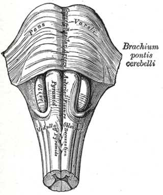
Although a coma patient may appear to be awake, they are unable to consciously feel, speak, hear, or move. For a patient to maintain consciousness, two important neurological components must function impeccably. The first is the cerebral cortex which is the gray matter covering the outer layer of the brain. The other is a structure located in the brainstem, called reticular activating system (RAS or ARAS). Injury to either or both of these components is sufficient to cause a patient to experience a coma.[ citation needed ]

Brain death

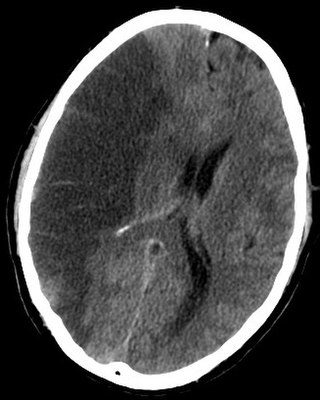
Brain death is the irreversible end of all brain activity, and function (including involuntary activity necessary to sustain life). The main cause is total necrosis of the cerebral neurons following loss of brain oxygenation. After brain death the patient lacks any sense of awareness; sleep-wake cycles or behavior, and typically look as if they are dead or are in a deep sleep-state or coma. Although visually similar to a comatose state such as persistent vegetative state, the two should not be confused. Criteria for brain death differ from country to country. However, the clinical assessments are the same and require the loss of all brainstem reflexes and the demonstration of continuing apnea in a persistently comatose patient (< 4 weeks). [18] Functional imaging using PET or CT scans, typically show a hollow skull phenomenon. This confirms the absence of neuronal function in the whole brain. Patients classified as brain dead are legally dead and can qualify as organ donors, in which their organs are surgically removed and prepared for a particular recipient.[ citation needed ]

Brain death is one of the deciding factors when pronouncing a trauma patient as dead. Determining function and presence of necrosis after trauma to the whole brain or brain-stem may be used to determine brain death, and is used in many states in the US.[ citation needed ]

Methodological problems

Metabolic studies are useful, but they are not able to identify neural activity within a specific region to specific cognitive processes. Functionality can only be identified at the most general level: Metabolism in cortical and subcortical regions that may contribute to cognitive processes.[ citation needed ]

At present, there is no established relation between cerebral metabolic rates of glucose or oxygen as measured by PET and patient outcome. The decrease of cerebral metabolism occurs also when patients are treated with anesthetics to the point of unresponsiveness. Lowest value (28% of normal range) have been reported during propofol anesthesia. Also, deep sleep represents a phase of decreased metabolism (down to 40% of the normal range) [19] [20] In general, quantitative PET studies and the assessment of cerebral metabolic rates depends on many assumptions. PET, for example, requires a correction factor, the lumped constant, which is stable in healthy brains. There are reports that a global decrease of this constant emerges after a traumatic brain injury. [21] But, not only the correction factors change due to TBI. Another issue is the possibility of anaerobic glycolysis that could occur after TBI. In such a case, the glucose levels measured by the PET are not tightly connected to the oxygen consumption of the patient's brain. [22] Third point regarding PET scans is the overall measurement per unit volume of brain tissue. The imaging can be affected by the inclusion of metabolically inactive spaces e.g. cerebrospinal fluid in the case of gross hydrocephalus, which artificially lowers the calculated metabolism. [23] Also, the issue of radiation exposure must be considered in patients with already severely damaged brains and preclude longitudinal or follow-up studies.[ citation needed ]

Ethical issues

Disorders of consciousness present a variety of ethical concerns. Most obvious is the lack of consent in any treatment decisions. Patients in PVS or MCS are not able to decide for the possibility of withdrawal of life-support. It is also a general question whether they should receive life-sustaining therapy and, if so, for what duration. The problems regarding a patient's consent also account for neuroimaging studies. Without patient's consent, such studies are perceived as unethical. [24] Additionally, only few patients have created advance directives before losing decision-making capacity.[ citation needed ] Typically, approval must be obtained from family or legal representatives depending on governmental and hospital guidelines but, even with the consent of representatives, researchers have been refused grants, ethics committee approval and publication.[ citation needed ]

Social issues arise from the enormous costs associated with people who have disorders of consciousness, especially chronic comatose and vegetative patients, when recovery is highly unlikely and treatment in the ICU is considered futile by clinicians.[ citation needed ] In addition to the aforementioned problems, the question rises why medical resources were being used not for the broader public good but for patients who seemed to have only little to gain from them. Nevertheless, the irreversibility of these conditions remains an open question. Some studies demonstrated that some patients with disorders of consciousness may be aware despite clinical unresponsiveness.[ citation needed ] These findings could have a major impact on ethical and social issues. [25]

See also

Related Research Articles

<span class="mw-page-title-main">Consciousness</span>  Awareness of internal and external existence

Consciousness, at its simplest, is awareness of internal and external existence. However, its nature has led to millennia of analyses, explanations and debates by philosophers, theologians, linguists, and scientists. Opinions differ about what exactly needs to be studied or even considered consciousness. In some explanations, it is synonymous with the mind, and at other times, an aspect of mind. In the past, it was one's "inner life", the world of introspection, of private thought, imagination and volition. Today, it often includes any kind of cognition, experience, feeling or perception. It may be awareness, awareness of awareness, or self-awareness either continuously changing or not. The disparate range of research, notions and speculations raises a curiosity about whether the right questions are being asked.

<span class="mw-page-title-main">Coma</span> State of unconsciousness

A coma is a deep state of prolonged unconsciousness in which a person cannot be awakened, fails to respond normally to painful stimuli, light, or sound, lacks a normal wake-sleep cycle and does not initiate voluntary actions. The person may experience respiratory and circulatory problems due to the body's inability to maintain normal bodily functions. People in a coma often require extensive medical care to maintain their health and prevent complications such as pneumonia or blood clots. Coma patients exhibit a complete absence of wakefulness and are unable to consciously feel, speak or move. Comas can be derived by natural causes, or can be medically induced.

<span class="mw-page-title-main">Head injury</span> Serious trauma to the cranium

A head injury is any injury that results in trauma to the skull or brain. The terms traumatic brain injury and head injury are often used interchangeably in the medical literature. Because head injuries cover such a broad scope of injuries, there are many causes—including accidents, falls, physical assault, or traffic accidents—that can cause head injuries.

<span class="mw-page-title-main">Locked-in syndrome</span> Condition in which a patient is aware but completely paralysed

Locked-in syndrome (LIS), also known as pseudocoma, is a condition in which a patient is aware but cannot move or communicate verbally due to complete paralysis of nearly all voluntary muscles in the body except for vertical eye movements and blinking. The individual is conscious and sufficiently intact cognitively to be able to communicate with eye movements. Electroencephalography results are normal in locked-in syndrome. Total locked-in syndrome, or completely locked-in state (CLIS), is a version of locked-in syndrome wherein the eyes are paralyzed as well. Fred Plum and Jerome B. Posner coined the term for this disorder in 1966.

Brain death is the permanent, irreversible, and complete loss of brain function which may include cessation of involuntary activity necessary to sustain life. It differs from persistent vegetative state, in which the person is alive and some autonomic functions remain. It is also distinct from comas as long as some brain and bodily activity and function remain, and it is also not the same as the condition locked-in syndrome. A differential diagnosis can medically distinguish these differing conditions.

A vegetative state (VS) or post-coma unresponsiveness (PCU), is a disorder of consciousness in which patients with severe brain damage are in a state of partial arousal rather than true awareness. After four weeks in a vegetative state, the patient is classified as being in a persistent vegetative state (PVS). This diagnosis is classified as a permanent vegetative state some months after a non-traumatic brain injury or one year after a traumatic injury. The term unresponsive wakefulness syndrome may be alternatively used, as "vegetative state" has some negative connotations among the public.

<span class="mw-page-title-main">Brain damage</span> Destruction or degeneration of brain cells

Neurotrauma, brain damage or brain injury (BI) is the destruction or degeneration of brain cells. Brain injuries occur due to a wide range of internal and external factors. In general, brain damage refers to significant, undiscriminating trauma-induced damage.

<span class="mw-page-title-main">Cerebral edema</span> Excess accumulation of fluid (edema) in the intracellular or extracellular spaces of the brain

Cerebral edema is excess accumulation of fluid (edema) in the intracellular or extracellular spaces of the brain. This typically causes impaired nerve function, increased pressure within the skull, and can eventually lead to direct compression of brain tissue and blood vessels. Symptoms vary based on the location and extent of edema and generally include headaches, nausea, vomiting, seizures, drowsiness, visual disturbances, dizziness, and in severe cases, coma and death.

An induced coma – also known as a medically induced coma (MIC), barbiturate-induced coma, or drug-induced coma – is a temporary coma brought on by a controlled dose of an anesthetic drug, often a barbiturate such as pentobarbital or thiopental. Other intravenous anesthetic drugs such as midazolam or propofol may be used.

<span class="mw-page-title-main">Minimally conscious state</span> Disorder of Consciousness where overt signs of awareness are preserved

A minimally conscious state or MCS is a disorder of consciousness distinct from persistent vegetative state and locked-in syndrome. Unlike persistent vegetative state, patients with MCS have partial preservation of conscious awareness. MCS is a relatively new category of disorders of consciousness. The natural history and longer term outcome of MCS have not yet been thoroughly studied. The prevalence of MCS was estimated to be 9 times of PVS cases, or between 112,000 and 280,000 in the US by year 2000.

<span class="mw-page-title-main">Central neurogenic hyperventilation</span> Abnormal pattern of breathing

Central neurogenic hyperventilation (CNH) is an abnormal pattern of breathing characterized by deep and rapid breaths at a rate of at least 25 breaths per minute. Increasing irregularity of this respiratory rate generally is a sign that the patient will enter into coma. CNH is unrelated to other forms of hyperventilation, like Kussmaul's respirations. CNH is the human body's response to reduced carbon dioxide levels in the blood. This reduction in carbon dioxide is caused by contraction of cranial arteries from damage caused by lesions in the brain stem. However, the mechanism by which CNH arises as a result from these lesions is still very poorly understood. Current research has yet to provide an effective means of treatment for the rare number of patients who are diagnosed with this condition.

<span class="mw-page-title-main">Brain herniation</span> Potentially deadly side effect of very high pressure within the skull

Brain herniation is a potentially deadly side effect of very high pressure within the skull that occurs when a part of the brain is squeezed across structures within the skull. The brain can shift across such structures as the falx cerebri, the tentorium cerebelli, and even through the foramen magnum. Herniation can be caused by a number of factors that cause a mass effect and increase intracranial pressure (ICP): these include traumatic brain injury, intracranial hemorrhage, or brain tumor.

<span class="mw-page-title-main">Brain ischemia</span> Medical condition

Brain ischemia is a condition in which there is insufficient bloodflow to the brain to meet metabolic demand. This leads to poor oxygen supply or cerebral hypoxia and thus leads to the death of brain tissue or cerebral infarction/ischemic stroke. It is a sub-type of stroke along with subarachnoid hemorrhage and intracerebral hemorrhage.

<span class="mw-page-title-main">Posterior cingulate cortex</span> Caudal part of the cingulate cortex of the brain

The posterior cingulate cortex (PCC) is the caudal part of the cingulate cortex, located posterior to the anterior cingulate cortex. This is the upper part of the "limbic lobe". The cingulate cortex is made up of an area around the midline of the brain. Surrounding areas include the retrosplenial cortex and the precuneus.

<span class="mw-page-title-main">Neural correlates of consciousness</span> Neuronal events sufficient for a specific conscious percept

The neural correlates of consciousness (NCC) refer to the relationships between mental states and neural states and constitute the minimal set of neuronal events and mechanisms sufficient for a specific conscious percept. Neuroscientists use empirical approaches to discover neural correlates of subjective phenomena; that is, neural changes which necessarily and regularly correlate with a specific experience. The set should be minimal because, under the materialist assumption that the brain is sufficient to give rise to any given conscious experience, the question is which of its components are necessary to produce it.

Traumatic brain injury can cause a variety of complications, health effects that are not TBI themselves but that result from it. The risk of complications increases with the severity of the trauma; however even mild traumatic brain injury can result in disabilities that interfere with social interactions, employment, and everyday living. TBI can cause a variety of problems including physical, cognitive, emotional, and behavioral complications.

<span class="mw-page-title-main">Steven Laureys</span> Belgian neurologist (born 1968)

Steven Laureys is a Belgian neurologist. He is principally known as a clinician and researcher in the field of neurology of consciousness.

<span class="mw-page-title-main">Altered level of consciousness</span> Measure of arousal other than normal

An altered level of consciousness is any measure of arousal other than normal. Level of consciousness (LOC) is a measurement of a person's arousability and responsiveness to stimuli from the environment. A mildly depressed level of consciousness or alertness may be classed as lethargy; someone in this state can be aroused with little difficulty. People who are obtunded have a more depressed level of consciousness and cannot be fully aroused. Those who are not able to be aroused from a sleep-like state are said to be stuporous. Coma is the inability to make any purposeful response. Scales such as the Glasgow coma scale have been designed to measure the level of consciousness.

Brainstem death is a clinical syndrome defined by the absence of reflexes with pathways through the brainstem – the "stalk" of the brain, which connects the spinal cord to the mid-brain, cerebellum and cerebral hemispheres – in a deeply comatose, ventilator-dependent patient. Identification of this state carries a very grave prognosis for survival; cessation of heartbeat often occurs within a few days, although it may continue for weeks if intensive support is maintained.

Fred Plum was an American neurologist who developed the terms "persistent vegetative state" and "locked-in syndrome" as part of his continuing research on consciousness and comas and care of the comatose.

References

  1. 1 2 3 Bernat JL (8 Apr 2006). "Chronic disorders of consciousness". Lancet. 367 (9517): 1181–1192. doi:10.1016/S0140-6736(06)68508-5. PMID   16616561. S2CID   13550675.
  2. Bernat JL (20 Jul 2010). "The natural history of chronic disorders of consciousness". Neurology. 75 (3): 206–207. doi:10.1212/WNL.0b013e3181e8e960. PMID   20554939. S2CID   30959964.
  3. Coleman MR, Davis MH, Rodd JM, Robson T, Ali A, Owen AM, Pickard JD (Sep 2009). "Towards the routine use of brain imaging to aid the clinical diagnosis of disorders of consciousness". Brain. 132 (9): 2541–2552. doi: 10.1093/brain/awp183 . PMID   19710182.
  4. Monti MM, Vanhaudenhuyse A, Coleman MR, Boly M, Pickard JD, Tshibanda L, Owen AM, Laureys S (18 Feb 2010). "Willful modulation of brain activity in disorders of consciousness". N Engl J Med. 362 (7): 579–589. doi: 10.1056/NEJMoa0905370 . PMID   20130250. S2CID   13358991.
  5. Seel RT, Sherer M, Whyte J, Katz DI, Giacino JT, Rosenbaum AM, Hammond FM, Kalmar K, Pape TL, et al. (Dec 2010). "Assessment scales for disorders of consciousness: evidence-based recommendations for clinical practice and research". Arch Phys Med Rehabil. 91 (12): 1795–1813. doi:10.1016/j.apmr.2010.07.218. PMID   21112421.
  6. Steriade M, Jones BG, McCormick D. Thalamus. New York Elesevier, 1997.
  7. Laureys S, Majerus S, Moonen G. Assessing consciousness in critically ill patients. In: Vincent JL, ed. 2002 Yearbook of Intensive Care and Emergency Medicine. Heidelberg: Springer-Verlag, 2002: 715-27
  8. Jain, Ritika (2020). "Electrophysiological and Neuroimaging Studies – During Resting State and Sensory Stimulation in Disorders of Consciousness: A Review". Front. Neurosci. 14:555093: 555093. doi: 10.3389/fnins.2020.555093 . PMC   7522478 . PMID   33041757.
  9. American Congress of Rehabilitation Medicine. Recommendations for use of uniform nomenclature pertinent to patients with severe alternations of consciousness. Arch Pyhs Med Rehabil 1995; 76;: 205-9
  10. Bauer, G. and Gerstenbrand, F. and Rumpl, E. (1979). "Varieties of the locked-in syndrome". Journal of Neurology. 221 (2): 77–91. doi:10.1007/BF00313105. PMID   92545. S2CID   10984425.{{cite journal}}: CS1 maint: multiple names: authors list (link)
  11. 1 2 Laureys, Steven, Adrian M. Owen, and Nicholas D. Schiff. "Brain function in coma, vegetative state, and related disorders." The Lancet Neurology 3.9 (2004): 537-546.
  12. Giacino JT. Disorders of consciousness: differential diagnosis and neuropathologic features. Semin Neurol 1997; 17:105-11
  13. Laureys S, Faymonville ME, Goldman S, et al. Impaired cerebral connectivity in vegetative state. In: Gjedde A, Hansen SB, Knudsen GM, Paulson OB, eds. Physiological imaging of the brain with PET. San Diego: Academic Press, 2000: 329-34.
  14. Tommasino C, Grana C, Lucignani G, Torri G, Fazio F. Regional cerebral metabolism of glucose in comatose and vegetative patients. J Neurosurg Anesthesiol 1995; 7: 109-16
  15. Menon DK, Owen AM, Williams EJ, et al. Cortical processing in persistent vegetative state. Lancet 1998; 352: 200.
  16. Bersneider M, Havda DA, Lee SM, et al. Dissociation of cerebral glucose metabolism and level of consciousness during the period of metabolic depression following human traumatic brain injury. J Neurotrauma 200; 17: 389-401
  17. Plum F, Posner JB. The diagnosis of stupor and coma (3rd edn). Philadelphia: FA Davis, 1983.
  18. Medical Consultants on the Diagnosis of Death. Guidelines for the determination of death: report of the medical consultants on the diagnosis of death to the President's commission for the Study of Ethical Problems in Medicine and Biomedical and Behavioral Research. JAMA 1981; 246: 2184-86
  19. Maquet P, Degueldre C, Delfiore G, et al. Functional neuroanatomy of human slow wave sleep. J Neurosci 1997; 17 2807-12
  20. Buchsbaum MS, Gillin JC, Wu J, et al. Regional cerebral glucose metabolic rate in human sleep assessed by positron emission tomography. Life Sci 1989; 45: 1349-56
  21. Wu HM, Huang SC, Hattori N, et al. Selective metabolic reduction in gray matter acutely following human traumatic brain injury. J Neurotrauma 2004; 21: 149-61
  22. Hovda DA, Becker DP, Katayama Y. Secondary injury and acidosis. J Neurotrauma 1992; 9 (suppl 1): S47-60
  23. Meltzer CC, Zubieta JK, Links JM, et al. MR-based correction of brain PET measurements for heterogeneous gray matter radioactivity distribution. J Cereb Blood Flow Metlab 1996; 16: 650-58
  24. Fins JJ. Constructing an ethical stereotaxy for severe brain injury: balancing risks, benefits and access. Nat Rev Neurosci 2003; 4: 323-27.
  25. Jox RJ, Bernat JL, Laureys S, Racine E. Disorders of consciousness: responding to request for novel diagnostic and therapeutic interventions. Lancet Neurol. 2012; 11(8): 732-38