Fusarium solani

Last updated

Fusarium solani
Fusarium solani (Mart.) Sacc. 5393379.jpg
Scientific classification OOjs UI icon edit-ltr.svg
Domain: Eukaryota
Kingdom: Fungi
Division: Ascomycota
Class: Sordariomycetes
Order: Hypocreales
Family: Nectriaceae
Genus: Fusarium
Species:
F. solani
Binomial name
Fusarium solani
(Mart.) Sacc. (1881)
Synonyms
  • Fusisporium solaniMart. (1842)
  • Fusarium solani(Mart.) Appel & Wollenw. (1910)
  • Neocosmospora solani(Martius) L. Lombard & Crous (2015)
  • Fusarium martiiAppel & Wollenw. (1910)
  • Nectria cancriRutgers (1913)
  • Fusarium striatumSherb. (1915)
  • Fusarium solani var. minusWollenw. (1916)
  • Cephalosporium keratoplasticumT. Morik. (1939)
  • Fusarium solani f. keratitisY.N. Ming & T.F. Yu (1966)
  • Cylindrocarpon vaginaeC. Booth, Y.M. Clayton & Usherw. (1985)
Effects of F. solani on Alfalfa Fusarium solani f.sp. pisi sintomi su cece.jpg
Effects of F. solani on Alfalfa
Hyphae of F. solani Fusarium solani (257 25) Cultured and stained deuteromycetes.jpg
Hyphae of F. solani

Fusarium solani is a species complex of at least 26 closely related filamentous fungi in the division Ascomycota, family Nectriaceae. [1] It is the anamorph of Nectria haematococca . [1] It is a common soil fungus and colonist of plant materials. [2] Fusarium solani is implicated in plant diseases as well as in serious human diseases such as fungal keratitis. [1]

Contents

History and taxonomy

The genus Fusarium was described in 1809 by Link. [3] In the 1930s, Wollenweber and Reinking organized the genus Fusarium into sections, including Martiella and Ventricosum, [3] which were collapsed together by Snyder and Hansen in the 1940s to form a single species, Fusarium solani; [3] one of nine Fusarium species they recognized based on morphological features. [2] The current concept of F. solani is as a species complex consisting of multiple, closely related and morphologically poorly distinguishable, "cryptic" species with characteristic genetic differences. [4] There is a proposed concept for the entire genus - widely subscribed by specialists - that would include this complex. [5] [6] However, there is a smaller counterproposal that radically refiles the genus including making this complex into a genus Neocosmospora. [7] The fungus is allied with the sexual species, Nectria haematococca, in the family Nectriaceae (phylum Ascomycota). [1]

Growth and morphology

Like other species in its genus, Fusarium solani produces colonies that are white and cottony. However, instead of developing a pink or violet centre like most Fusarium species, [8] F. solani becomes blue-green or bluish brown. [1] [8] [9] On the underside, they may be pale, tea-with-milk-brown, or red-brown. [1] However, some clinical isolates have been blue-green or ink-blue on the underside. [1] F. solani colonies are low-floccose, loose, slimy, and sporadic. [1] When grown on potato dextrose agar (PDA), this fungus grows rapidly, but not as rapidly as Fusarium oxysporum . [9] In PDA, F. solani colonies reach a diameter of 64–70 mm in 7 days. [1]

F. solani has aerial hyphae that give rise to conidiophores laterally. [1] The conidiophores branch into thin, elongated monophialides that produce conidia. [1] [9] Phialides that produce macroconidia are shorter than those that produce microconidia. [1] The macroconidia produced by F. solani are slightly curved, hyaline, and broad, [1] often aggregating in fascicles. [10] Typically the macroconidia of this species have 3 septa but may have as many as 4–5. [1] [10] Microconidia have thickened basal cells and tapered, rounded apical cells. [1] However, some F. solani isolates have pointed, rather than rounded, macroconidia. [1] Microconidia are oval or cylindrical, hyaline, and smooth. [1] Some microconidia may be curved. [1] Microconidia typically lack septa, but occasionally they may have up to two. [1] Fusarium solani also forms chlamydospores most commonly under suboptimal growth conditions. [10] These may be produced in pairs or individually. [9] [10] They are abundant, have rough walls, and are 6-11 μm. [1] F. solani chlamydospores are also brown and round. [11]

Ecology

F. solani is found in soil worldwide. [2] However, a given species within the complex may not be as widespread [2] and may not have the same ecology as others in the complex. [10] In general, as a soil fungus, F. solani is associated with the roots of plants [1] and may be found as deep in the ground as 80 cm. [10] It is frequently isolated in tropic, subtropic, and temperate locations, and less frequently isolated from alpine habitats. [10] The pH of soil does not have a significant effect on F. solani, however, soil fumigation causes an increase in occurrence. [10] F. solani is typically sensitive to soil fungicides. [10] F. solani has been found in ponds, rivers, sewage facilities, and water pipes. [1] It has also been found in larvae and adults of the picnic beetle, is a symbiote of the ambrosia beetle. [10]

Life cycle

F. solani can be found in soils worldwide, where its chlamydospores overwinter on plant tissue/seed or as mycelium in the soil. [12] The pathogen enters hosts through developing roots, where it can infect the host. After infection, F. solani produces asexual macro and microconidia which are dispersed through wind and rain. [13] The pathogen can persist in the soil for a decade, and if left unchecked can cause complete crop loss.

Physiology and biochemistry

F. solani have 5-13 chromosomes, [3] [10] with a genome size of about 40 Mb. [3] The GC-content of its DNA is 50%. [10] Mycelium of F. solani is rich in the amino acid alanine, as well as a range of fatty acids including δ-aminobutyric-, palmitic-, oleic-, and linolenic acids. [10] Fusarium solani requires potassium for growth, [10] and develops a feathery pattern when potassium levels are below 3 mM. [3] In culture the following disaccharides are utilized (from most- to least preferential): mannose, rhamnose and sorbose. [10] This species can decompose cellulose at an optimal pH of 6.5 and temperature of 30 °C. [10] It can also metabolise steroids and lignin, [3] and reduce Fe3+ to Fe2+. [10] Fusarium solani produces mycotoxins like Fusaric acid and naphthoquinones. [1] [14] Other toxins have also been isolated from F. solani, including:

Pathology

Humans

F. solani is largely resistant to typical antifungal agents. [3] The most effective antifungals in treating F. solani infections are amphotericin B and natamycin; however, these agents have only modest success in the treatment of serious systemic infection. [3]

As of 2006, there has been increasing evidence that F. solani  can act as a causal agent of mycoses in humans. [15] F. solani has been implicated in the following diseases: disseminated disease, osteomyelitis, skin infection, fungemia, and endophthalmitis. [16] Half of human disease involving Fusarium is caused by F. solani and it is involved in most cases of systemic fusariosis and corneal infections. [4] In immunocompromised patients, F. solani is one of the most common agents in disseminated and cutaneous infections. [1]

In the southern USA, fungal keratitis has been most commonly caused by F. solani, as well as F. oxysporum. Cases occur most frequently during harvest season as a result of corneal trauma from dust or plant material. Fungal spores come into contact with the damaged cornea and grow. Without treatment, the hyphae can grow into the cornea and into the anterior chamber of the eye. [17] F. solani is also a major cause of fungal keratitis in HIV positive patients in Africa. [1]

As of 2011, F. solani was implicated in cases of fungal keratitis involving the Bausch and Lomb ReNu contact lens solution. [4] Some strains of F. solani can produce a biofilm on soft contact lenses. However, when lenses are cleaned correctly with solution, these biofilms are prevented. [4] Prevention also includes leaving lenses in polyhexanide biguanide solution overnight to inhibit F. solani. [1] Other risk factors of contact lens-related Fusarium keratitis include use of daily-wear lenses beyond the recommended timeline and overnight wear. [4]

An investigation into a meningitis outbreak of 79 cases since October 2022, which had killed 35 people (34 of them women who had undergone cesarean section) in Durango (city) revealed contamination of bupivacaine with Fusarium solani in 4 batches, used by an anesthesiologist. [18] US news reported however, that the anesthesiologist used multi-dose vials of morphine, which he would administer in more than one patient for his anesthesias in the 4 private hospitals. [19] As of May 26, 2023 WHO had been asked to declare a public health emergency. [20]

As of June 1, 2023, a multistate outbreak of meningitis due to F. solani was ongoing among patients who underwent epidural anesthesia at two clinics in the Mexican city of Matamoros, Tamaulipas, with a total of 212 residents in 25 US states identified as being at risk, two of whom had died. [21] [22]

Other animals

F. solani is implicated in cutaneous infections of young turtles as well as infections of turtle egg shells. [1] It has also caused infections in Australian crocodile farms, sea lions and grey seals. [1] F. solani is a facultative pathogen of the castor bean tick. It is also lethal to southern pine beetles. [10]

Plants

F. solani rots the roots of its host plant. [23] It also causes soft rot of plant tissues by penetrating plant cell walls and destroying the torus. [10] It is implicated, along with Pythium myriotylum , in pod rot of the pods of groundnuts. [10] F. solani can cause damping off, corn rot, and root rot, as well as sudden death of soybeans (SDS). It is a very generalistic fungal species and has been known to infect peas, beans, potatoes, and many types of cucurbits. [24] Symptoms include general plant decline, wilting, and large necrotic spots on tap roots.

Recently the pathogen has also done serious damage to olive trees throughout the mediterranean.[ citation needed ]

Virulence of this agent in plants is controlled by the cutinase genes cut1 and cut2. These genes are upregulated by exposure to the plant's cutin monomers. [25] F. solani is known to cause sudden death syndrome in soybeans, and it is also known to cause disease in other economically important crops such as avocado, citrus, orchids, passion fruit, peas, peppers, potato, and squash. [3]

Management

Agriculture

The ubiquitous nature of  F. solani gives rise to a plethora of management practices developed independently. One particular method is the use of the bacterial complex Burkholderia cepacia,  which is a registered control method. This bacterial complex has been shown to produce several types of antibiotics (depending on the strain), and can act as a substitute for chemical pesticides. [26] Precautionary methods include planting during warm/dry weather, 3 plus years of crop rotation of non host species, and avoiding dense seed planting. [12]

Humans

In the 2023 Matamoros outbreak of F. solani meningitis CDC recommended liposomal amphotericin B and voriconazole, [27] however, disease progressed on this regimen, [28] and patients were trialed on fosmanogepix through a compassionate use authorization.

Biotechnology

F. solani has been investigated as a biological control for certain plants including leafy spurge, morning glory, striga, gourd, and water hyacinth. [3]

Related Research Articles

<span class="mw-page-title-main">Fusarium ear blight</span> Fungal disease of cereals

Fusarium ear blight (FEB), is a fungal disease of cereals, including wheat, barley, oats, rye and triticale. FEB is caused by a range of Fusarium fungi, which infects the heads of the crop, reducing grain yield. The disease is often associated with contamination by mycotoxins produced by the fungi already when the crop is growing in the field. The disease can cause severe economic losses as mycotoxin-contaminated grain cannot be sold for food or feed.

<i>Fusarium oxysporum</i> Species of fungus

Fusarium oxysporum, an ascomycete fungus, comprises all the species, varieties and forms recognized by Wollenweber and Reinking within an infrageneric grouping called section Elegans. It is part of the family Nectriaceae.

<span class="mw-page-title-main">Fusarium wilt</span> Fungal plant disease

Fusarium wilt is a common vascular wilt fungal disease, exhibiting symptoms similar to Verticillium wilt. This disease has been investigated extensively since the early years of this century. The pathogen that causes Fusarium wilt is Fusarium oxysporum. The species is further divided into formae speciales based on host plant.

<span class="mw-page-title-main">Panama disease</span> Plant disease of bananas

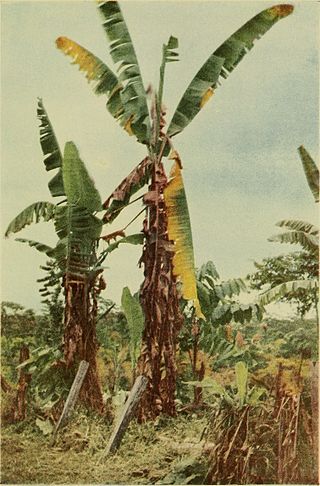
Panama disease is a plant disease that infects banana plants. It is a wilting disease caused by the fungus Fusarium oxysporum f. sp. cubense (Foc). The pathogen is resistant to fungicides and its control is limited to phytosanitary measures.

<i>Fusarium</i> Genus of fungi

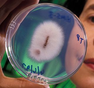
Fusarium is a large genus of filamentous fungi, part of a group often referred to as hyphomycetes, widely distributed in soil and associated with plants. Most species are harmless saprobes, and are relatively abundant members of the soil microbial community. Some species produce mycotoxins in cereal crops that can affect human and animal health if they enter the food chain. The main toxins produced by these Fusarium species are fumonisins and trichothecenes. Despite most species apparently being harmless, some Fusarium species and subspecific groups are among the most important fungal pathogens of plants and animals.

<span class="mw-page-title-main">Conidium</span> Asexual, non-motile spore of a fungus

A conidium, sometimes termed an asexual chlamydospore or chlamydoconidium, is an asexual, non-motile spore of a fungus. The word conidium comes from the Ancient Greek word for dust, κόνις (kónis). They are also called mitospores due to the way they are generated through the cellular process of mitosis. They are produced exogenously. The two new haploid cells are genetically identical to the haploid parent, and can develop into new organisms if conditions are favorable, and serve in biological dispersal.

Fungal keratitis is a fungal infection of the cornea, which can lead to blindness. It generally presents with a red, painful eye and blurred vision. There is also increased sensitivity to light, and excessive tears or discharge.

<i>Rhizoctonia solani</i> Species of fungus

Rhizoctonia solani is a species of fungus in the order Cantharellales. Basidiocarps are thin, effused, and web-like, but the fungus is more typically encountered in its anamorphic state, as hyphae and sclerotia. The name Rhizoctonia solani is currently applied to a complex of related species that await further research. In its wide sense, Rhizoctonia solani is a facultative plant pathogen with a wide host range and worldwide distribution. It causes various plant diseases such as root rot, damping off, and wire stem. It can also form mycorrhizal associations with orchids.

<i>Fusarium culmorum</i> Fungal disease, head blight of wheat

Fusarium culmorum is a fungal plant pathogen and the causal agent of seedling blight, foot rot, ear blight, stalk rot, common root rot and other diseases of cereals, grasses, and a wide variety of monocots and dicots. In coastal dunegrass, F. culmorum is a nonpathogenic symbiont conferring both salt and drought tolerance to the plant.

<i>Gibberella zeae</i> Species of fungus

Gibberella zeae, also known by the name of its anamorph Fusarium graminearum, is a fungal plant pathogen which causes fusarium head blight (FHB), a devastating disease on wheat and barley. The pathogen is responsible for billions of dollars in economic losses worldwide each year. Infection causes shifts in the amino acid composition of wheat, resulting in shriveled kernels and contaminating the remaining grain with mycotoxins, mainly deoxynivalenol (DON), which inhibits protein biosynthesis; and zearalenone, an estrogenic mycotoxin. These toxins cause vomiting, liver damage, and reproductive defects in livestock, and are harmful to humans through contaminated food. Despite great efforts to find resistance genes against F. graminearum, no completely resistant variety is currently available. Research on the biology of F. graminearum is directed towards gaining insight into more details about the infection process and reveal weak spots in the life cycle of this pathogen to develop fungicides that can protect wheat from scab infection.

Ceratocystis paradoxa or Black Rot of Pineapple is a plant pathogen that is a fungus, part of the phylum Ascomycota. It is characterized as the teleomorph or sexual reproduction stage of infection. This stage contains ascocarps, or sacs/fruiting bodies, which contain the sexually produced inoculating ascospores. These are the structures which are used primarily to survive long periods of time or overwinter to prepare for the next growing season of its host. Unfortunately, the sexual stage is not often seen in the natural field but instead the anamorph, or asexual stage is more commonly seen. This asexual stage name is Thielaviopsis paradoxa and is the common cause of Black rot or stem-end rot of its hosts.

Fusarium incarnatum is a fungal pathogen in the genus Fusarium, family Nectriaceae. It is usually associated with over 40 phylogenetic species in the natural environment to form the Fusarium incarnatum-equiseti species complex(FIESC). This complex is widespread across the globe in subtropical and temperate regions, resulting in many reported cases of crop diseases. It produces various mycotoxins including trichothecenes zearalenone, causing both plant and animal diseases.

<i>Fusarium oxysporum</i> f.sp. <i>ciceris</i> Fungal plant pathogen

Fusarium oxysporum f.sp. ciceris is a fungal plant pathogen that causes fusarium wilt of chickpea.

Fusarium redolens is a species of fungus in the genus Fusarium and family Nectriaceae. This species is a soil-borne plant pathogen in temperate prairies. It causes diseases such as root, crown, and spear rot, seedling damping-off, and wilting disease. It is a known producer of the alkaloids peimisine and imperialine-3β-d-glucoside, which has implications for traditional Chinese medicine.

<i>Fusarium oxysporum</i> f.sp. <i>cubense</i> Fungus, causes banana wilt/Panama disease

Fusarium oxysporum f. sp. cubense is a fungal plant pathogen that causes Panama disease of banana, also known as Fusarium wilt. The fungi and the related disease are responsible for widespread pressure on banana growing regions, destroying the economic viability of several commercially important banana varieties.

<i>Fusarium circinatum</i> Species of fungus

Fusarium circinatum is a fungal plant pathogen that causes the serious disease pitch canker on pine trees and Douglas firs. The most common hosts of the pathogen include slash pine, loblolly pine, Monterey pine, Mexican weeping pine, and Douglas fir. Like other Fusarium species in the phylum Ascomycota, it is the asexual reproductive state of the fungus and has a teleomorph, Gibberella circinata.

<span class="mw-page-title-main">Fungal contamination of contact lenses</span>

Microbial corneal infection is the most serious and "most common vision threatening" complication of contact lens wear, which is believed to be strongly associated with contact lens cases. Such infections "are being increasingly recognized as an important cause of morbidity and blindness" and "may even be life-threatening." While the cornea is believed to be the most common site for fungal eye infections, other parts of the eye such as the orbit, sclera, eyelids, and more may also be involved. Contact lens cases are recognized as a "potential source of pathogens associated with corneal ulcers" and according to Moorfields Eye Hospital, contact lens wear is “the most prevalent risk factor for new cases of corneal ulcers.” Contaminants "isolated from contact lens associated corneal ulcers have often been shown to be" the same as found in the patient's contact lens case, thus providing evidence contaminated contact lens cases may be a "replenishable source of pathogenic microbes."

<i>Fusarium mangiferae</i> Species of fungus

Fusarium mangiferae is a fungal plant pathogen that infects mango trees. Its aerial mycelium is white and floccose. Conidiophores on aerial mycelium originating erect and prostrate from substrate; they are sympodially branched bearing mono and polyphialides. Polyphialides have 2–5 conidiogenous openings. Phialides on the aerial conidiophores mono- and polyphialidic. Sterile hyphae are absent. Microconidia are variable in shape, obovoid conidia are the most abundant type, oval to allantoid conidia occurring occasionally. Microconidia mostly 0-septate with 1-septate conidia occurring less abundantly. Sporodochia are present. Macroconidia are long and slender, usually 3–5 septate. Chlamydospores are absent.

Sudden death syndrome (SDS), a disease in soybean plants, quickly spread across the southern United States in the 1970s, eventually reaching most agricultural areas of the US. SDS is caused by multiple Fusarium fungi in the Fusariumsolani complex. Fusarium virguliforme is the sole causal agent in North America. In South America, Fusarium brasiliense, F. cuneirostrum, F. tucumaniae, and F. virguliforme are all causal agents. Losses could exceed hundreds of millions of dollars in US soybean markets alone making it one of the most important diseases found in Soybeans across the US.

<i>Epidermophyton floccosum</i> Species of fungus

Epidermophyton floccosum is a filamentous fungus that causes skin and nail infections in humans. This anthropophilic dermatophyte can lead to diseases such as tinea pedis, tinea cruris, tinea corporis and onychomycosis. Diagnostic approaches of the fungal infection include physical examination, culture testing, and molecular detection. Topical antifungal treatment, such as the use of terbinafine, itraconazole, voriconazole, and ketoconazole, is often effective.

References

  1. 1 2 3 4 5 6 7 8 9 10 11 12 13 14 15 16 17 18 19 20 21 22 23 24 25 26 27 28 Summerbell, Richard (2003). "Ascomycetes: Aspergillus, Fusarium, Sporothrix, Piedraia, and Their Relatives". In Howard, Dexter H. (ed.). Pathogenic Fungi in Humans and Animals (2 ed.). New York: Marcel Dekker. pp. 400–425. ISBN   978-0824706838.
  2. 1 2 3 4 Summerell, Brett A.; Laurence, Matthew H.; Liew, Edward C. Y.; Leslie, John F. (14 September 2010). "Biogeography and phylogeography of Fusarium: a review". Fungal Diversity. 44 (1): 3–13. doi:10.1007/s13225-010-0060-2. S2CID   37051295.
  3. 1 2 3 4 5 6 7 8 9 10 11 12 Leslie, John F.; Summerell, Brett A. (2006). The Fusarium Laboratory Manual. Ames: Blackwell. pp. 250–254. ISBN   978-0813819198.
  4. 1 2 3 4 5 Manikandon, Palanisamy; Galgoczy, Laszlo; Selvam, Kanesan Panneer; Shobana, Coimbatore Subramanian; Kocsube, Sandor; Vagvolgyi, Csaba; Narendran, Venkatapathy; Kredics, Laszlo (2011). "Fusarium". In Liu, Dongyou (ed.). Molecular Detection of Human Fungal Pathogens. Boca Raton: CRC Press. pp. 418–424. ISBN   9781439812402.
  5. Geiser, David M; Al-Hatmi, Abdullah; Aoki, Takayuki; Arie, Tsutomu (2020-11-17). "Phylogenomic analysis of a 55.1 kb 19-gene dataset resolves a monophyletic Fusarium that includes the Fusarium solani Species Complex". Phytopathology . 111 (7). American Phytopathological Society: 1064–1079. doi:10.1094/phyto-08-20-0330-le. hdl: 2434/797012 . ISSN   0031-949X. PMID   33200960. S2CID   226991166.
  6. Geiser, David M.; Aoki, Takayuki; Bacon, Charles W.; Baker, Scott E. (2013). "One Fungus, One Name: Defining the Genus Fusarium in a Scientifically Robust Way That Preserves Longstanding Use". Phytopathology . 103 (5). American Phytopathological Society: 400–408. doi: 10.1094/phyto-07-12-0150-le . hdl: 2263/31751 . ISSN   0031-949X. PMID   23379853.
  7. Lombard, L.; van der Merwe, N.A.; Groenewald, J.Z.; Crous, P.W. (2015). "Generic concepts in Nectriaceae". Studies in Mycology . 80. Elsevier BV: 189–245. doi: 10.1016/j.simyco.2014.12.002 . ISSN   0166-0616. PMC   4779799 . PMID   26955195.
  8. 1 2 Larone, Davise H. (2011). "Thermally Monomorphic Moulds". Medically Important Fungi: A Guide to Identification. Washington: ASM Press. p. 305. ISBN   9781555816605.
  9. 1 2 3 4 Sigler, Lynne (1997). "Lesser Known Fungi of Clinical Importance". In Jacobs, Paul H.; Nall, Lexie (eds.). Fungal Disease: Biology, Immunology, and Diagnosis. New York: Marcel Dekker. p. 90. ISBN   978-0824794026.
  10. 1 2 3 4 5 6 7 8 9 10 11 12 13 14 15 16 17 18 19 20 21 22 23 24 25 26 27 28 29 Domsch, K.H.; Gams, W.; Anderson, Traute-Heidi (1980). "Fusarium". Compendium of Soil Fungi. Vol. 28. London: Academic Press. pp.  333–337. Bibcode:1982Geode..28...63M. doi:10.1016/0016-7061(82)90042-8. ISBN   978-0122204012.{{cite book}}: |journal= ignored (help)
  11. Watanabe, Tsuneo (2011). "Anamorphic Fungi". Pictorial Atlas of Soil and Seed Fungi (3rd ed.). Boca Raton: CRC Press. p. 185. ISBN   9781439804193.
  12. 1 2 "American Phytopathological Society". American Phytopathological Society. Retrieved 2018-12-12.
  13. "Fusarium solani". projects.ncsu.edu. Archived from the original on 2020-05-13. Retrieved 2018-12-12.
  14. Ciegler, A.; Burmeister, H.R.; Vesonder, R.F. (1983). "Poisonous Fungi: Mycotoxins and Mycotoxicoses". In Howard, Dexter H. (ed.). Fungi Pathogenic for Humans and Animals. New York: Marcel Dekker. p. 439. ISBN   978-0824711443.
  15. Zhang, Ning; O'Donnell, Kerry; Sutton, Deanna A.; Nalim, F. Ameena; Summerbell, Richard C.; Padhye, Arvind A.; Geiser, David M. (June 2006). "Members of the Fusarium solani species complex that cause infections in both humans and plants are common in the environment". Journal of Clinical Microbiology. 44 (6): 2186–2190. doi:10.1128/JCM.00120-06. ISSN   0095-1137. PMC   1489407 . PMID   16757619.
  16. Kwon-Chung, K.J.; Bennett, John E. (1992). "Infections Due to Miscellaneous Molds". Medical Mycology. Philadelphia: Lea & Febiger. p. 746. ISBN   9780812114638.
  17. Pearlman, E; Leal, S; Tarabishy, A; Sun, Y; Szczotka-Flynn, L; Imamura, Y; Mukherjee, P; Chandra, J; Momany, M; Hastings-Cowden, S; Ghannoum, M (2011). "Pathogenesis of Fungal Keratitis". In Dartt, Darlene A.; D'Amore, Patricia; Dana, Reza; Niederkorn, Jerry Y. (eds.). Immunology, Inflammation and Disease of the Eye. San Diego: Academic Press. p. 110. ISBN   9780123819741.
  18. Cid, Alejandro Santos (2023-02-07). "Detenidos los tres primeros presuntos responsables del brote de meningitis en Durango". El Pais (in Mexican Spanish). Retrieved 2023-05-15.
  19. "Mexico Blames Anesthesiologist for 35 Meningitis Deaths". US News. 2023-02-07.
  20. Alexander Tin (2023-05-26). "U.S., Mexico ask WHO for emergency declaration over deadly fungal outbreak". CBS News. Retrieved 2023-05-31.
  21. Health Alert Network (HAN) - 00492 (2023-06-01). "Important Updates on Outbreak of Fungal Meningitis in U.S. Patients Who Underwent Surgical Procedures under Epidural Anesthesia in Matamoros, Mexico". emergency.cdc.gov. Retrieved 2023-06-04.{{cite web}}: CS1 maint: numeric names: authors list (link)
  22. Strong, Nora; Meeks, Grant; Sheth, Sunil A.; McCullough, Louise; Villalba, Julian A.; Tan, Chunfeng; Barreto, Andrew; Wanger, Audrey; McDonald, Michelle; Kan, Peter; Shaltoni, Hashem; Campo Maldonado, Jose; Parada, Victoria; Hassan, Ameer E.; Reagan-Steiner, Sarah (2024-02-08). "Neurovascular Complications of Iatrogenic Fusarium solani Meningitis". New England Journal of Medicine. 390 (6): 522–529. doi:10.1056/NEJMoa2308192. ISSN   0028-4793.
  23. Summerell, Brett Anthony; Leslie, John F. (7 September 2011). "Fifty years of Fusarium: how could nine species have ever been enough?". Fungal Diversity. 50 (1): 135–144. doi:10.1007/s13225-011-0132-y. S2CID   45420289.
  24. Wrather, J. Allen; Koenning, Steve R. (June 2006). "Estimates of Disease Effects on Soybean Yields in the United States 2003 to 2005". Journal of Nematology. 38 (2): 173–180. ISSN   0022-300X. PMC   2586459 . PMID   19259444.
  25. Bignell, Elaine; Rogers, Tom; Haynes, Ken (2004). "Host Recognition by Fungal Pathogens". In San-Blas, Gioconda; Calderone, Richard A. (eds.). Pathogenic Fungi: Host Interactions and Emerging Strategies for Control. Wymondham: Caister Academic Press. p. 17. ISBN   9780954246488.
  26. "Fusarium Root Rot - Bugwoodwiki". wiki.bugwood.org. Retrieved 2018-12-12.
  27. Tom Chiller,Dallas Smith,Luis Ostrosky-Zeichner (2023-06-08). "Interim Recommendations for Diagnosing and Managing Suspected Fungal Meningitis Associated with Epidural Anesthesia Administered in Matamoros, Mexico". emergency.cdc.gov. Retrieved 2023-06-08.{{cite web}}: CS1 maint: multiple names: authors list (link)
  28. Strong, Nora; Meeks, Grant; Sheth, Sunil A.; McCullough, Louise; Villalba, Julian A.; Tan, Chunfeng; Barreto, Andrew; Wanger, Audrey; McDonald, Michelle; Kan, Peter; Shaltoni, Hashem; Campo Maldonado, Jose; Parada, Victoria; Hassan, Ameer E.; Reagan-Steiner, Sarah (2024-02-08). "Neurovascular Complications of Iatrogenic Fusarium solani Meningitis". New England Journal of Medicine. 390 (6): 522–529. doi:10.1056/NEJMoa2308192. ISSN   0028-4793.