Leukocoria

Last updated
Leucocoria
Other namesLeucocoria, [1] white pupillary reflex
Rb whiteeye.PNG
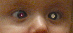
A child with leukocoria due to retinoblastoma in the left eye
Specialty Ophthalmology, pediatrics

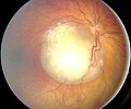
Leukocoria (also white pupillary reflex) is an abnormal white reflection from the retina of the eye. Leukocoria resembles eyeshine, but leukocoria can also occur in animals that lack eyeshine because their retina lacks a tapetum lucidum .

Contents

Leukocoria is a medical sign for a number of conditions, including Coats disease, congenital cataract, corneal scarring, melanoma of the ciliary body, [2] Norrie disease, ocular toxocariasis, persistence of the tunica vasculosa lentis (PFV/PHPV), retinoblastoma, and retrolental fibroplasia.

Because of the potentially life-threatening nature of retinoblastoma, a cancer, that condition is usually considered in the evaluation of leukocoria. In some rare cases (1%) the leukocoria is caused by Coats' disease (leaking retinal vessels).

Diagnosis

On photographs taken using a flash, instead of the familiar red-eye effect, leukocoria can cause a bright white reflection in an affected eye. Leukocoria may appear also in low indirect light, similar to eyeshine.

Leukocoria can be detected by a routine eye exam (see Ophthalmoscopy). For screening purposes, the red reflex test is used. In this test, when a light is shone briefly through the pupil, an orange red reflection is normal. A white reflection is leukocoria.

Society

Related Research Articles

Red-eye effect

The red-eye effect in photography is the common appearance of red pupils in color photographs of the eyes of humans and several other animals. It occurs when using a photographic flash that is very close to the camera lens in ambient low light.

Iris (anatomy) Part of an eye

In humans and most mammals and birds, the iris is a thin, annular structure in the eye, responsible for controlling the diameter and size of the pupil, thus the amount of light reaching the retina. Eye color is defined by that of the iris. In optical terms, the pupil is the eye's aperture, while the iris is the diaphragm.

Optic nerve

The optic nerve, also known as cranial nerve II, or simply as CN II, is a paired cranial nerve that transmits visual information from the retina to the brain. In humans, the optic nerve is derived from optic stalks during the seventh week of development and is composed of retinal ganglion cell axons and glial cells; it extends from the optic disc to the optic chiasma and continues as the optic tract to the lateral geniculate nucleus, pretectal nuclei, and superior colliculus.

<i>Tapetum lucidum</i> Layer of eye tissue which aids in night vision

The tapetum lucidum is a layer of tissue in the eye of many vertebrates. Lying immediately behind the retina, it is a retroreflector. It reflects visible light back through the retina, increasing the light available to the photoreceptors. The tapetum lucidum contributes to the superior night vision of some animals. Many of these animals are nocturnal, especially carnivores, while others are deep sea animals.

Retinoblastoma Medical condition

Retinoblastoma (Rb) is a rare form of cancer that rapidly develops from the immature cells of a retina, the light-detecting tissue of the eye. It is the most common primary malignant intraocular cancer in children, and it is almost exclusively found in young children.

Choroid

The choroid, also known as the choroidea or choroid coat, is the vascular layer of the eye, containing connective tissues, and lying between the retina and the sclera. The human choroid is thickest at the far extreme rear of the eye, while in the outlying areas it narrows to 0.1 mm. The choroid provides oxygen and nourishment to the outer layers of the retina. Along with the ciliary body and iris, the choroid forms the uveal tract.

Pupillary light reflex

The pupillary light reflex (PLR) or photopupillary reflex is a reflex that controls the diameter of the pupil, in response to the intensity (luminance) of light that falls on the retinal ganglion cells of the retina in the back of the eye, thereby assisting in adaptation of vision to various levels of lightness/darkness. A greater intensity of light causes the pupil to constrict, whereas a lower intensity of light causes the pupil to dilate. Thus, the pupillary light reflex regulates the intensity of light entering the eye. Light shone into one eye will cause both pupils to constrict.

Uvea

The uvea, also called the uveal layer, uveal coat, uveal tract, vascular tunic or vascular layer is the pigmented middle of the three concentric layers that make up an eye.

Fluorescein angiography Technique for examining the circulation of the retina and choroid of the eye

Fluorescein angiography (FA), fluorescent angiography (FAG), or fundus fluorescein angiography (FFA) is a technique for examining the circulation of the retina and choroid using a fluorescent dye and a specialized camera. Sodium fluorescein is added into the systemic circulation, the retina is illuminated with blue light at a wavelength of 490 nanometers, and an angiogram is obtained by photographing the fluorescent green light that is emitted by the dye. The fluorescein is administered intravenously in intravenous fluorescein angiography (IVFA) and orally in oral fluorescein angiography (OFA). The test is a dye tracing method.

Uveitis Inflammation of the uvea

Uveitis is the inflammation of the uvea, the pigmented layer that lies between the inner retina and the outer fibrous layer composed of the sclera and cornea. The uvea consists of the middle layer of pigmented vascular structures of the eye and includes the iris, ciliary body, and choroid. Uveitis is an ophthalmic emergency and requires a thorough examination by an ophthalmologist or optometrist and urgent treatment to control the inflammation. It is commonly associated with other ocular disorders, such as glaucoma, retinal detachment, optic nerve damage, cataracts, and, in some cases, a permanent loss of vision.

Eye examination A series of tests assessing vision and pertaining to the eyes

An eye examination is a series of tests performed to assess vision and ability to focus on and discern objects. It also includes other tests and examinations pertaining to the eyes. Eye examinations are primarily performed by an optometrist, ophthalmologist, orthoptist, or an optician. Health care professionals often recommend that all people should have periodic and thorough eye examinations as part of routine primary care, especially since many eye diseases are asymptomatic.

Accommodation (eye)

Accommodation is the process by which the vertebrate eye changes optical power to maintain a clear image or focus on an object as its distance varies. In this, distances vary for individuals from the far point—the maximum distance from the eye for which a clear image of an object can be seen, to the near point—the minimum distance for a clear image. Accommodation usually acts like a reflex, including part of the accommodation-vergence reflex, but it can also be consciously controlled. Mammals, birds and reptiles vary their eyes' optical power by changing the form of the elastic lens using the ciliary body. Fish and amphibians vary the power by changing the distance between a rigid lens and the retina with muscles.

Red eye (medicine) Eye that appears red due to illness or injury

A red eye is an eye that appears red due to illness or injury. It is usually injection and prominence of the superficial blood vessels of the conjunctiva, which may be caused by disorders of these or adjacent structures. Conjunctivitis and subconjunctival hemorrhage are two of the less serious but more common causes.

Progressive retinal atrophy (PRA) is a group of genetic diseases seen in certain breeds of dogs and, more rarely, cats. Similar to retinitis pigmentosa in humans, it is characterized by the bilateral degeneration of the retina, causing progressive vision loss culminating in blindness. The condition in nearly all breeds is inherited as an autosomal recessive trait, with the exception of the Siberian Husky and the Bullmastiff. There is no treatment.

Coats disease Human eye disease causing full or partial blindness

Coats' disease, is a rare congenital, nonhereditary eye disorder, causing full or partial blindness, characterized by abnormal development of blood vessels behind the retina. Coats' disease can also fall under glaucoma.

Fundus (eye)

The fundus of the eye is the interior surface of the eye opposite the lens and includes the retina, optic disc, macula, fovea, and posterior pole. The fundus can be examined by ophthalmoscopy and/or fundus photography.

Eye neoplasm Medical condition

Eye neoplasms can affect all parts of the eye, and can be a benign tumor or a malignant tumor (cancer). Eye cancers can be primary or metastatic cancer. The two most common cancers that spread to the eye from another organ are breast cancer and lung cancer. Other less common sites of origin include the prostate, kidney, thyroid, skin, colon and blood or bone marrow.

The red reflex refers to the reddish-orange reflection of light from the back of the eye, or fundus, observed when using an ophthalmoscope or retinoscope. The reflex relies on the transparency of optical media and reflects off the fundus back through media into the aperture of the ophthalmoscope. The red reflex is considered abnormal if there is any asymmetry between the eyes, dark spots, or white reflex (Leukocoria).

Mammalian eye

Mammals normally have a pair of eyes. Although mammalian vision is not so excellent as bird vision, it is at least dichromatic for most of mammalian species, with certain families possessing a trichromatic color perception.

Diktyoma Type of eye tumor

Diktyoma, or ciliary body medulloepithelioma, or teratoneuroma, is a rare tumor arising from primitive medullary epithelium in the ciliary body of the eye. Almost all diktyomas arise in the ciliary body, although, rarely, they may arise from the optic nerve head or retina.

References

  1. thefreedictionary.com
  2. Demirci H, Shields CL, Shields JA, Honavar SG, Eagle RC (January 2001). "Leucocoria as the presenting sign of a ciliary body melanoma in a child". The British Journal of Ophthalmology. 85 (1): 115–6. doi:10.1136/bjo.85.1.110f. PMC   1723667 . PMID   11201946.
Classification
D
External resources