Linear energy transfer

Last updated
Diffusion cloud chamber with tracks of ionizing radiation (alpha particles) that are made visible as strings of droplets Cloud chamber ani bionerd.gif
Diffusion cloud chamber with tracks of ionizing radiation (alpha particles) that are made visible as strings of droplets

In dosimetry, linear energy transfer (LET) is the amount of energy that an ionizing particle transfers to the material traversed per unit distance. It describes the action of radiation into matter.

Contents

It is identical to the retarding force acting on a charged ionizing particle travelling through the matter. [1] By definition, LET is a positive quantity. LET depends on the nature of the radiation as well as on the material traversed.

A high LET will slow down the radiation more quickly, generally making shielding more effective and preventing deep penetration. On the other hand, the higher concentration of deposited energy can cause more severe damage to any microscopic structures near the particle track. If a microscopic defect can cause larger-scale failure, as is the case in biological cells and microelectronics, the LET helps explain why radiation damage is sometimes disproportionate to the absorbed dose. Dosimetry attempts to factor in this effect with radiation weighting factors.

Linear energy transfer is closely related to stopping power, since both equal the retarding force. The unrestricted linear energy transfer is identical to linear electronic stopping power, as discussed below. But the stopping power and LET concepts are different in the respect that total stopping power has the nuclear stopping power component, [2] and this component does not cause electronic excitations. Hence nuclear stopping power is not contained in LET.

The appropriate SI unit for LET is the newton, but it is most typically expressed in units of kiloelectronvolts per micrometre (keV/μm) or megaelectronvolts per centimetre (MeV/cm). While medical physicists and radiobiologists usually speak of linear energy transfer, most non-medical physicists talk about stopping power.

Restricted and unrestricted LET

The secondary electrons produced during the process of ionization by the primary charged particle are conventionally called delta rays, if their energy is large enough so that they themselves can ionize. [3] Many studies focus upon the energy transferred in the vicinity of the primary particle track and therefore exclude interactions that produce delta rays with energies larger than a certain value Δ. [1] This energy limit is meant to exclude secondary electrons that carry energy far from the primary particle track, since a larger energy implies a larger range. This approximation neglects the directional distribution of secondary radiation and the non-linear path of delta rays, but simplifies analytic evaluation. [4]

In mathematical terms, Restricted linear energy transfer is defined by

where is the energy loss of the charged particle due to electronic collisions while traversing a distance , excluding all secondary electrons with kinetic energies larger than Δ. If Δ tends toward infinity, then there are no electrons with larger energy, and the linear energy transfer becomes the unrestricted linear energy transfer which is identical to the linear electronic stopping power . [1] Here, the use of the term "infinity" is not to be taken literally; it simply means that no energy transfers, however large, are excluded.

Application to radiation types

During his investigations of radioactivity, Ernest Rutherford coined the terms alpha rays, beta rays and gamma rays for the three types of emissions that occur during radioactive decay.

Alpha particles and other positive ions

Bragg curve of 5.49 MeV alpha particles in air. This radiation is produced by the decay of radon ( Rn); its range is 4.14 cm. Stopping power (which is essentially identical to LET) is plotted here versus path length; its peak is the "Bragg peak" Bragg Curve for Alphas in Air-PT-en.svg
Bragg curve of 5.49 MeV alpha particles in air. This radiation is produced by the decay of radon ( Rn); its range is 4.14 cm. Stopping power (which is essentially identical to LET) is plotted here versus path length; its peak is the "Bragg peak"

Linear energy transfer is best defined for monoenergetic ions, i.e. protons, alpha particles, and the heavier nuclei called HZE ions found in cosmic rays or produced by particle accelerators. These particles cause frequent direct ionizations within a narrow diameter around a relatively straight track, thus approximating continuous deceleration. As they slow down, the changing particle cross section modifies their LET, generally increasing it to a Bragg peak just before achieving thermal equilibrium with the absorber, i.e., before the end of range. At equilibrium, the incident particle essentially comes to rest or is absorbed, at which point LET is undefined.

Since the LET varies over the particle track, an average value is often used to represent the spread. Averages weighted by track length or weighted by absorbed dose are present in the literature, with the latter being more common in dosimetry. These averages are not widely separated for heavy particles with high LET, but the difference becomes more important in the other type of radiations discussed below. [4]

Often overlooked for alpha particles is the recoil-nucleus of the alpha emitter, which has significant ionization energy of roughly 5% of the alpha particle, but because of its high electric charge and large mass, has an ultra-short range of only a few Angstroms. This can skew results significantly if one is examining the Relative Biological Effectiveness of the alpha particle in the cytoplasm, while ignoring the recoil nucleus contribution, which alpha-parent being one of numerous heavy metals, is typically adhered to chromatic material such as chromosomes.

Beta particles

Electrons produced in nuclear decay are called beta particles. Because of their low mass relative to atoms, they are strongly scattered by nuclei (Coulomb or Rutherford scattering), much more so than heavier particles. Beta particle tracks are therefore crooked. In addition to producing secondary electrons (delta rays) while ionizing atoms, they also produce bremsstrahlung photons. A maximum range of beta radiation can be defined experimentally [5] which is smaller than the range that would be measured along the particle path.

Gamma rays

Gamma rays are photons, whose absorption cannot be described by LET. When a gamma quantum passes through matter, it may be absorbed in a single process (photoelectric effect, Compton effect or pair production), or it continues unchanged on its path. (Only in the case of the Compton effect, another gamma quantum of lower energy proceeds). Gamma ray absorption therefore obeys an exponential law (see Gamma rays); the absorption is described by the absorption coefficient or by the half-value thickness.

LET has therefore no meaning when applied to photons. However, many authors speak of "gamma LET" anyway, [6] where they are actually referring to the LET of the secondary electrons, i.e., mainly Compton electrons, produced by the gamma radiation. [7] The secondary electrons will ionize far more atoms than the primary photon. This gamma LET has little relation to the attenuation rate of the beam, but it may have some correlation to the microscopic defects produced in the absorber. Even a monoenergetic gamma beam will produce a spectrum of electrons, and each secondary electron will have a variable LET as it slows down, as discussed above. The "gamma LET" is therefore an average.

The transfer of energy from an uncharged primary particle to charged secondary particles can also be described by using the mass energy-transfer coefficient. [1]

Biological effects

The ICRP used to recommend quality factors as a generalized approximation of RBE based on LET. QF vs LET.jpg
The ICRP used to recommend quality factors as a generalized approximation of RBE based on LET.

Many studies have attempted to relate linear energy transfer to the relative biological effectiveness (RBE) of radiation, with inconsistent results. The relationship varies widely depending on the nature of the biological material, and the choice of endpoint to define effectiveness. Even when these are held constant, different radiation spectra that shared the same LET have significantly different RBE. [4]

Despite these variations, some overall trends are commonly seen. The RBE is generally independent of LET for any LET less than 10 keV/µm, so a low LET is normally chosen as the reference condition where RBE is set to unity. Above 10 keV/µm, some systems show a decline in RBE with increasing LET, while others show an initial increase to a peak before declining. Mammalian cells usually experience a peak RBE for LET's around 100 keV/µm. [4] These are very rough numbers; for example, one set of experiments found a peak at 30 keV/µm.

The International Commission on Radiation Protection (ICRP) proposed a simplified model of RBE-LET relationships for use in dosimetry. They defined a quality factor of radiation as a function of dose-averaged unrestricted LET in water, and intended it as a highly uncertain, but generally conservative, approximation of RBE. Different iterations of their model are shown in the graph to the right. The 1966 model was integrated into their 1977 recommendations for radiation protection in ICRP 26. This model was largely replaced in the 1991 recommendations of ICRP 60 by radiation weighting factors that were tied to the particle type and independent of LET. ICRP 60 revised the quality factor function and reserved it for use with unusual radiation types that did not have radiation weighting factors assigned to them. [8]

Application fields

When used to describe the dosimetry of ionizing radiation in the biological or biomedical setting, the LET (like linear stopping power) is usually expressed in units of keV/µm.

In space applications, electronic devices can be disturbed by the passage of energetic electrons, protons or heavier ions that may alter the state of a circuit, producing "single event effects". [9] The effect of the radiation is described by the LET (which is here taken as synonymous with stopping power), typically expressed in units of MeV·cm²/mg of material, the units used for mass stopping power (the material in question is usually Si for MOS devices). The units of measurement arise from a combination of the energy lost by the particle to the material per unit path length (MeV/cm) divided by the density of the material (mg/cm³). [10]

"Soft errors" of electronic devices due to cosmic rays on earth are, however, mostly due to neutrons which do not directly interact with the material and whose passage can therefore not be described by LET. Rather, one measures their effect in terms of neutrons per cm2 per hour, see Soft error.

Related Research Articles

<span class="mw-page-title-main">Geiger counter</span> Instrument used for measuring ionizing radiation

A Geiger counter is an electronic instrument used for detecting and measuring ionizing radiation. It is widely used in applications such as radiation dosimetry, radiological protection, experimental physics, and the nuclear industry.

<span class="mw-page-title-main">Radiation</span> Waves or particles moving through space

In physics, radiation is the emission or transmission of energy in the form of waves or particles through space or through a material medium. This includes:

<span class="mw-page-title-main">Beta particle</span> Ionizing radiation

A beta particle, also called beta ray or beta radiation, is a high-energy, high-speed electron or positron emitted by the radioactive decay of an atomic nucleus during the process of beta decay. There are two forms of beta decay, β decay and β+ decay, which produce electrons and positrons respectively.

<i>Bremsstrahlung</i> Electromagnetic radiation due to deceleration of charged particles

Bremsstrahlung, from bremsen "to brake" and Strahlung "radiation"; i.e., "braking radiation" or "deceleration radiation", is electromagnetic radiation produced by the deceleration of a charged particle when deflected by another charged particle, typically an electron by an atomic nucleus. The moving particle loses kinetic energy, which is converted into radiation, thus satisfying the law of conservation of energy. The term is also used to refer to the process of producing the radiation. Bremsstrahlung has a continuous spectrum, which becomes more intense and whose peak intensity shifts toward higher frequencies as the change of the energy of the decelerated particles increases.

<span class="mw-page-title-main">Geiger–Müller tube</span> Part of a Geiger counter

The Geiger–Müller tube or G–M tube is the sensing element of the Geiger counter instrument used for the detection of ionizing radiation. It is named after Hans Geiger, who invented the principle in 1908, and Walther Müller, who collaborated with Geiger in developing the technique further in 1928 to produce a practical tube that could detect a number of different radiation types.

<span class="mw-page-title-main">Sievert</span> SI unit of equivalent dose of ionizing radiation

The sievert is a unit in the International System of Units (SI) intended to represent the stochastic health risk of ionizing radiation, which is defined as the probability of causing radiation-induced cancer and genetic damage. The sievert is important in dosimetry and radiation protection. It is named after Rolf Maximilian Sievert, a Swedish medical physicist renowned for work on radiation dose measurement and research into the biological effects of radiation.

Ionizing radiation, including nuclear radiation, consists of subatomic particles or electromagnetic waves that have sufficient energy to ionize atoms or molecules by detaching electrons from them. Some particles can travel up to 99% of the speed of light, and the electromagnetic waves are on the high-energy portion of the electromagnetic spectrum.

<span class="mw-page-title-main">External beam radiotherapy</span> Treatment of cancer with ionized radiation

External beam radiotherapy (EBRT) is the most common form of radiotherapy. The patient sits or lies on a couch and an external source of ionizing radiation is pointed at a particular part of the body. In contrast to brachytherapy and unsealed source radiotherapy, in which the radiation source is inside the body, external beam radiotherapy directs the radiation at the tumour from outside the body. Orthovoltage ("superficial") X-rays are used for treating skin cancer and superficial structures. Megavoltage X-rays are used to treat deep-seated tumours, whereas megavoltage electron beams are typically used to treat superficial lesions extending to a depth of approximately 5 cm. X-rays and electron beams are by far the most widely used sources for external beam radiotherapy. A small number of centers operate experimental and pilot programs employing beams of heavier particles, particularly protons, owing to the rapid dropoff in absorbed dose beneath the depth of the target.

The gray is the unit of ionizing radiation dose in the International System of Units (SI), defined as the absorption of one joule of radiation energy per kilogram of matter.

Radiation dosimetry in the fields of health physics and radiation protection is the measurement, calculation and assessment of the ionizing radiation dose absorbed by an object, usually the human body. This applies both internally, due to ingested or inhaled radioactive substances, or externally due to irradiation by sources of radiation.

Radiation protection, also known as radiological protection, is defined by the International Atomic Energy Agency (IAEA) as "The protection of people from harmful effects of exposure to ionizing radiation, and the means for achieving this". Exposure can be from a source of radiation external to the human body or due to internal irradiation caused by the ingestion of radioactive contamination.

Equivalent dose is a dose quantity H representing the stochastic health effects of low levels of ionizing radiation on the human body which represents the probability of radiation-induced cancer and genetic damage. It is derived from the physical quantity absorbed dose, but also takes into account the biological effectiveness of the radiation, which is dependent on the radiation type and energy. In the SI system of units, the unit of measure is the sievert (Sv).

<span class="mw-page-title-main">Health physics</span>

Health physics, also referred to as the science of radiation protection, is the profession devoted to protecting people and their environment from potential radiation hazards, while making it possible to enjoy the beneficial uses of radiation. Health physicists normally require a four-year bachelor’s degree and qualifying experience that demonstrates a professional knowledge of the theory and application of radiation protection principles and closely related sciences. Health physicists principally work at facilities where radionuclides or other sources of ionizing radiation are used or produced; these include research, industry, education, medical facilities, nuclear power, military, environmental protection, enforcement of government regulations, and decontamination and decommissioning—the combination of education and experience for health physicists depends on the specific field in which the health physicist is engaged.

Scintillation is the physical process where a material, called a scintillator, emits UV or visible light under excitation from high energy photons or energetic particles. See scintillator and scintillation counter for practical applications.

In radiation physics, kerma is an acronym for "kinetic energy released per unit mass", defined as the sum of the initial kinetic energies of all the charged particles liberated by uncharged ionizing radiation in a sample of matter, divided by the mass of the sample. It is defined by the quotient .

The ionization chamber is the simplest type of gaseous ionisation detector, and is widely used for the detection and measurement of many types of ionizing radiation, including X-rays, gamma rays, and beta particles. Conventionally, the term "ionization chamber" refers exclusively to those detectors which collect all the charges created by direct ionization within the gas through the application of an electric field. It uses the discrete charges created by each interaction between the incident radiation and the gas to produce an output in the form of a small direct current. This means individual ionising events cannot be measured, so the energy of different types of radiation radiation cannot be differentiated, but it gives a very good measurement of overall ionising effect.

<span class="mw-page-title-main">Fast neutron therapy</span>

Fast neutron therapy utilizes high energy neutrons typically between 50 and 70 MeV to treat cancer. Most fast neutron therapy beams are produced by reactors, cyclotrons (d+Be) and linear accelerators. Neutron therapy is currently available in Germany, Russia, South Africa and the United States. In the United States, one treatment center is operational, in Seattle, Washington. The Seattle center uses a cyclotron which produces a proton beam impinging upon a beryllium target.


In radiobiology, the relative biological effectiveness is the ratio of biological effectiveness of one type of ionizing radiation relative to another, given the same amount of absorbed energy. The RBE is an empirical value that varies depending on the type of ionizing radiation, the energies involved, the biological effects being considered such as cell death, and the oxygen tension of the tissues or so-called oxygen effect.

<span class="mw-page-title-main">Gamma ray</span> Energetic electromagnetic radiation arising from radioactive decay of atomic nuclei

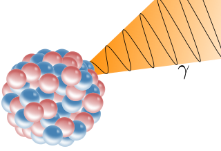
A gamma ray, also known as gamma radiation (symbol γ or ), is a penetrating form of electromagnetic radiation arising from the radioactive decay of atomic nuclei. It consists of the shortest wavelength electromagnetic waves, typically shorter than those of X-rays. With frequencies above 30 exahertz (3×10¹⁹ Hz), it imparts the highest photon energy. Paul Villard, a French chemist and physicist, discovered gamma radiation in 1900 while studying radiation emitted by radium. In 1903, Ernest Rutherford named this radiation gamma rays based on their relatively strong penetration of matter; in 1900 he had already named two less penetrating types of decay radiation (discovered by Henri Becquerel) alpha rays and beta rays in ascending order of penetrating power.

Internal dosimetry is the science and art of internal ionising radiation dose assessment due to radionuclides incorporated inside the human body.

References

  1. 1 2 3 4 International Commission on Radiation Units and Measurements (October 2011). Seltzer, Stephen M. (ed.). "Report 85: Fundamental Quantities and Units for Ionizing Radiation". Journal of the International Commission on Radiation Units and Measurements (Revised ed.). 11 (1): 1–31. doi:10.1093/jicru/ndr012. PMID   24174259. ICRU report 85a.
  2. Smith, Roger (1997). Atomic & ion collisions in solids and at surfaces: theory, simulation and applications. Cambridge, UK: Cambridge University Press.
  3. "Delta ray" in Encyclopedia britannica online, retrieved 22 Dec. 2012
  4. 1 2 3 4 International Commission on Radiation Units and Measurements (1970). Linear Energy Transfer. Washington D.C. doi:10.1093/jicru/os9.1.Report16. ISBN   978-0913394090. ICRU report 16.
  5. G. Knop and W. Paul: Interaction of electrons in Alpha- Beta- and Gamma-Ray Spectroscopy edited by K. Siegbahn, North-Holland, Amsterdam, 1966
  6. ICRP (International Commission on Radiation Protection) publication 103, ICRP 37 (2-4) (2007): "(116) Photons, electrons, and muons are radiations with LET values of less than 10 keV/microm."
  7. Chabot, George. "Radiation Basics — Radiation Quantities and Units". Ask the Experts FAQ. Health Physics Society. Retrieved 12 December 2012. When the term "stopping power" is used in reference to photons, as seems to be the case for the example you give, it is not really being used for the photons themselves, but for the electrons set free by the photon interactions.
  8. Sinclair, Dr. W. K.; et al. (January 2003). "Relative biological effectiveness (RBE), quality factor (Q) and radiation weighting factor (Wr)". Annals of the ICRP. 33 (4). ISBN   978-0-08-044311-9. ICRP Publication 92.
  9. V. Zajic and P. Thieberger, "Heavy Ion Linear Energy Transfer Measurements during Single Event Upset Testing of Electronic Devices," IEEE Transactions on Nuclear Science 46, pp. 59-69, (1999)
  10. Radiation Effects & Analysis Home Page of NASA