Preimplantation factor

Last updated
Preimplantation factor
Preimplantation factor.png
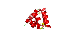
Simulated structure of preimplantation factor
Identifiers
SymbolPIF
Alt. namesPreImplantation factor, PIF, sPIF, PIF*

Preimplantation factor(PIF) is a peptide secreted by trophoblast cells prior to placenta formation in early embryonic development. [1] Human embryos begin to express PIF at the 4-cell stage, with expression increasing by the morula stage and continuing to do so throughout the first trimester. [2] [1] [3] Expression of preimplantation factor in the blastocyst was discovered as an early correlate of the viability of the eventual pregnancy. [1] [4] Preimplantation factor was identified in 1994 by a lymphocyte platelet-binding assay, where it was thought to be an early biomarker of pregnancy. [5] It has a simple primary structure with a short sequence of fifteen amino acids without any known quaternary structure. [6] A synthetic analogue of preimplantation factor (commonly abbreviated in studies as sPIF or PIF*) that has an identical amino acid sequence and mimics the normal biological activity of PIF has been developed and is commonly used in research studies, particularly those that aim to study potential adult therapeutics. [7] [8] [9]

Contents

Preimplantation factor acts by paracrine signaling; that is to say trophoblast cells, which collectively form extra-embryonic tissues, secrete it onto the surface of the endometrium. PIF is known to influence many events in the implantation process, the process by which an early embryo implants into the uterine wall. A crucial event in human implantation is when trophoblast cells expressing preimplantation factor invade the uterine wall and found the placenta, an organ that connects maternal blood supply, and along with it, nutrients, to the growing fetus. This requires changes to the histology of the endometrium; a process called decidualisation. Upregulated expression of PIF increases the presence of integrins on the endometrium wall, promoting the embryo's adhesion to the uterine wall. [10] PIF is thought to modulate and facilitate the depth of the trophoblast's invasion into the uterus at physiological doses. [1]

Maternal immune system regulation is also a critical event in implantation as the early embryo is essentially a partial allograft, that is a tissue that is recognised as fully identical to that of the mother. [11] [12] Consequently, the embryo may be rejected and attacked if it is not recognised, an event that normally causes spontaneous miscarriage. [11] [12] Preimplantation factor regionally modulates the mother's immune system, decreasing the activity of peripheral maternal leukocytes, reducing inflammation and consequently also increasing the chance that the embryo will be tolerated. [13] Preimplantation factor is also an anti-apoptotic effector, maintaining the trophoblast cell integrity through the intrinsic p53 signalling pathway. [14] Moreover, preimplantation factor protects the central nervous system by downregulating pathways that promote neurone death and promoting neurogenesis. [7] [9] PIF is also known to signal against neonatal prematurity and rescues embryos from toxic uterine environments. [7] [11] [15]

Due to its multiple autoimmune and neuroprotective effects in the embryonic environment, preimplantation factor has been studied in clinical environments as a potential novel therapy for reproductive, autoimmune and neurodegenerative diseases. PIF has been successfully studied as a therapy for recurrent pregnancy loss, as it is able to rescue non-viable embryos from a hostile maternal environment. [16] It has also been shown to prevent diabetes mellitus type 1 in mice due to its ability to modulate immunological tolerance in the pancreas. [8] Finally, it reverses paralysis and neuroinflammation whilst promoting neurogenesis in adult patients with neurodegenerative diseases. [11] [17] It also may be able to decrease the severity of brain injuries by modulating the behaviour of supporting cells in the nervous system. [9]

Discovery and structure

Preimplantation factor has a simple primary peptide structure with a 15 amino acid sequence (MVRIKPGSANKPSDD). [18]

As the regulation of the maternal immune system is a requisite for successful implantation, the immune system shows different characteristics in pregnant women and non-pregnant women. In 1994, preimplantation factor was isolated by a lymphocyte platelet-binding assay that compared immune responses and proteins found in pregnant women and non-pregnant women. [5] The assay also compared immune responses with men to verify if the proteins were specific to female reproductive tissues. [5] Results generated in the preliminary study showed that "a preimplantation factor" was being expressed exclusively in pregnant women. [5] On the fourth day after embryo transfer in women who had undergone successful in-vitro fertilisation, this protein was also found, suggesting that it had a role in the determination of the viability of the embryo. [5] Subsequent studies, most seminally including a 1996 study that partially characterised the biological activity of PIF, adopted and established the current term "preimplantation factor" as the name for this novel peptide. [6]

Functions

The outer layer of trophoblast cells invade the endometrium Diagram of Blastocyst stage.png
The outer layer of trophoblast cells invade the endometrium

Trophoblast invasion and adhesion

Trophoblast cells form the outer lining of the blastocyst in preimplantation development, eventually forming more differentiated extra-embryonic tissues including the placenta. [19] Before this differentiation can occur the embryo's invasion and infiltration into the uterine wall must be tightly regulated by both maternal and foetal signals, including secretion of PIF by trophoblast cells. [20] In particular, preimplantation factor is thought to have a paracrine effect on the decidualisation process, which ultimately primes trophoblast cells to invade appropriately into the endometrium. [1] When compared to non-functional short peptides at the same concentration, application of PIF to the endometrium at the implantation stage promoted deeper invasion of the embryo. [1] This effect was not observed to occur indefinitely with successive increases of concentration and any artificial increases of PIF above the human physiological concentration (approximately 50 nmol/L) did not meaningfully increase the invasion of the embryo. [1] Consequently, it is thought that PIF is limited in its promotion of trophoblast invasion by maternal signals. [1] [12]

The outermost layer of the uterine wall is an epithelial tissue called the endometrium that requires cell surface adhesion molecules called integrins to adhere the embryo. This additional paracrine effect of PIF has been shown to increase the expression of the integrin molecule α2β3 on the cell membranes of cells in the endometrium. [10] Integrins are a broad class of cell adhesion molecules that allow cells to bind to extracellular matrix. [10] In this way, they assist the entire embryo in binding to the uterine wall, an important event in successfully generating a placenta. [10]

Maternal immune tolerance

The embryo is immunologically characterised as a partial allograft as it is not a maternal tissue. [3] [11] During fertilisation, a paternal spermatozoon fuses with a maternal oocyte producing a zygote. Phenotypically, the zygote expresses certain epitopes that are controlled by genes inherited from the father, making the embryo a foreign material. In order for successful implantation to occur, the maternal immune system must tolerate the presence of the embryo while not completely inactivating its innate responsiveness to foreign pathogens. This process is not always successful; indeed maternal immune rejection of the embryo is a common and well-characterised cause of recurrent pregnancy loss. [16]

Preimplantation factor has a significant role in signalling this grafting behaviour; it has been, for instance shown to signal an anti-inflammatory response in a broad range of peripheral blood mononuclear cells. [3] PIF also impacts similar cytoskeletal proteins in CD14+, CD8+ and CD4+ cells suggesting that they have a broad and integrative role in modulating the immune system of the mother. [21] In particular, PIF inhibits the process of platelet aggregation in helper T lymphocytes and skeletal proteins in cytotoxic T cells. [21] While PIF attenuates or modulates the immune system, it does not effect the response to other pathogens or foreign material. [11] This modulatory effect on immunological tolerance is responsible for a strong correlation between PIF expression and the viability of pregnancy. [4]

Viability of pregnancy

The expression of preimplantation factor in the embryo is strongly correlated with the likelihood of a live birth. [4] [21] This observed viability is not solely due to PIF's ability to mediate the implantation and allografting process but also due to its ability to promote the upregulation and integrity of certain intracellular targets that are positively associated with normal developmental processes. [21] For instance, PIF is known to target the enzyme disulfide isomerase, which reduces intracellular oxidative stress and also heat-shock proteins, which are molecular chaperones that ensure proteins produced by a cell will fold into the correct conformation for their function. [22] Additionally, PIF is known to promote the production of vital cytoskeletal proteins including actin and tubulin that are required for the current morphological development of nerve axons and the viscera of vital organs. [15] Axons use circular tubulin polymers called microtubules to transport intracellular material between the cell body and the axon terminal and require actin to form synapses. [23] They are hence important for the organisation and function of the growing immune system.

Additionally, when uterine serum from patients with recurrent pregnancy loss is applied to embryos that are positive for PIF, they display the capacity to resist the toxin and are able to survive. [22] Combined, these observations and combination of intracellular effects suggest that PIF has multifaceted impacts directed towards viable pregnancy.

Neurogenic and anti-apoptotic effects

In the prenatal environment, PIF has neuroprotective impacts. It protects the growing fetus against neonatal prematurity, preventing the fetus from being delivered before adequate neural development has taken place. [7] [11] The neurogenic effects of PIF are not isolated to the prenatal environment; in fact PIF is thought to have impacts throughout life. In adult models, PIF has multiple neurogenic effects: it promotes the growth of neurons and reduces neuroinflammation. [7] [11] [17] It is thought to have these impacts by modulating signalling through the ubiquitous protein kinase A and protein kinase C intracellular signalling pathways. [7] PIF also inhibits microRNA let-7, a sequence that is highly upregulated in the central nervous system. The Let-7 system has been associated with cell death in neurons, and PIF is known to inhibit this process from occurring. [9] In rats that were induced to have a hypoxic-ischemic brain injury, PIF was able to promote neuron growth, reduced detrimental responses by neuroglia and was able to generate a significant cerebral cortex volume, suggesting it could rescue rats from side effects of brain damage. [9]

Preimplantation factor deactivates p53, preventing apoptosis P53 pathways.jpg
Preimplantation factor deactivates p53, preventing apoptosis

PIF also has a series of anti-apoptotic impacts in human extravillous trophoblasts, mediated by the TP53 gene. [14] Apoptosis is a controlled cell death process that must not occur if a cell is to proliferate. PIF has specific anti-apoptotic impacts by reducing the phosphorylation of the p53 protein at the serine-15 residue. Without phosphorylation p53 is unstable and undergoes ubiquitylation, signalling the trophoblast and endometrial cells to degrade it in proteasomes and attenuating downstream apoptotic effects. PIF, in particular, has been correlated with increasing the expression of anti-apoptotic effector BCL2 and decreasing the expression of pro-apoptotic effector BAX. [14] BCL2, which is upregulated by PIF, ensures that cytochrome c remains within the inner mitochondrial membrane and hence does not trigger the production of an apoptosome in the cell cytosol. BAX, which is downregulated by PIF, produces transmembrane transport channels that liberate cytochrome c, triggering apoptosis. Collectively, these biochemical effects show that PIF signals against the internal mechanisms of apoptosis in extravillous trophoblast cells, allowing them to proliferate before they implant into the uterine wall.

Therapeutic uses

Given its multifaceted functionality, including autoimmune, neuroprotective and anti-apoptotic effects, preimplantation factor has been extensively studied as a potential therapeutic agent in both reproductive and non-reproductive medical contexts. PIF is also advantageous because of its easily replicable biochemical structure. [6] In reproductive contexts, PIF has been studied as a treatment for infertility. In women with recurrent pregnancy loss, treatment with PIF is able to rescue a non-viable embryo and promotes a successful implantation and pregnancy. [16] It does this by mitigating the toxic influence of certain factors that naturally occur in the uterus, such as acidity. [16]

PIF has also been studied in a range of other non-reproductive contexts. Due to the ability of PIF to attenuate the attack mechanisms of mononuclear immune cells, it has been implicated as a successful treatment for autoimmune diseases including diabetes mellitus type 1 in mice studies. Diabetes mellitus type 1 is characterised by the misrecognition of pancreatic beta islet cells as foreign material. [8] These studies show that PIF is able to preserve the pancreatic beta islet cell's integrity, rescuing them from the autoimmune attacks which cause diabetes. [8] In adult models, PIF also reverses the pathological neuroinflammation caused by autoimmune diseases such as multiple sclerosis. [17] It also reverses paralysis and promotes growth of neurons in patients with neurodegeneration. [11]

Related Research Articles

<span class="mw-page-title-main">Endometrium</span> Inner mucous membrane of the mammalian uterus

The endometrium is the inner epithelial layer, along with its mucous membrane, of the mammalian uterus. It has a basal layer and a functional layer: the basal layer contains stem cells which regenerate the functional layer. The functional layer thickens and then is shed during menstruation in humans and some other mammals, including apes, Old World monkeys, some species of bat, the elephant shrew and the Cairo spiny mouse. In most other mammals, the endometrium is reabsorbed in the estrous cycle. During pregnancy, the glands and blood vessels in the endometrium further increase in size and number. Vascular spaces fuse and become interconnected, forming the placenta, which supplies oxygen and nutrition to the embryo and fetus. The speculated presence of an endometrial microbiota has been argued against.

<span class="mw-page-title-main">Blastulation</span> Sphere of cells formed during early embryonic development in animals

Blastulation is the stage in early animal embryonic development that produces the blastula. In mammalian development the blastula develops into the blastocyst with a differentiated inner cell mass and an outer trophectoderm. The blastula is a hollow sphere of cells known as blastomeres surrounding an inner fluid-filled cavity called the blastocoel. Embryonic development begins with a sperm fertilizing an egg cell to become a zygote, which undergoes many cleavages to develop into a ball of cells called a morula. Only when the blastocoel is formed does the early embryo become a blastula. The blastula precedes the formation of the gastrula in which the germ layers of the embryo form.

<span class="mw-page-title-main">Blastocyst</span> Structure formed around day 5 of mammalian embryonic development

The blastocyst is a structure formed in the early embryonic development of mammals. It possesses an inner cell mass (ICM) also known as the embryoblast which subsequently forms the embryo, and an outer layer of trophoblast cells called the trophectoderm. This layer surrounds the inner cell mass and a fluid-filled cavity known as the blastocoel. In the late blastocyst the trophectoderm is known as the trophoblast. The trophoblast gives rise to the chorion and amnion, the two fetal membranes that surround the embryo. The placenta derives from the embryonic chorion and the underlying uterine tissue of the mother.

<span class="mw-page-title-main">Trophoblast</span> Early embryonic structure that gives rise to the placenta

The trophoblast is the outer layer of cells of the blastocyst. Trophoblasts are present four days after fertilization in humans. They provide nutrients to the embryo and develop into a large part of the placenta. They form during the first stage of pregnancy and are the first cells to differentiate from the fertilized egg to become extraembryonic structures that do not directly contribute to the embryo. After blastulation, the trophoblast is contiguous with the ectoderm of the embryo and is referred to as the trophectoderm. After the first differentiation, the cells in the human embryo lose their totipotency because they can no longer form a trophoblast. They become pluripotent stem cells.

<span class="mw-page-title-main">GroES</span> Protein-coding gene in the species Homo sapiens

Heat shock 10 kDa protein 1 (Hsp10), also known as chaperonin 10 (cpn10) or early-pregnancy factor (EPF), is a protein that in humans is encoded by the HSPE1 gene. The homolog in E. coli is GroES that is a chaperonin which usually works in conjunction with GroEL.

<span class="mw-page-title-main">Relaxin</span> Protein hormone

Relaxin is a protein hormone of about 6000 Da, first described in 1926 by Frederick Hisaw.

<span class="mw-page-title-main">Decidua</span> Part of uterus modified in pregnancy

The decidua is the modified mucosal lining of the uterus that forms every month, in preparation for pregnancy. It is shed off each month when there is no fertilised egg to support. The decidua is under the influence of progesterone. Endometrial cells become highly characteristic. The decidua forms the maternal part of the placenta and remains for the duration of the pregnancy. After birth the decidua is shed together with the placenta.

<span class="mw-page-title-main">Syncytiotrophoblast</span> Embryonic cell of the placental surface

Syncytiotrophoblast is the epithelial covering of the highly vascular embryonic placental villi, which invades the wall of the uterus to establish nutrient circulation between the embryo and the mother. It is a multinucleate, terminally differentiated syncytium, extending to 13 cm.

<span class="mw-page-title-main">Placentation</span> Formation and structure of the placenta

Placentation refers to the formation, type and structure, or arrangement of the placenta. The function of placentation is to transfer nutrients, respiratory gases, and water from maternal tissue to a growing embryo, and in some instances to remove waste from the embryo. Placentation is best known in live-bearing mammals (theria), but also occurs in some fish, reptiles, amphibians, a diversity of invertebrates, and flowering plants. In vertebrates, placentas have evolved more than 100 times independently, with the majority of these instances occurring in squamate reptiles.

<span class="mw-page-title-main">Cytotrophoblast</span>

"Cytotrophoblast" is the name given to both the inner layer of the trophoblast or the cells that live there. It is interior to the syncytiotrophoblast and external to the wall of the blastocyst in a developing embryo.

<span class="mw-page-title-main">Implantation (embryology)</span> First stage of pregnancy

Implantation, also known as nidation, is the stage in the embryonic development of mammals in which the blastocyst hatches, attaches, adheres, and invades into the wall of the female's uterus. Implantation is the first stage of gestation, and, when successful, the female is considered to be pregnant. An implanted embryo is detected by the presence of increased levels of human chorionic gonadotropin (hCG) in a pregnancy test. The implanted embryo will receive oxygen and nutrients in order to grow.

<span class="mw-page-title-main">Decidualization</span>

Decidualization is a process that results in significant changes to cells of the endometrium in preparation for, and during, pregnancy. This includes morphological and functional changes to endometrial stromal cells (ESCs), the presence of decidual white blood cells (leukocytes), and vascular changes to maternal arteries. The sum of these changes results in the endometrium changing into a structure called the decidua. In humans, the decidua is shed during childbirth.

Reproductive immunology refers to a field of medicine that studies interactions between the immune system and components related to the reproductive system, such as maternal immune tolerance towards the fetus, or immunological interactions across the blood-testis barrier. The concept has been used by fertility clinics to explain fertility problems, recurrent miscarriages and pregnancy complications observed when this state of immunological tolerance is not successfully achieved. Immunological therapy is a method for treating many cases of previously "unexplained infertility" or recurrent miscarriage.

Immune tolerance in pregnancy or maternal immune tolerance is the immune tolerance shown towards the fetus and placenta during pregnancy. This tolerance counters the immune response that would normally result in the rejection of something foreign in the body, as can happen in cases of spontaneous abortion. It is studied within the field of reproductive immunology.

Hormonal regulation occurs at every stage of development. A milieu of hormones simultaneously affects development of the fetus during embryogenesis and the mother, including human chorionic gonadotropin (hCG) and progesterone (P4).

<span class="mw-page-title-main">Chorionic gonadotropin beta</span> Protein-coding gene in the species Homo sapiens

Choriogonadotropin subunit beta (CG-beta) also known as chorionic gonadotrophin chain beta is a protein that in humans is encoded by the CGB gene.

Embryo quality is the ability of an embryo to perform successfully in terms of conferring a high pregnancy rate and/or resulting in a healthy person. Embryo profiling is the estimation of embryo quality by qualification and/or quantification of various parameters. Estimations of embryo quality guides the choice in embryo selection in in vitro fertilization.

Embryokines are regulatory molecules produced by the oviduct and endometrium in the reproductive tract that modulate embryonic growth and development.

<span class="mw-page-title-main">Uterine natural killer cells</span> Maternal lymphocytes that make up 70% of the total found during pregnancy

Uterine natural killer cells make up approximately 70% of maternal lymphocytes during pregnancy, occupying both the decidua basalis of the endometrium at the implantation site and the mesometrial lymphoid aggregate of pregnancy (MLAp) that surrounds the blood vessels supplying the placenta. This number is at its peak in early pregnancy but declines at parturition.

<span class="mw-page-title-main">Maternal recognition of pregnancy</span> Crucial aspect of carrying a pregnancy to full term

Maternal recognition of pregnancy is a crucial aspect of carrying a pregnancy to full term. Without maternal recognition to maintain pregnancy, the initial messengers which stop luteolysis and promote foetal implantation, growth and uterine development finish with nothing to replace them and the pregnancy is lost.

References

  1. 1 2 3 4 5 6 7 8 Duzyj CM, Barnea ER, Li M, Huang SJ, Krikun G, Paidas MJ (October 2010). "Preimplantation factor promotes first trimester trophoblast invasion". American Journal of Obstetrics and Gynecology. 203 (4): 402.e1–4. doi:10.1016/j.ajog.2010.06.060. PMC   2947608 . PMID   20708167.
  2. Zare F, Seifati SM, Dehghan-Manshadi M, Fesahat F (May 2020). "Preimplantation Factor (PIF): a peptide with various functions". JBRA Assisted Reproduction. 24 (2): 214–218. doi:10.5935/1518-0557.20190082. PMC   7169918 . PMID   32202400.
  3. 1 2 3 Barnea ER, Kirk D, Ramu S, Rivnay B, Roussev R, Paidas MJ (October 2012). "PreImplantation Factor (PIF) orchestrates systemic antiinflammatory response by immune cells: effect on peripheral blood mononuclear cells". American Journal of Obstetrics and Gynecology. 207 (4): 313.e1–11. doi:10.1016/j.ajog.2012.07.017. PMID   23021695.
  4. 1 2 3 Stamatkin CW, Roussev RG, Stout M, Absalon-Medina V, Ramu S, Goodman C, Coulam CB, Gilbert RO, Godke RA, Barnea ER (May 2011). "PreImplantation Factor (PIF) correlates with early mammalian embryo development-bovine and murine models". Reproductive Biology and Endocrinology. 9 (1): 63. doi: 10.1186/1477-7827-9-63 . PMC   3112407 . PMID   21569635.
  5. 1 2 3 4 5 Barnea ER, Lahijani KI, Roussev R, Barnea JD, Coulam CB (October 1994). "Use of lymphocyte platelet binding assay for detecting a preimplantation factor: a quantitative assay". American Journal of Reproductive Immunology. 32 (3): 133–8. doi:10.1111/j.1600-0897.1994.tb01103.x. PMID   7880393. S2CID   20230576.
  6. 1 2 3 Roussev RG, Coulam CB, Kaider BD, Yarkoni M, Leavis PC, Barnea ER (November 1996). "Embryonic origin of preimplantation factor (PIF): biological activity and partial characterization". Molecular Human Reproduction. 2 (11): 883–7. doi: 10.1093/molehr/2.11.883 . PMID   9237230.
  7. 1 2 3 4 5 6 Mueller M, Schoeberlein A, Zhou J, Joerger-Messerli M, Oppliger B, Reinhart U, Bordey A, Surbek D, Barnea ER, Huang Y, Paidas M (December 2015). "PreImplantation Factor bolsters neuroprotection via modulating Protein Kinase A and Protein Kinase C signaling". Cell Death and Differentiation. 22 (12): 2078–86. doi:10.1038/cdd.2015.55. PMC   4816111 . PMID   25976303.
  8. 1 2 3 4 Weiss L, Bernstein S, Jones R, Amunugama R, Krizman D, Jebailey L, Almogi-Hazan O, Hazan O, Yekhtin Z, Yachtin J, Shiner R, Reibstein I, Triche E, Slavin S, Or R, Barnea ER (August 2011). "Preimplantation factor (PIF) analog prevents type I diabetes mellitus (TIDM) development by preserving pancreatic function in NOD mice". Endocrine. 40 (1): 41–54. doi:10.1007/s12020-011-9438-5. PMID   21424847. S2CID   21571195.
  9. 1 2 3 4 5 Mueller M, Zhou J, Yang L, Gao Y, Wu F, Schoeberlein A, Surbek D, Barnea ER, Paidas M, Huang Y (September 2014). "PreImplantation factor promotes neuroprotection by targeting microRNA let-7". Proceedings of the National Academy of Sciences of the United States of America. 111 (38): 13882–7. Bibcode:2014PNAS..11113882M. doi: 10.1073/pnas.1411674111 . PMC   4183321 . PMID   25205808.
  10. 1 2 3 4 Barnea ER, Kirk D, Paidas MJ (July 2012). "Preimplantation factor (PIF) promoting role in embryo implantation: increases endometrial integrin-α2β3, amphiregulin and epiregulin while reducing betacellulin expression via MAPK in decidua". Reproductive Biology and Endocrinology. 10 (1): 50. doi: 10.1186/1477-7827-10-50 . PMC   3444419 . PMID   22788113.
  11. 1 2 3 4 5 6 7 8 9 Barnea ER, Almogi-Hazan O, Or R, Mueller M, Ria F, Weiss L, Paidas MJ (December 2015). "Immune regulatory and neuroprotective properties of preimplantation factor: From newborn to adult". Pharmacology & Therapeutics. 156: 10–25. doi:10.1016/j.pharmthera.2015.10.008. PMID   26546485.
  12. 1 2 3 Hearn JP (March 1986). "The embryo-maternal dialogue during early pregnancy in primates". Journal of Reproduction and Fertility. 76 (2): 809–19. doi: 10.1530/jrf.0.0760809 . PMID   3517317.
  13. Nash DM, Paddison J, Davies Morel MC, Barnea ER (November 2018). "Preimplantation factor modulates acute inflammatory responses of equine endometrium". Veterinary Medicine and Science. 4 (4): 351–356. doi:10.1002/vms3.126. PMC   6236140 . PMID   30273998.
  14. 1 2 3 Moindjie H, Santos ED, Gouesse RJ, Swierkowski-Blanchard N, Serazin V, Barnea ER, Vialard F, Dieudonné MN (December 2016). "Preimplantation factor is an anti-apoptotic effector in human trophoblasts involving p53 signaling pathway". Cell Death & Disease. 7 (12): e2504. doi:10.1038/cddis.2016.382. PMC   5261002 . PMID   27906186.
  15. 1 2 Duzyj CM, Paidas MJ, Jebailey L, Huang JS, Barnea ER (2014). "PreImplantation factor (PIF*) promotes embryotrophic and neuroprotective decidual genes: effect negated by epidermal growth factor". Journal of Neurodevelopmental Disorders. 6 (1): 36. doi: 10.1186/1866-1955-6-36 . PMC   4470351 . PMID   26085845.
  16. 1 2 3 4 Barnea ER, Barder TJ, Stamatkin C, Coulam CB, Roussev RG, Absalon-Medina V, Gilbert R, Lubman DM, Liu Y, Paidas MJ (2011). "Preimplantation factor (PIF*) directly targets and rescues embryos from adverse environment: relevance for recurrent pregnancy loss". Journal of Reproductive Immunology. 90 (2): 141–142. doi:10.1016/j.jri.2011.06.023. ISSN   0165-0378.
  17. 1 2 3 Weiss L, Or R, Jones RC, Amunugama R, JeBailey L, Ramu S, Bernstein SA, Yekhtin Z, Almogi-Hazan O, Shainer R, Reibstein I, Vortmeyer AO, Paidas MJ, Zeira M, Slavin S, Barnea ER (January 2012). "Preimplantation factor (PIF*) reverses neuroinflammation while promoting neural repair in EAE model". Journal of the Neurological Sciences. 312 (1–2): 146–57. doi:10.1016/j.jns.2011.07.050. PMID   21996270. S2CID   12460162.
  18. Paidas MJ, Krikun G, Huang SJ, Jones R, Romano M, Annunziato J, Barnea ER (May 2010). "A genomic and proteomic investigation of the impact of preimplantation factor on human decidual cells". American Journal of Obstetrics and Gynecology. 202 (5): 459.e1–8. doi:10.1016/j.ajog.2010.03.024. PMC   2867836 . PMID   20452489.
  19. Duzyj C, Heller D, Mannion C, Koenig C, Zamudio S, Illsley N (2016). "Evidence that extravillous trophoblast fusion into multinuclear trophoblast giant cells involves a mesenchymal-epithelial transition". Placenta. 45: 96–97. doi:10.1016/j.placenta.2016.06.124. ISSN   0143-4004.
  20. Jauniaux E, Barnea ER, Edwards RG (1997). Embryonic medicine and therapy. Oxford University Press. ISBN   978-0-19-262729-2. OCLC   37444013.
  21. 1 2 3 4 Barnea ER, Hayrabedyan S, Todorova K, Almogi-Hazan O, Or R, Guingab J, McElhinney J, Fernandez N, Barder T (July 2016). "PreImplantation factor (PIF*) regulates systemic immunity and targets protective regulatory and cytoskeleton proteins". Immunobiology. 221 (7): 778–93. doi:10.1016/j.imbio.2016.02.004. PMID   26944449.
  22. 1 2 Stamatkin CW, Roussev RG, Stout M, Coulam CB, Triche E, Godke RA, Barnea ER (October 2011). "Preimplantation factor negates embryo toxicity and promotes embryo development in culture". Reproductive Biomedicine Online. 23 (4): 517–24. doi: 10.1016/j.rbmo.2011.06.009 . PMID   21900046.
  23. Dent EW, Baas PW (April 2014). "Microtubules in neurons as information carriers". Journal of Neurochemistry. 129 (2): 235–9. doi:10.1111/jnc.12621. PMC   3979999 . PMID   24266899.