Related Research Articles

The central nervous system (CNS) is the part of the nervous system consisting primarily of the brain and spinal cord. The CNS is so named because the brain integrates the received information and coordinates and influences the activity of all parts of the bodies of bilaterally symmetric animals—i.e., all multicellular animals except sponges and jellyfish. It is a structure composed of nervous tissue positioned along the rostral to caudal axis of the body and may have an enlarged section at the rostral end which is a brain. Not all animals with a central nervous system have a brain, although the large majority do.

A nerve is an enclosed, cable-like bundle of fibers in the peripheral nervous system.

The peripheral nervous system (PNS) is one of two components that make up the nervous system of bilateral animals, with the other part being the central nervous system (CNS). The PNS consists of the nerves and ganglia outside the brain and spinal cord. The main function of the PNS is to connect the CNS to the limbs and organs, essentially serving as a relay between the brain and spinal cord and the rest of the body. Unlike the CNS, the PNS is not protected by the vertebral column and skull, or by the blood–brain barrier, which leaves it exposed to toxins and mechanical injuries.

Syringomyelia is a generic term referring to a disorder in which a cyst or cavity forms within the spinal cord. Often, syringomyelia is used as a generic term before an etiology is determined. This cyst, called a syrinx, can expand and elongate over time, destroying the spinal cord. The damage may result in loss of feeling, paralysis, weakness, and stiffness in the back, shoulders, and extremities. Syringomyelia may also cause a loss of the ability to feel extremes of hot or cold, especially in the hands. It may also lead to a cape-like bilateral loss of pain and temperature sensation along the upper chest and arms. Each patient experiences a different combination of symptoms. These symptoms typically vary depending on the extent and, often more critically, on the location of the syrinx within the spinal cord.
Cranial nerves are the nerves that emerge directly from the brain, of which there are conventionally considered twelve pairs. Cranial nerves relay information between the brain and parts of the body, primarily to and from regions of the head and neck, including the special senses of vision, taste, smell, and hearing.

Chiari malformation (CM) is a structural defect in the cerebellum, characterized by a downward displacement of one or both cerebellar tonsils through the foramen magnum. CMs can cause headaches, difficulty swallowing, vomiting, dizziness, neck pain, unsteady gait, poor hand coordination, numbness and tingling of the hands and feet, and speech problems. Less often, people may experience ringing or buzzing in the ears, weakness, slow heart rhythm, or fast heart rhythm, curvature of the spine (scoliosis) related to spinal cord impairment, abnormal breathing, such as central sleep apnea, characterized by periods of breathing cessation during sleep, and, in severe cases, paralysis.

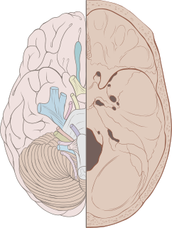
The brainstem is the posterior stalk-like part of the brain that connects the cerebrum with the spinal cord. In the human brain the brainstem is composed of the midbrain, the pons, and the medulla oblongata. The midbrain is continuous with the thalamus of the diencephalon through the tentorial notch, and sometimes the diencephalon is included in the brainstem.
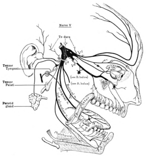
The trigeminal nerve (the fifth cranial nerve, or simply CN V) is a nerve responsible for sensation in the face and motor functions such as biting and chewing; it is the most complex of the cranial nerves. Its name ("trigeminal" = tri-, or three, and - geminus, or twin: thrice-twinned) derives from each of the two nerves (one on each side of the pons) having three major branches: the ophthalmic nerve (V1), the maxillary nerve (V2), and the mandibular nerve (V3). The ophthalmic and maxillary nerves are purely sensory, whereas the mandibular nerve supplies motor as well as sensory (or "cutaneous") functions. Adding to the complexity of this nerve is that autonomic nerve fibers as well as special sensory fibers (taste) are contained within it.

Arachnoiditis is an inflammatory condition of the arachnoid mater or 'arachnoid', one of the membranes known as meninges that surround and protect the nerves of the central nervous system, including the brain and spinal cord. The arachnoid can become inflamed because of adverse reactions to chemicals, infection from bacteria or viruses, as the result of direct injury to the spine, chronic compression of spinal nerves, complications from spinal surgery or other invasive spinal procedures, or the accidental intrathecal injection of steroids intended for the epidural space. Inflammation can sometimes lead to the formation of scar tissue and adhesion that can make the spinal nerves "stick" together, a condition where such tissue develops in and between the leptomeninges. The condition is extremely painful, especially when progressing to adhesive arachnoiditis. Another form of the condition is arachnoiditis ossificans, in which the arachnoid becomes ossified, or turns to bone, and is thought to be a late-stage complication of the adhesive form of arachnoiditis.

The sternohyoid muscle is a thin, narrow muscle attaching the hyoid bone to the sternum. It is one of the paired strap muscles of the infrahyoid muscles. It is supplied by the ansa cervicalis. It depresses the hyoid bone.

The sternothyroid muscle, or sternothyroideus, is an infrahyoid muscle in the neck. It acts to depress the hyoid bone. It is below the sternohyoid muscle. It is shorter and wider than the sternohyoid.

The central canal is the cerebrospinal fluid-filled space that runs through the spinal cord. The central canal lies below and is connected to the ventricular system of the brain, from which it receives cerebrospinal fluid, and shares the same ependymal lining. The central canal helps to transport nutrients to the spinal cord as well as protect it by cushioning the impact of a force when the spine is affected.
Dissociated sensory loss is a pattern of neurological damage caused by a lesion to a single tract in the spinal cord which involves preservation of fine touch and proprioception withselective loss of pain and temperature
A syrinx is a rare, fluid-filled neuroglial cavity within the spinal cord (syringomyelia), in the brain stem (syringobulbia), or in the nerves of the elbow, usually in a young age.
Hypoesthesia or numbness is a common side effect of various medical conditions which manifests as a reduced sense of touch or sensation, or a partial loss of sensitivity to sensory stimuli. In everyday speech this is generally referred to as numbness.

A neurological disorder is any disorder of the nervous system. Structural, biochemical or electrical abnormalities in the brain, spinal cord or other nerves can result in a range of symptoms. Examples of symptoms include paralysis, muscle weakness, poor coordination, loss of sensation, seizures, confusion, pain and altered levels of consciousness. There are many recognized neurological disorders, some relatively common, but many rare. They may be assessed by neurological examination, and studied and treated within the specialities of neurology and clinical neuropsychology.
The spinal cord is a long, thin, tubular structure made up of nervous tissue, which extends from the medulla oblongata in the brainstem to the lumbar region of the vertebral column. It encloses the central canal of the spinal cord, which contains cerebrospinal fluid. The brain and spinal cord together make up the central nervous system (CNS). In humans, the spinal cord begins at the occipital bone, passing through the foramen magnum and entering the spinal canal at the beginning of the cervical vertebrae. The spinal cord extends down to between the first and second lumbar vertebrae, where it ends. The enclosing bony vertebral column protects the relatively shorter spinal cord. It is around 45 cm (18 in) long in adult men and around 43 cm (17 in) long in adult women. The diameter of the spinal cord ranges from 13 mm in the cervical and lumbar regions to 6.4 mm in the thoracic area.
Basilar invagination is invagination (infolding) of the base of the skull that occurs when the top of the C2 vertebra migrates upward. It can cause narrowing of the foramen magnum. It also may press on the lower brainstem.
Chiari-like malformation (CM) the most common cause of foramen magnum obstruction and syringomyelia in dogs. Syringomyelia (SM) is a disease of the spinal cord typified by fluid filled cavities, or syrinxes, within the spinal cord substance but it can cause pain by disrupting the cerebrospinal fluid (CSF), in the brain CM is a condition characterized by the mismatch of size between the brain and the skull. CM is very widespread in many Toy breed dogs and has been studied in the Cavalier King Charles Spaniel and the Griffon Bruxellois and Chihuahua. As many as 95% of Cavalier King Charles Spaniels may have CM. It is worldwide in scope and not limited to any country, breeding line, or kennel, and experts report that it is believed to be inherited CM is so widespread in the Cavalier that it may be an inherent part of the CKCS's breed standard. This disease not only affects thousands of dogs, but a similar condition affects over three hundred thousand children yearly. Therefore, canines are an appropriate model for the treatment of the human condition.

Cervicocranial syndrome or is a neurological illness. It is a combination of symptoms that are caused by an abnormality in the neck. The bones of the neck that are affected are cervical vertebrae. This syndrome can be identified by confirming cervical bone shifts, collapsed cervical bones or misalignment of the cervical bone leading to improper functioning of cervical spinal nerves. Cervicocranial syndrome is either congenital or acquired. Some examples of diseases that could result in cervicocranial syndrome are Chiari disease, Klippel-Feil malformation osteoarthritis, and trauma. Treatment options include neck braces, pain medication and surgery. The quality of life for individuals suffering from CCJ syndrome can improve through surgery.
References
- 1 2 3 4 5 6 7 8 9 10 Boivie, Jörgen (2003). "20 - Central pain". Handbook of Pain Management - A Clinical Companion to Wall and Melzack's textbook of pain. Churchill Livingstone. pp. 305–327. doi:10.1016/B978-0-443-07201-7.50024-2. ISBN 978-0-443-07201-7.
- ↑ Chokroverty, Sudhansa; Montagna, Pasquale (2009). "29 - Sleep, Breathing, and Neurologic Disorders". Sleep Disorders Medicine - Basic Science, Technical Considerations, and Clinical Aspects (3rd ed.). Saunders. pp. 436–498. doi:10.1016/B978-0-7506-7584-0.00029-X. ISBN 978-0-7506-7584-0.
- ↑ Sgouros, S. (2009). "Syringomyelia". Encyclopedia of Neuroscience. Academic Press. pp. 839–847. doi:10.1016/B978-008045046-9.00614-8. ISBN 978-0-08-045046-9.
- ↑ "Syringobulbia".