Related Research Articles

A finger is a prominent digit on the forelimbs of most tetrapod vertebrate animals, especially those with prehensile extremities such as humans and other primates. Most tetrapods have five digits (pentadactyly), and short digits are typically referred to as toes, while those that are notably elongated are called fingers. In humans, the fingers are flexibly articulated and opposable, serving as an important organ of tactile sensation and fine movements, which are crucial to the dexterity of the hands and the ability to grasp and manipulate objects.

The radial nerve is a nerve in the human body that supplies the posterior portion of the upper limb. It innervates the medial and lateral heads of the triceps brachii muscle of the arm, as well as all 12 muscles in the posterior osteofascial compartment of the forearm and the associated joints and overlying skin.
Wrist drop is a medical condition in which the wrist and the fingers cannot extend at the metacarpophalangeal joints. The wrist remains partially flexed due to an opposing action of flexor muscles of the forearm. As a result, the extensor muscles in the posterior compartment remain paralyzed.

The upper limbs or upper extremities are the forelimbs of an upright-postured tetrapod vertebrate, extending from the scapulae and clavicles down to and including the digits, including all the musculatures and ligaments involved with the shoulder, elbow, wrist and knuckle joints. In humans, each upper limb is divided into the arm, forearm and hand, and is primarily used for climbing, lifting and manipulating objects.

The extensor digiti minimi is a slender muscle of the forearm, placed on the ulnar side of the extensor digitorum communis, with which it is generally connected.

The extensor digitorum muscle is a muscle of the posterior forearm present in humans and other animals. It extends the medial four digits of the hand. Extensor digitorum is innervated by the posterior interosseous nerve, which is a branch of the radial nerve.

In human anatomy, the palmar or volar interossei are four muscles, one on the thumb that is occasionally missing, and three small, unipennate, central muscles in the hand that lie between the metacarpal bones and are attached to the index, ring, and little fingers. They are smaller than the dorsal interossei of the hand.

The posterior interosseous nerve is a nerve in the forearm. It is the continuation of the deep branch of the radial nerve, after this has crossed the supinator muscle. It is considerably diminished in size compared to the deep branch of the radial nerve. The nerve fibers originate from cervical segments C7 and C8 in the spinal column.
In human anatomy, the extensor indicis [proprius] is a narrow, elongated skeletal muscle in the deep layer of the dorsal forearm, placed medial to, and parallel with, the extensor pollicis longus. Its tendon goes to the index finger, which it extends.
The extensor retinaculum is a thickened portion of the antebrachial fascia that holds the tendons of the extensor muscles in place. It is located on the back of the forearm, just proximal to the hand. It is continuous with the palmar carpal ligament.
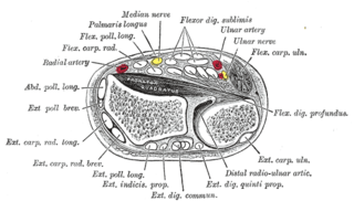
The posterior compartment of the forearm contains twelve muscles which primarily extend the wrist and digits. It is separated from the anterior compartment by the interosseous membrane between the radius and ulna.

The muscles of the hand are the skeletal muscles responsible for the movement of the hand and fingers. The muscles of the hand can be subdivided into two groups: the extrinsic and intrinsic muscle groups. The extrinsic muscle groups are the long flexors and extensors. They are called extrinsic because the muscle belly is located on the forearm. The intrinsic group are the smaller muscles located within the hand itself. The muscles of the hand are innervated by the radial, median, and ulnar nerves from the brachial plexus.

The extrinsic extensor muscles of the hand are located in the back of the forearm and have long tendons connecting them to bones in the hand, where they exert their action. Extrinsic denotes their location outside the hand. Extensor denotes their action which is to extend, or open flat, joints in the hand. They include the extensor carpi radialis longus (ECRL), extensor carpi radialis brevis (ECRB), extensor digitorum (ED), extensor digiti minimi (EDM), extensor carpi ulnaris (ECU), abductor pollicis longus (APL), extensor pollicis brevis (EPB), extensor pollicis longus (EPL), and extensor indicis (EI).

The extensor medii proprius is a rare anatomical variant in the extensor compartment of the forearm. The aberrant muscle is analogous to the extensor indicis with the insertion being the middle finger instead of the index finger.

The extensor indicis et medii communis is a rare anatomical variant in the extensor compartment of forearm. This additional muscle lies in the deep extensor layer next to the extensor indicis proprius and the extensor pollicis longus. The characteristics of this anomalous muscle resemble those of the extensor indicis proprius, with split tendons to the index and the middle finger. This muscle can also be considered as a variation of the aberrant extensor medii proprius.
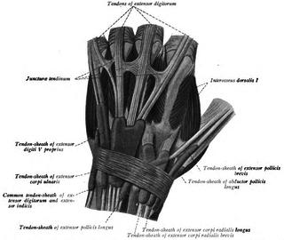
In human anatomy, juncturae tendinum or connexus intertendinei refers to the connective tissues that link the tendons of the extensor digitorum communis, and sometimes, to the tendon of the extensor digiti minimi. Juncturae tendinum are located on the dorsal aspect of the hand in the first, second and third inter-metacarpal spaces proximal to the metacarpophalangeal joint.

Palmaris profundus is a rare anatomical variant in the anterior compartment of forearm. It was first described in 1908. It is usually found incidentally in cadaveric dissection or surgery.

In human anatomy, the extensor pollicis et indicis communis is an aberrant muscle in the posterior compartment of forearm. It was first described in 1863. The muscle has a prevalence from 0.5% to 4%.
Linburg–Comstock variation is an occasional tendinous connection between the flexor pollicis longus and the flexor digitorum profundus of the index, the middle finger or both. It is found in around 21% of the population. It is an anatomical variation in human, which may be viewed as a pathology if causes symptoms. It was recognised as early as the 1800s, but was first described by Linburg and Comstock in 1979.
References
- ↑ Albinus, Bernhardus-Siegefridus (1758). "De extensore digitorum brevis manus" [On the extensor of the fingers of the short hand]. Academicarum Annotationum (in Latin). 4: 28–29.
- 1 2 3 4 Ranade, Anu V.; Rai, Rajalakshmi; Prabhu, Latha V.; Rajanigandha, V.; Janardhanan, Jiji P.; Ramanathan, Lakshmi; Prameela, M. D. (December 2008). "Incidence of Extensor Digitorum Brevis Manus Muscle". HAND. 3 (4): 320–323. doi:10.1007/s11552-008-9111-5. PMC 2584220 . PMID 18780016.
- 1 2 3 Yammine, Kaissar (January 2015). "The prevalence of extensor digitorum brevis manus and its variants in humans: a systematic review and meta-analysis". Surgical and Radiologic Anatomy. 37 (1): 3–9. doi:10.1007/s00276-014-1312-8. PMID 24849464. S2CID 8863347.
- 1 2 Çavdar, Safiye; Doğan, Teoman; Bayramiçli, Mehmet; Şehirli, Ümit; Yüksel, Mehtap (January 1998). "An unusual variation of extensor digitorum brevis manus: A case report and literature review". The Journal of Hand Surgery. 23 (1): 173–177. doi:10.1016/S0363-5023(98)80108-1. PMID 9523974.
- 1 2 Ogura, Takashi; Inoue, Hajime; Tanabe, Gozo (January 1987). "Anatomic and clinical studies of the extensor digitorum brevis manus". The Journal of Hand Surgery. 12 (1): 100–107. doi:10.1016/s0363-5023(87)80171-5. PMID 3805622.
- 1 2 Paraskevas, G.; Papaziogas, B.; Spanidou, S.; Papadopoulos, A. (January 2002). "Unusual variation of the extensor digitorum brevis manus: A case report". European Journal of Orthopaedic Surgery & Traumatology. 12 (3): 158–160. doi:10.1007/s00590-002-0035-4. PMID 24573895. S2CID 207024098.
- ↑ Le Double, Anatole-Félix (1897). Traité des variations du système musculaire de l'homme et de leur signification au point de vue de l'anthropologie zoologique [Treatise on the variations of the muscular system of man and their significance from the point of view of zoological anthropology] (in French). Schleicher frères.[ page needed ]
- 1 2 Suwannakhan, Athikhun; Tawonsawatruk, Tulyapruek; Meemon, Krai (November 2016). "Extensor tendons and variations of the medial four digits of hand: a cadaveric study". Surgical and Radiologic Anatomy. 38 (9): 1083–1093. doi:10.1007/s00276-016-1673-2. PMID 27056052. S2CID 8544399.
- ↑ Straus, William (February 1941). "The Phylogeny of the Human Forearm Extensors". Human Biology. 13 (1): 23. ProQuest 1301827777.
- ↑ Souter, W A (8 December 2005). "The extensor digitorum brevis manus". British Journal of Surgery. 53 (9): 821–823. doi:10.1002/bjs.1800530923. PMID 5911774. S2CID 42145116.
- ↑ Dixit, Daksha; Kubavant, Dharati M.; Lakhani, Chintan J.; Patel, Mital M.; Rathod, Suresh P.; Singel, T.C. (2012). "A morphometric evaluation of Extensor digitorum brevis manus by dissection: A rare atavistic muscle of the dorsum of hand" (PDF). Medica Innovatica. 1 (2): 59–63.