Extensor indicis et medii communis muscle

Last updated
Extensor indicis et medii communis

Extensor indicis et medii communis 2014.jpeg

Extensor indicis et medii communis (EIMC) and extensor indicis proprius (EIP)
Details
Origin posterior distal third of ulna and interosseous membrane
Insertion index finger and middle finger
Artery posterior interosseous artery
Nerve posterior interosseous nerve
Actions extends index finger and middle finger
Identifiers
Latin musculus extensor indicis et medii communis

Anatomical terms of muscle

The extensor indicis et medii communis is a rare anatomical variant in the extensor compartment of forearm. This additional muscle lies in the deep extensor layer next to the extensor indicis proprius and the extensor pollicis longus. The characteristics of this anomalous muscle resemble those of the extensor indicis proprius, with split tendons to the index and the middle finger. This muscle can also be considered as a variation of the aberrant extensor medii proprius.

The forearm is the region of the upper limb between the elbow and the wrist. The term forearm is used in anatomy to distinguish it from the arm, a word which is most often used to describe the entire appendage of the upper limb, but which in anatomy, technically, means only the region of the upper arm, whereas the lower "arm" is called the forearm. It is homologous to the region of the leg that lies between the knee and the ankle joints, the crus.

Extensor indicis muscle

In human anatomy, the extensor indicis [proprius] is a narrow, elongated skeletal muscle in the deep layer of the dorsal forearm, placed medial to, and parallel with, the extensor pollicis longus. Its tendon goes to the index finger, which it extends.

In human anatomy, the extensor pollicis longus muscle (EPL) is a skeletal muscle located dorsally on the forearm. It is much larger than the extensor pollicis brevis, the origin of which it partly covers and acts to stretch the thumb together with this muscle.

Contents

Structure

The extensor indicis et medii communis originates from the distal third of ulna next to the extensor indicis proprius. [1] After passing the wrist joint through the fourth extensor compartment, the tendon splits into two to insert to the extensor expansion of the index and the middle finger. [2]

Ulna long bone of the forearm

The ulna is a long bone found in the forearm that stretches from the elbow to the smallest finger, and when in anatomical position, is found on the medial side of the forearm. It runs parallel to the radius, the other long bone in the forearm, and is the larger and longer of the two.

The posterior compartment of the forearm contains twelve muscles which are chiefly responsible for extension of the wrist and digits, and supination of the forearm. It is separated from the anterior compartment by the interosseous membrane between the radius and ulna.

Extensor expansion

An extensor expansion is the special connective attachments by which the extensor tendons insert into the phalanges.

Prevalence

The extensor indicis et medii communis has an incidence between 0% and 6.5%. [2] [3] Meta-analysis showed that the muscle was present in average of 1.6% of the total 3,760 hands, and was more prevalent in North American populations. [3]

Incidence (epidemiology) measure of the probability of occurrence of a given medical condition in a population within a specified period of time

Incidence in epidemiology is a measure of the probability of occurrence of a given medical condition in a population within a specified period of time. Although sometimes loosely expressed simply as the number of new cases during some time period, it is better expressed as a proportion or a rate with a denominator.

Meta-analysis statistical method that summarizes data from multiple sources

A meta-analysis is a statistical analysis that combines the results of multiple scientific studies.

Function

The extensor indicis et medii communis extends the index and the middle finger.

See also

Extensor medii proprius muscle

The extensor medii proprius is a rare anatomical variant in the extensor compartment of the forearm. The aberrant muscle is analogous to the extensor indicis with the insertion being the middle finger instead of the index finger.

Related Research Articles

Finger organ of manipulation and sensation found in the hands of humans and other primates

A finger is a limb of the human body and a type of digit, an organ of manipulation and sensation found in the hands of humans and other primates. Normally humans have five digits, the bones of which are termed phalanges, on each hand, although some people have more or fewer than five due to congenital disorders such as polydactyly or oligodactyly, or accidental or medical amputations. The first digit is the thumb, followed by index finger, middle finger, ring finger, and little finger or pinky. According to different definitions, the thumb can be called a finger, or not.

Radial nerve nerve in the human body that supplies the posterior portion of the upper limb

The radial nerve is a nerve in the human body that supplies the posterior portion of the upper limb. It innervates the medial and lateral heads of the triceps brachii muscle of the arm, as well as all 12 muscles in the posterior osteofascial compartment of the forearm and the associated joints and overlying skin.

The flexor digitorum profundus is a muscle in the forearm of humans that flexes the fingers. It is considered an extrinsic hand muscle because it acts on the hand while its muscle belly is located in the forearm.

Extensor digiti minimi muscle muscle of the forearm

The extensor digiti minimi is a slender muscle of the forearm, placed on the ulnar side of the extensor digitorum communis, with which it is generally connected.

Extensor digitorum muscle

The extensor digitorum muscle is a muscle of the posterior forearm present in humans and other animals. It extends the medial four digits of the hand. Extensor digitorum is innervated by the posterior interosseous nerve, which is a branch of the radial nerve.

Palmar interossei muscles

In human anatomy, the palmar or volar interossei are three small, unipennate muscles in the hand that lie between the metacarpal bones and are attached to the index, ring, and little fingers. They are smaller than the dorsal interossei of the hand.

Dorsal interossei of the hand

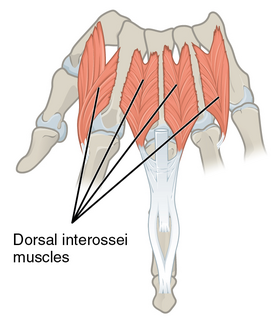
In human anatomy, the dorsal interossei (DI) are four muscles in the back of the hand that act to abduct (spread) the index, middle, and ring fingers away from hand's midline and assist in flexion at the metacarpophalangeal joints and extension at the interphalangeal joints of the index, middle and ring fingers.

Extensor digitorum brevis manus is an extra or accessory muscle on the backside (dorsum) of the hand. It was first described by Albinus in 1758. The muscles lies in the fourth extensor compartment of the wrist, and is relatively rare. It has a prevalence of 4% in the general population according to a meta-analysis. This muscle is commonly misdiagnosed as a ganglion cysta, synovial nodule or cyst.

Mucous sheaths on back of wrist

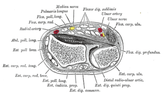
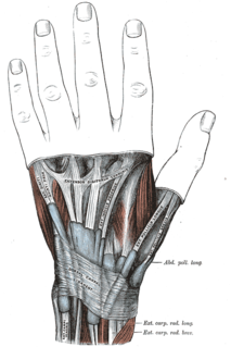
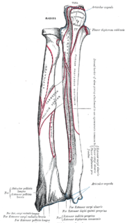
The mucous sheaths of the tendons on the back of the wrist are protective coverings for tendons in the wrist. Between the dorsal carpal ligament and the bones six compartments are formed for the passage of tendons, each compartment having a separate mucous sheath. One is found in each of the following positions:

  1. on the lateral side of the radial styloid process, for the tendons of the Abductor pollicis longus and Extensor pollicis brevis;
  2. behind the styloid process, for the tendons of the Extensores carpi radialis longus and brevis;
  3. about the middle of the dorsal surface of the radius, for the tendon of the Extensor pollicis longus;
  4. to the medial side of the latter, for the tendons of the Extensor digitorum communis and Extensor indicis proprius;
  5. opposite the interval between the radius and ulna, for the Extensor digiti quinti proprius;
  6. between the head and styloid process of the ulna, for the tendon of the Extensor carpi ulnaris.
Extensor tendon compartments of the wrist

Extensor tendon compartments of the wrist are anatomical tunnels on the back of the wrist that contain tendons of muscles that extend the wrist and the digits.

Extrinsic extensor muscles of the hand

The extrinsic extensor muscles of the hand are located in the back of the forearm and have long tendons connecting them to bones in the hand, where they exert their action. Extrinsic denotes their location outside the hand. Extensor denotes their action which is to extend, or open flat, joints in the hand. They include the extensor carpi radialis longus (ECRL), extensor carpi radialis brevis (ECRB), extensor digitorum (ED), extensor digiti minimi (EDM), extensor carpi ulnaris (ECU), abductor pollicis longus (APL), extensor pollicis brevis (EPB), extensor pollicis longus (EPL), and extensor indicis (EI).

Juncturae tendinum

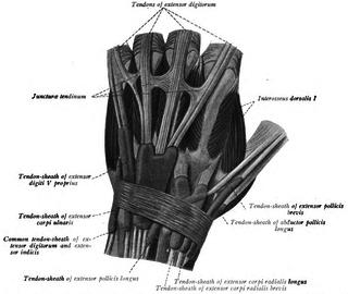
In human anatomy, juncturae tendinum or connexus intertendinei refers to the connective tissues that link the tendons of the extensor digitorum communis, and sometimes, to the tendon of the extensor digiti minimi. Juncturae tendinum are located on the dorsal aspect of the hand in the first, second and third inter-metacarpal spaces proximal to the metacarpophalangeal joint.

Palmaris profundus muscle

Palmaris profundus is a rare anatomical variant in the anterior compartment of forearm. It was first described in 1908. It is usually found incidentally in cadaveric dissection or surgery. The prevalence of the palmaris longus was reported to be one in 1600 upper limbs.

Extensor pollicis et indicis communis muscle

In human anatomy, the extensor pollicis et indicis communis is an aberrant muscle in the posterior compartment of forearm. It was first described in 1863. The muscle has a prevalence from 0.5% to 4%.

Linburg–Comstock variation is an occasional tendinous connection between the flexor pollicis longus and the flexor digitorum profundus of the index, the middle finger or both. It is found in around 21% of the population. It is an anatomical variation in human, which may be viewed as a pathology if causes symptoms. It was recognised as early as the 1800s, but was first described by Linburg and Comstock in 1979.

References

  1. Tan, S. T.; Smith, P. J. (May 1999). "Anomalous extensor muscles of the hand: a review". The Journal of Hand Surgery. 24 (3): 449–455. doi:10.1053/jhsu.1999.0449. ISSN   0363-5023. PMID   10357521.
  2. 1 2 Yalçin, Bülent; Kutoglu, Tunç; Ozan, Hasan; Gürbüz, Hülya (Mar 2006). "The extensor indicis et medii communis". Clinical Anatomy. 19 (2): 112–114. doi:10.1002/ca.20173. ISSN   0897-3806. PMID   16283647.
  3. 1 2 Yammine, Kaissar (Apr 2015). "The prevalence of the extensor indicis tendon and its variants: a systematic review and meta-analysis". Surgical and radiologic anatomy: SRA. 37 (3): 247–254. doi:10.1007/s00276-014-1352-0. ISSN   1279-8517. PMID   25096501.