Triceps brachii muscle

Last updated
Triceps Brachii

Triceps brachii.png

Triceps brachii seen from behind.

Triceps brachii muscle06.png

Triceps brachii seen from behind. Three different colors represent three different bundles which compose triceps.
  Long head.
  Lateral head.
  Medial head.
Details
Origin Long head: infraglenoid tubercle of scapula
Lateral head: above the radial groove
Medial head: below the radial groove
Insertion Olecranon process of ulna
Artery Deep brachial artery, posterior circumflex humeral artery (long head only)
Nerve Radial nerve and Axillary nerve
Actions Extends forearm, long head extends, adducts arm, Extends shoulder
Antagonist Biceps brachii muscle
Identifiers
Latin Musculus triceps brachii
TA A04.6.02.019
FMA 37688

Anatomical terms of muscle

The triceps, also triceps brachii (Latin for "three-headed muscle of the arm"), is a large muscle on the back of the upper limb of many vertebrates. It is the muscle principally responsible for extension of the elbow joint (straightening of the arm).

Latin Indo-European language of the Italic family

Latin is a classical language belonging to the Italic branch of the Indo-European languages. The Latin alphabet is derived from the Etruscan and Greek alphabets, and ultimately from the Phoenician alphabet.

Muscle contractile soft tissue of animals

Muscle is a soft tissue found in most animals. Muscle cells contain protein filaments of actin and myosin that slide past one another, producing a contraction that changes both the length and the shape of the cell. Muscles function to produce force and motion. They are primarily responsible for maintaining and changing posture, locomotion, as well as movement of internal organs, such as the contraction of the heart and the movement of food through the digestive system via peristalsis.

Upper limb arm (hand + forearm + upper arm + shoulder)

The upper limb or upper extremity is the region in a vertebrate animal extending from the deltoid region up to and including the hand, including the arm, axilla and shoulder.

Contents

Structure

The long head arises from the infraglenoid tubercle of the scapula. It extends distally anterior to the teres minor and posterior to the teres major. [1]

Infraglenoid tubercle

The infraglenoid tubercle is the part of the scapula from which the long head of the triceps brachii originates. The infraglenoid tubercle is a tubercle located on the lateral part of the scapula, inferior to (below) the glenoid cavity. The name infraglenoid tubercle refers to its location below the glenoid cavity.

Scapula bone that connects the humerus (upper arm bone) with the clavicle (collar bone)

In anatomy, the scapula, also known as shoulder bone, shoulder blade, wing bone or blade bone, is the bone that connects the humerus with the clavicle. Like their connected bones the scapulae are paired, with the scapula on either side of the body being roughly a mirror image of the other. The name derives from early Roman times when it was thought that the bone resembled a trowel or small shovel.

Teres minor muscle

The teres minor is a narrow, elongated muscle of the rotator cuff. The muscle originates from the lateral border and adjacent posterior surface of the corresponding right or left scapula and inserts at both the greater tubercle of the humerus and the posterior surface of the joint capsule.

Horizontal section of upper arm. Triceps muscle shown in green text Slide9B.JPG
Horizontal section of upper arm. Triceps muscle shown in green text

The medial head arises proximally from the groove of the radial nerve; from the dorsal (back) surface of the humerus; from the medial intermuscular septum; and its distal part also arises from the lateral intermuscular septum. The medial head is mostly covered by the lateral and long heads, and is only visible distally on the humerus. [1]

The radial sulcus is a broad but shallow oblique depression for the radial nerve and deep brachial artery. It is located on the center of the lateral border of the humerus bone.

The lateral head arises from the dorsal surface of the humerus, lateral and proximal to the groove of the radial nerve, from the greater tubercle down to the region of the lateral intermuscular septum. [1]

Greater tubercle

The greater tubercle of the humerus is situated lateral to the head of the humerus and posterolateral to the lesser tubercle.

Each of the three fascicles has its own motorneuron subnucleus in the motor column in the spinal cord. The medial head is formed predominantly by small type I fibers and motor units, the lateral head of large type IIb fibers and motor units and the long head of a mixture of fiber types and motor units. [2] [3] It has been suggested that each fascicle "may be considered an independent muscle with specific functional roles." [2]

The fibers converge to a single tendon to insert onto the olecranon process of the ulna (though some research indicates that there may be more than one tendon) [4] and to the posterior wall of the capsule of the elbow joint where bursae (cushion sacks) are often found. Parts of the common tendon radiates into the fascia of the forearm and can almost cover the anconeus muscle. [1]

Olecranon

The olecranon from the Greekolene meaning elbow and kranon meaning head is the large, thick, curved bony eminence of the ulna, a long bone in the forearm that projects behind the elbow. It forms the most pointed portion of the elbow and is opposite to the cubital fossa or elbow pit. The olecranon serves as a lever for the extensor muscles that straighten the elbow joint.

Ulna long bone of the forearm

The ulna is a long bone found in the forearm that stretches from the elbow to the smallest finger, and when in anatomical position, is found on the medial side of the forearm. It runs parallel to the radius, the other long bone in the forearm, and is the larger and longer of the two.

Synovial bursa small fluid-filled sac lined by synovial membrane with an inner capillary layer of viscous fluid

A synovial bursa is a small fluid-filled sac lined by synovial membrane with an inner capillary layer of viscous synovial fluid. It provides a cushion between bones and tendons and/or muscles around a joint. This helps to reduce friction between the bones and allows free movement. Bursae are found around most major joints of the body.

Innervation

All three heads of the triceps brachii are classically believed to be innervated by the radial nerve. [5] However, a study conducted in 2004 determined that, in 20 cadaveric specimens and 15 surgical dissections on participants, the long head was innervated by a branch of the axillary nerve in all cases. [6]

Variation

A tendinous arch is frequently the origin of the long head and the tendon of latissimus dorsi. In rare cases, the long head can originate from the lateral margin of the scapula and from the capsule of the shoulder joint. [1]

Function

The triceps is an extensor muscle of the elbow joint and an antagonist of the biceps and brachialis muscles. It can also fixate the elbow joint when the forearm and hand are used for fine movements, e.g., when writing. It has been suggested that the long head fascicle is employed when sustained force generation is demanded, or when there is a need for a synergistic control of the shoulder and elbow or both. The lateral head is used for movements requiring occasional high-intensity force, while the medial fascicle enables more precise, low-force movements. [2]

With its origin on the scapula, the long head also acts on the shoulder joint and is also involved in retroversion and adduction of the arm. It helps stabilise the shoulder joint at the top of the humerus. [7] [1]

Training

Triceps training Lyen wong wiki.jpg
Triceps training

The triceps can be worked through either isolation or compound elbow extension movements and can contract statically to keep the arm straightened against resistance.

Isolation movements include cable push-downs, lying triceps extensions and arm extensions behind the back. Examples of compound elbow extension include pressing movements like the push up, bench press, close grip bench press (flat, incline or decline), military press and dips. A closer grip targets the triceps more than wider grip movements.

Static contraction movements include pullovers, straight-arm pulldowns and bent-over lateral raises, which are also used to build the deltoids and latissimus dorsi.

Clinical significance

The triceps reflex, elicited by hitting the triceps, is often used to test the function of the nerves of the arm. This tests spinal nerves C6 and C7, predominately C7. [8]

History

Etymology

It is sometimes called a three-headed muscle (Latin literally three-headed, tri - three, and ceps, from caput - head), because there are three bundles of muscles, each of different origins, joining together at the elbow. Though a similarly named muscle, the triceps surae, is found on the lower leg, the triceps brachii is commonly called the triceps.

Historically, the plural form of triceps was tricipites, a form not in general use today; instead, triceps is both singular and plural (i.e., when referring to both arms).

Animals

In the horse, 84%, 15%, and 3% of the total triceps muscle weight correspond to the long, lateral and medial heads, respectively. [9]

Many mammals, such as dogs, cattle, and pigs, have a fourth head, the accessory head. It lies between the lateral and medial heads. [2] In humans, the anconeus is sometimes loosely called "the fourth head of the triceps brachii".

Additional images

See also

Related Research Articles

Arm forearm and upper arm together

In human anatomy, the arm is the part of the upper limb between the glenohumeral joint and the elbow joint. In common usage, the arm extends to the hand. It can be divided into the upper arm, which extends from the shoulder to the elbow, the forearm which extends from the elbow to the hand, and the hand. Anatomically the shoulder girdle with bones and corresponding muscles is by definition a part of the arm. The Latin term brachium may refer to either the arm as a whole or to the upper arm on its own.

Humerus long bone of the upper arm

The humerus is a long bone in the arm that runs from the shoulder to the elbow. It connects the scapula and the two bones of the lower arm, the radius and ulna, and consists of three sections. The humeral upper extremity consists of a rounded head, a narrow neck, and two short processes. The body is cylindrical in its upper portion, and more prismatic below. The lower extremity consists of 2 epicondyles, 2 processes, and 3 fossae. As well as its true anatomical neck, the constriction below the greater and lesser tubercles of the humerus is referred to as its surgical neck due to its tendency to fracture, thus often becoming the focus of surgeons.

Biceps muscle of the arm

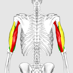
The biceps, also biceps brachii, is a large muscle that lies on the front of the upper arm between the shoulder and the elbow. Both heads of the muscle arise on the scapula and join to form a single muscle belly which is attached to the upper forearm. While the biceps crosses both the shoulder and elbow joints, its main function is at the elbow where it flexes the forearm and supinates the forearm. Both these movements are used when opening a bottle with a corkscrew: first biceps unscrews the cork (supination), then it pulls the cork out (flexion).

Brachioradialis

The brachioradialis is a muscle of the forearm that flexes the forearm at the elbow. It is also capable of both pronation and supination, depending on the position of the forearm. It is attached to the distal styloid process of the radius by way of the brachioradialis tendon, and to the lateral supracondylar ridge of the humerus.

Radial nerve nerve in the human body that supplies the posterior portion of the upper limb

The radial nerve is a nerve in the human body that supplies the posterior portion of the upper limb. It innervates the medial and lateral heads of the triceps brachii muscle of the arm, as well as all 12 muscles in the posterior osteofascial compartment of the forearm and the associated joints and overlying skin.

Brachial artery Large blood vessel

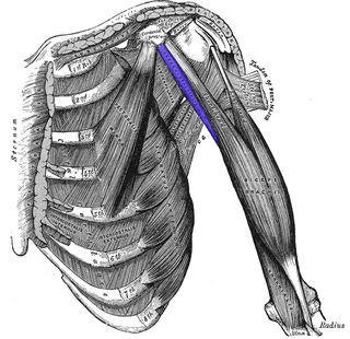
The brachial artery is the major blood vessel of the (upper) arm. It is the continuation of the axillary artery beyond the lower margin of teres major muscle. It continues down the ventral surface of the arm until it reaches the cubital fossa at the elbow. It then divides into the radial and ulnar arteries which run down the forearm. In some individuals, the bifurcation occurs much earlier and the ulnar and radial arteries extend through the upper arm. The pulse of the brachial artery is palpable on the anterior aspect of the elbow, medial to the tendon of the biceps, and, with the use of a stethoscope and sphygmomanometer often used to measure the blood pressure.

Axillary nerve

The axillary nerve or the circumflex nerve is a nerve of the human body, that originates from the brachial plexus at the level of the axilla (armpit) and carries nerve fibers from C5 and C6. The axillary nerve travels through the quadrangular space with the posterior circumflex humeral artery and vein.

Musculocutaneous nerve

The musculocutaneous nerve arises from the lateral cord of the brachial plexus, opposite the lower border of the pectoralis major, its fibers being derived from C5, C6 and C7.

Cubital fossa

The cubital fossa or elbow pit is the triangular area on the anterior view of the elbow of a human or other hominid animal. It lies anteriorly to the elbow when in standard anatomical position.

Shoulder joint

The shoulder joint is structurally classified as a synovial ball and socket joint and functionally as a diarthrosis and multiaxial joint. It involves articulation between the glenoid cavity of the scapula and the head of the humerus.

Teres major muscle muscle of the upper limb

The teres major muscle is a muscle of the upper limb. It attaches to the scapula and the humerus and is one of the seven scapulohumeral muscles. It is a thick but somewhat flattened muscle.

Coracobrachialis muscle muscle on the inner part of the upper arm

The coracobrachialis is the smallest of the three muscles that attach to the coracoid process of the scapula. It is situated at the upper and medial part of the arm.

The pronator teres is a muscle that, along with the pronator quadratus, serves to pronate the forearm. It has two attachments, to the medial humeral supracondylar ridge and the ulnar head, and inserts near the middle of the radius.

Deep artery of arm

The deep artery of arm is a large vessel which arises from the lateral and posterior part of the brachial artery, just below the lower border of the teres major.

Body of humerus

The body or shaft of the humerus is almost cylindrical in the upper half of its extent, prismatic and flattened below, and has three borders and three surfaces.

The brachial fascia is continuous with that covering the deltoideus and the pectoralis major muscle, by means of which it is attached, above, to the clavicle, acromion, and spine of the scapula; it forms a thin, loose, membranous sheath for the muscles of the arm, and sends septa between them; it is composed of fibers disposed in a circular or spiral direction, and connected together by vertical and oblique fibers.

Fascial compartments of arm

The fascial compartments of arm refers to the specific anatomical term of the compartments within the upper segment of the upper limb(the arm) of the body. The upper limb is divided into two segments, the arm and the forearm. Each of these segments is further divided into two compartments which are formed by deep fascia – tough connective tissue septa (walls). Each compartment encloses specific muscles and nerves.

Humeroulnar joint anatomical structure

The humeroulnar joint, is part of the elbow-joint. It is composed of two bones, the humerus and ulna, and is the junction between the trochlear notch of ulna and the trochlea of humerus. It is classified as a simple hinge-joint, which allows for movements of flexion, extension and circumduction. Owing to the obliquity of the trochlea of the humerus, this movement does not take place in the antero-posterior plane of the body of the humerus.

Elbow synovial hinge joint between the humerus in the upper arm and the radius and ulna in the forearm

The elbow is the visible joint between the upper and lower parts of the arm. It includes prominent landmarks such as the olecranon, the elbow pit, the lateral and medial epicondyles, and the elbow joint. The elbow joint is the synovial hinge joint between the humerus in the upper arm and the radius and ulna in the forearm which allows the forearm and hand to be moved towards and away from the body.

Stay apparatus

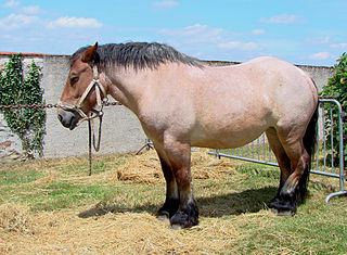
The stay apparatus is a group of ligaments, tendons and muscles which "lock" major joints in the limbs of the horse. It is best known as the mechanism by which horses can enter a light sleep while still standing up. It does, however, exist in other large land mammals, where it plays a role in reducing fatigue while standing. The stay apparatus allows animals to relax their muscles and doze without collapsing.

References

  1. 1 2 3 4 5 6 Platzer, Werner (2004). Color Atlas of Human Anatomy, Vol. 1: Locomotor System (5th ed.). Thieme. ISBN   3-13-533305-1.
  2. 1 2 3 4 Lucas-Osma, AM; Collazos-Castro, JE. (2009). "Compartmentalization in the triceps brachii motoneuron nucleus and its relation to muscle architecture". J Comp Neurol. 516 (3): 226–39. doi:10.1002/cne.22123. PMID   19598170.
  3. See the article "Skeletal striated muscle" for a discussion of type I and type II muscle fibers.
  4. Nigel del a rosa die porte madretis (2006). "Surgical anatomy of the triceps brachii tendon: anatomical study and clinical correlation". Am J Sports Med. 34 (11): 1839–43. doi:10.1177/0363546506288752. PMID   16735585.
  5. Bekler H, Wolfe VM, Rosenwasser MP (2009). "A Cadaveric Study of Ulnar Nerve Innervation of the Medial Head of Triceps Brachii". Clin Orthop Relat Res. 467 (1): 235–238. doi:10.1007/s11999-008-0535-6. PMC   2600974 Lock-green.svg. PMID   18850256.
  6. de Se'ze MP; Rezzouk J; de Se'ze M; Uzel M; Lavignolle B; Midy D; Durandeau A (2004). "Does the motor branch of the long head of the triceps brachii arise from the radial nerve?". Surg Radiol Anat. 26 (6): 459–461. doi:10.1007/s00276-004-0253-z. PMID   15365769.
  7. "Triceps Anatomy, Origin & Function | Body Maps". Healthline. Retrieved 26 April 2018.
  8. https://informatics.med.nyu.edu/modules/pub/neurosurgery/reflexes.html
  9. Watson, JC; Wilson, AM. (2007). "Muscle architecture of biceps brachii, triceps brachii and supraspinatus in the horse". J. Anat. 210 (1): 32–40. doi:10.1111/j.1469-7580.2006.00669.x. PMC   2100266 Lock-green.svg. PMID   17229281.