| Lagena | |
|---|---|
Lagena is #12, and is labeled at upper right. | |
Lagena is #12, and is labeled at upper left and lower left. | |
| Identifiers | |


The lagena (from Greek for flask) is a structure found in humans and in animals.
| Lagena | |
|---|---|
Lagena is #12, and is labeled at upper right. | |
Lagena is #12, and is labeled at upper left and lower left. | |
| Identifiers | |
The lagena (from Greek for flask) is a structure found in humans and in animals.
In the ear, the extremities of the ductus cochlearis are closed; the upper is termed the lagena and is attached to the cupula at the upper part of the helicotrema; the lower is lodged in the recessus cochlearis of the vestibule.

The cochlear cupula is a structure in the cochlea. It is the apex of the cochlea. The bony canal of the cochlea takes two and three-quarter turns around the modiolus. The modiolus is about 30 mm. in length, and diminishes gradually in diameter from the base to the summit, where it terminates in the cupula.
The helicotrema is the part of the cochlear labyrinth where the scala tympani and the scala vestibuli meet. It is the main component of the cochlear apex. The hair cells near this area best detect low frequency sounds.

The vestibule is the central part of the bony labyrinth in the inner ear, and is situated medial to the eardrum, behind the cochlea, and in front of the three semicircular canals.
The lagena is part of the vestibular system in fish and amphibians. It contains the otoliths asterisci. In fish, the lagena is implicated in hearing and the registration of vertical linear acceleration, [1] in amphibians is the latter only.
The vestibular system, in most mammals, is the sensory system that provides the leading contribution to the sense of balance and spatial orientation for the purpose of coordinating movement with balance. Together with the cochlea, a part of the auditory system, it constitutes the labyrinth of the inner ear in most mammals. As movements consist of rotations and translations, the vestibular system comprises two components: the semicircular canals which indicate rotational movements; and the otoliths which indicate linear accelerations. The vestibular system sends signals primarily to the neural structures that control eye movements, and to the muscles that keep an animal upright and in general control posture. The projections to the former provide the anatomical basis of the vestibulo-ocular reflex, which is required for clear vision; while the projections to the latter provide the anatomical means required to enable an animal to maintain its desired position in space.

An otolith, also called statoconium or otoconium or statolith, is a calcium carbonate structure in the saccule or utricle of the inner ear, specifically in the vestibular system of vertebrates. The saccule and utricle, in turn, together make the otolith organs. These organs are what allows an organism, including humans, to perceive linear acceleration, both horizontally and vertically (gravity). They have been identified in both extinct and extant vertebrates.

The urinary bladder is a hollow muscular organ in humans and some other animals that collects and stores urine from the kidneys before disposal by urination. In the human the bladder is a hollow muscular, and distensible organ, that sits on the pelvic floor. Urine enters the bladder via the ureters and exits via the urethra. The typical human bladder will hold between 300 and 500 mL before the urge to empty occurs, but can hold considerably more.
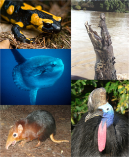
Vertebrates comprise all species of animals within the subphylum Vertebrata. Vertebrates represent the overwhelming majority of the phylum Chordata, with currently about 69,276 species described. Vertebrates include the jawless fishes and jawed vertebrates, which include the cartilaginous fishes and the bony fishes. The bony fishes in turn, cladistically speaking, also include the tetrapods, which include amphibians, reptiles, birds and mammals.

The inner ear is the innermost part of the vertebrate ear. In vertebrates, the inner ear is mainly responsible for sound detection and balance. In mammals, it consists of the bony labyrinth, a hollow cavity in the temporal bone of the skull with a system of passages comprising two main functional parts:

Tetrapods (from Greek: τετρα- "four" and πούς "foot") are four-limbed animals constituting the superclass Tetrapoda. It includes existing and extinct amphibians, reptiles, and mammals. Tetrapods evolved from a group of animals known as the Tetrapodomorpha which, in turn, evolved from ancient Sarcopterygii around 390 million years ago in the middle Devonian period; their forms were transitional between lobe-finned fishes and the four-limbed tetrapods. The first tetrapods appeared by the late Devonian, 367.5 million years ago; the specific aquatic ancestors of the tetrapods, and the process by which they colonized Earth's land after emerging from water remains unclear. The change from a body plan for breathing and navigating in water to a body plan enabling the animal to move on land is one of the most profound evolutionary changes known. The first tetrapods were primarily aquatic. Modern amphibians, which evolved from earlier groups, are generally semiaquatic; the first stage of their lives is as fish-like tadpoles, and later stages are partly terrestrial and partly aquatic. However, most tetrapod species today are amniotes, most of those are terrestrial tetrapods whose branch evolved from earlier tetrapods about 340 million years ago. The key innovation in amniotes over amphibians is laying of eggs on land or having further evolved to retain the fertilized egg(s) within the mother.

The clavicle or collarbone is a long bone that serves as a strut between the shoulder blade and the sternum or breastbone. There are two clavicles, one on the left and one on the right. The clavicle is the only long bone in the body that lies horizontally. Together with the shoulder blade it makes up the shoulder girdle. It is a touchable bone and in people who have less fat in this region, the location of the bone is clearly visible, as it creates a bulge in the skin. It receives its name from the Latin: clavicula because the bone rotates along its axis like a key when the shoulder is abducted. The clavicle is the most commonly fractured bone. It can easily be fractured due to impacts to the shoulder from the force of falling on outstretched arms or by a direct hit.

The maxilla in animals is the upper fixed bone of the jaw formed from the fusion of two maxillary bones. The upper jaw includes the hard palate in the front of the mouth. The two maxillary bones are fused at the intermaxillary suture, forming the anterior nasal spine. This is similar to the mandible, which is also a fusion of two mandibular bones at the mandibular symphysis. The mandible is the movable part of the jaw.

The jaw is any opposable articulated structure at the entrance of the mouth, typically used for grasping and manipulating food. The term jaws is also broadly applied to the whole of the structures constituting the vault of the mouth and serving to open and close it and is part of the body plan of most animals.

In the human skull, the zygomatic bone is a paired irregular bone which articulates with the maxilla, the temporal bone, the sphenoid bone and the frontal bone. It is situated at the upper and lateral part of the face and forms the prominence of the cheek, part of the lateral wall and floor of the orbit, and parts of the temporal fossa and the infratemporal fossa. It presents a malar and a temporal surface; four processes, and four borders.
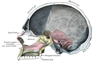
The palatine bones are two irregular bones of the facial skeleton in many animal species. Together with the maxillae they comprise the hard palate.

The frontal bone is a bone in the human skull. The bone consists of two portions. These are the vertically oriented squamous part, and the horizontally oriented orbital part, making up the bony part of the forehead, part of the bony orbital cavity holding the eye, and part of the bony part of the nose respectively. The name comes from the Latin word frons.

The lacrimal bone is a small and fragile bone of the facial skeleton; it is roughly the size of the little fingernail. It is situated at the front part of the medial wall of the orbit. It has two surfaces and four borders. Several bony landmarks of the lacrimal bone function in the process of lacrimation or crying. Specifically, the lacrimal bone helps form the nasolacrimal canal necessary for tear translocation. A depression on the anterior inferior portion of the bone, the lacrimal fossa, houses the membranous lacrimal sac. Tears or lacrimal fluid, from the lacrimal glands, collect in this sac during excessive lacrimation. The fluid then flows through the nasolacrimal duct and into the nasopharynx. This drainage results in what is commonly referred to a runny nose during excessive crying or tear production. Injury or fracture of the lacrimal bone can result in posttraumatic obstruction of the lacrimal pathways.

The vomer is one of the unpaired facial bones of the skull. It is located in the midsagittal line, and articulates with the sphenoid, the ethmoid, the left and right palatine bones, and the left and right maxillary bones. The vomer forms the inferior part of the nasal septum, with the superior part formed by the perpendicular plate of the ethmoid bone. The name is derived from the Latin word for a ploughshare and the shape of the bone.

Fish anatomy is the study of the form or morphology of fishes. It can be contrasted with fish physiology, which is the study of how the component parts of fish function together in the living fish. In practice, fish anatomy and fish physiology complement each other, the former dealing with the structure of a fish, its organs or component parts and how they are put together, such as might be observed on the dissecting table or under the microscope, and the latter dealing with how those components function together in living fish.

The Caudata are a group of amphibians containing the salamanders (Urodela) and all extinct species of salamander-like amphibians more closely related to salamanders than to frogs. They are typically characterized by a superficially lizard-like appearance, with slender bodies, blunt snouts, short limbs projecting at right angles to the body, and the presence of a tail in both larvae and adults.
The term paradidymis is applied to a small collection of convoluted tubules, situated in front of the lower part of the spermatic cord, above the head of the epididymis.
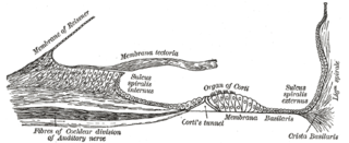
The periosteum, forming the outer wall of the ductus cochlearis, is greatly thickened and altered in character, and is called the spiral ligament.

In animal anatomy, the mouth, also known as the oral cavity, buccal cavity, or in Latin cavum oris, is the opening through which many animals take in food and issue vocal sounds. It is also the cavity lying at the upper end of the alimentary canal, bounded on the outside by the lips and inside by the pharynx and containing in higher vertebrates the tongue and teeth. This cavity is also known as the buccal cavity, from the Latin bucca ("cheek").

The skull roof, or the roofing bones of the skull, are a set of bones covering the brain, eyes and nostrils in bony fishes and all land-living vertebrates. The bones are derived from dermal bone and are part of the dermatocranium.
Lagena, a term derived from the Greek word meaning flask, refer to:
This article incorporates text in the public domain from page 1054 of the 20th edition of Gray's Anatomy (1918)
The public domain consists of all the creative works to which no exclusive intellectual property rights apply. Those rights may have expired, been forfeited, expressly waived, or may be inapplicable.

Gray's Anatomy is an English language textbook of human anatomy originally written by Henry Gray and illustrated by Henry Vandyke Carter. Earlier editions were called Anatomy: Descriptive and Surgical, Anatomy of the Human Body and Gray's Anatomy: Descriptive and Applied, but the book's name is commonly shortened to, and later editions are titled, Gray's Anatomy. The book is widely regarded as an extremely influential work on the subject, and has continued to be revised and republished from its initial publication in 1858 to the present day. The latest edition of the book, the 41st, was published in September 2015.
| This anatomy article is a stub. You can help Wikipedia by expanding it. |