Laryngeal tube

Last updated
Laryngeal tube
Laryngeal Tube.JPG
Standard Laryngeal Tube size 5 (VBM Medizintechnik, Sulz, Germany) with the syringe for cuff inflation
Synonyms King LT
Specialty Anesthesiology, Emergency medicine
Intervention Airway management
Invention date1999
Manufacturer VBM Medizintechnik
Related items Laryngeal mask airway

The laryngeal tube (also known as the King LT) [1] is an airway management device designed as an alternative to other airway management techniques such as mask ventilation, laryngeal mask airway, and tracheal intubation. This device can be inserted blindly through the oropharynx into the hypopharynx to create an airway during anaesthesia and cardiopulmonary resuscitation so as to enable mechanical ventilation of the lungs. [2]

Contents

Medical use

Various studies have shown that insertion and use of the standard tracheal tube is easy, providing a clear airway in the majority of cases. [2] [3] [4] [5] [6] Comparative studies indicate that the standard laryngeal tube is generally as effective as the laryngeal mask airway, [2] [7] [8] [9] [10] while some studies indicate that the Pro-seal laryngeal mask may be more effective than the standard laryngeal tube under controlled ventilation conditions in general anaesthesia. [11] [12] The indications and contraindications for use of the laryngeal tube are similar to those of the laryngeal mask airway and include the use in general anaesthesia for minor surgical operations. [2] Several studies describe the usefulness of the device in securing a difficult airway, even in cases where insertion of the laryngeal mask had failed. [13] [14] The double-lumen laryngeal tube-Suction II, with the possibility of placing a gastric tube, has been found to have distinct advantages over the standard laryngeal tube and has been recommended as a first-line device to secure the airway in emergency situations when direct laryngoscopy fails in neonates and infants. [15] The laryngeal tube is also recommended for medical personnel not experienced in tracheal intubation, and as a rescue device when intubation has failed in adults. [16] According to the manufacturer the use of Laryngeal tubes is contraindicated in people with an intact gag reflex, known oesophageal disease, and people who have ingested caustic substances. [17]

Description

In its basic (standard) version, the laryngeal tube is made up of a tube with a larger balloon cuff in the middle (oropharyngeal cuff) and a smaller balloon cuff at the end (oesophageal cuff). The tube is kinked at an angle of 30-45° in the middle; the kink is located in the larger cuff. There are two apertures, located between the two cuffs, through which ventilation takes place. Both cuffs are inflated through a single small lumen line and pilot balloon. The cuffs are high-volume, low-pressure cuffs with inflating volume ranging from 10 ml (size 0) to 90 ml (size 5). A large bore syringe, which is marked with the required volume for each size, is used to inflate the cuffs. A cuff inflator can also be used, in which case the cuffs should be inflated to a pressure of 60 cm H2O. Three black lines on the tube indicate the depth of insertion when aligned with the teeth. [2]

History

Laryngeal tube placement in glass model Larynxtubus.jpg
Laryngeal tube placement in glass model

The laryngeal tube was developed in Germany and introduced to the European market by VBM Medizintechnik in the autumn of 1999. [18] Since then the design has been modified several times. Currently four different models are used: the standard tube as single use or re-use models and the modified tube (laryngeal tube-Suction II) as single use or re-use models. The re-usable models can be autoclaved up to 50 times, while the modified laryngeal tube (Suction) incorporates an extra lumen for inserting a gastric tube or suction system. There are six sizes of the laryngeal tube, ranging from newborn (size 0) to large adult (size 5). The connector of the tube is color-coded for each size. The different sizes are calibrated according to weight or height. [2]

The laryngeal tube was licensed for use during cardiopulmonary resuscitation in Japan in 2002, [2] and approved for use in the United States by the Food and Drug Administration in 2003. [19] The European Resuscitation Council, in its 2005 guidelines for advanced life support (ALS), accepts its use as an alternate airway device for medical personnel who are not experienced in tracheal intubation. [20]

See also

Related Research Articles

<span class="mw-page-title-main">Tracheal intubation</span> Placement of a tube into the trachea

Tracheal intubation, usually simply referred to as intubation, is the placement of a flexible plastic tube into the trachea (windpipe) to maintain an open airway or to serve as a conduit through which to administer certain drugs. It is frequently performed in critically injured, ill, or anesthetized patients to facilitate ventilation of the lungs, including mechanical ventilation, and to prevent the possibility of asphyxiation or airway obstruction.

<span class="mw-page-title-main">Mechanical ventilation</span> Method to mechanically assist or replace spontaneous breathing

Mechanical ventilation, assisted ventilation or intermittent mandatory ventilation (IMV), is the medical term for using a machine called a ventilator to fully or partially provide artificial ventilation. Mechanical ventilation helps move air into and out of the lungs, with the main goal of helping the delivery of oxygen and removal of carbon dioxide. Mechanical ventilation is used for many reasons, including to protect the airway due to mechanical or neurologic cause, to ensure adequate oxygenation, or to remove excess carbon dioxide from the lungs. Various healthcare providers are involved with the use of mechanical ventilation and people who require ventilators are typically monitored in an intensive care unit.

<span class="mw-page-title-main">Tracheotomy</span> Temporary surgical incision to create an airway into the trachea

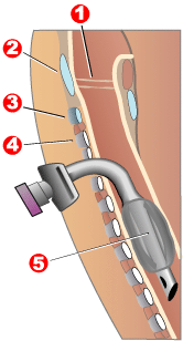
Tracheotomy, or tracheostomy, is a surgical airway management procedure which consists of making an incision (cut) on the anterior aspect (front) of the neck and opening a direct airway through an incision in the trachea (windpipe). The resulting stoma (hole) can serve independently as an airway or as a site for a tracheal tube or tracheostomy tube to be inserted; this tube allows a person to breathe without the use of the nose or mouth.

<span class="mw-page-title-main">Laryngoscopy</span> Endoscopy of the larynx

Laryngoscopy is endoscopy of the larynx, a part of the throat. It is a medical procedure that is used to obtain a view, for example, of the vocal folds and the glottis. Laryngoscopy may be performed to facilitate tracheal intubation during general anaesthesia or cardiopulmonary resuscitation or for surgical procedures on the larynx or other parts of the upper tracheobronchial tree.

A tracheal tube is a catheter that is inserted into the trachea for the primary purpose of establishing and maintaining a patent airway and to ensure the adequate exchange of oxygen and carbon dioxide.

<span class="mw-page-title-main">Laryngeal mask airway</span>

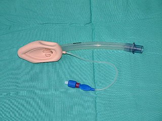
A laryngeal mask airway (LMA), also known as laryngeal mask, is a medical device that keeps a patient's airway open during anaesthesia or while they are unconscious. It is a type of supraglottic airway device. They are most commonly used by anaesthetists to channel oxygen or inhalational anaesthetic to the lungs during surgery and in the pre-hospital setting for unconscious patients.

<span class="mw-page-title-main">Airway management</span> Medical procedure ensuring an unobstructed airway

Airway management includes a set of maneuvers and medical procedures performed to prevent and relieve airway obstruction. This ensures an open pathway for gas exchange between a patient's lungs and the atmosphere. This is accomplished by either clearing a previously obstructed airway; or by preventing airway obstruction in cases such as anaphylaxis, the obtunded patient, or medical sedation. Airway obstruction can be caused by the tongue, foreign objects, the tissues of the airway itself, and bodily fluids such as blood and gastric contents (aspiration).

<span class="mw-page-title-main">Respiratory arrest</span> Medical condition

Respiratory arrest is a sickness caused by apnea or respiratory dysfunction severe enough it will not sustain the body. Prolonged apnea refers to a patient who has stopped breathing for a long period of time. If the heart muscle contraction is intact, the condition is known as respiratory arrest. An abrupt stop of pulmonary gas exchange lasting for more than five minutes may permanently damage vital organs, especially the brain. Lack of oxygen to the brain causes loss of consciousness. Brain injury is likely if respiratory arrest goes untreated for more than three minutes, and death is almost certain if more than five minutes.

<span class="mw-page-title-main">Artificial ventilation</span> Assisted breathing to support life

Artificial ventilation is a means of assisting or stimulating respiration, a metabolic process referring to the overall exchange of gases in the body by pulmonary ventilation, external respiration, and internal respiration. It may take the form of manually providing air for a person who is not breathing or is not making sufficient respiratory effort, or it may be mechanical ventilation involving the use of a mechanical ventilator to move air in and out of the lungs when an individual is unable to breathe on their own, for example during surgery with general anesthesia or when an individual is in a coma or trauma.

In advanced airway management, rapid sequence induction (RSI) – also referred to as rapid sequence intubation or as rapid sequence induction and intubation (RSII) or as crash induction – is a special process for endotracheal intubation that is used where the patient is at a high risk of pulmonary aspiration. It differs from other techniques for inducing general anesthesia in that several extra precautions are taken to minimize the time between giving the induction drugs and securing the tube, during which period the patient's airway is essentially unprotected.

<span class="mw-page-title-main">Arytenoid cartilage</span> Part of the larynx, to which the vocal folds (vocal cords) are attached

The arytenoid cartilages are a pair of small three-sided pyramids which form part of the larynx. They are the site of attachment of the vocal cords. Each is pyramidal or ladle-shaped and has three surfaces, a base, and an apex. The arytenoid cartilages allow for movement of the vocal cords by articulating with the cricoid cartilage. They may be affected by arthritis, dislocations, or sclerosis.

<span class="mw-page-title-main">Oropharyngeal airway</span>

An oropharyngeal airway is a medical device called an airway adjunct used in airway management to maintain or open a patient's airway. It does this by preventing the tongue from covering the epiglottis, which could prevent the person from breathing. When a person becomes unconscious, the muscles in their jaw relax and allow the tongue to obstruct the airway.

<span class="mw-page-title-main">Combitube</span> Device used to provide an airway

The Combitube—also known as the esophageal tracheal airway or esophageal tracheal double-lumen airway—is a blind insertion airway device (BIAD) used in the pre-hospital and emergency setting. It is designed to provide an airway to facilitate the mechanical ventilation of a patient in respiratory distress.

Cricoid pressure, also known as the Sellick manoeuvre or Sellick maneuver, is a technique used in endotracheal intubation to try to reduce the risk of regurgitation. The technique involves the application of pressure to the cricoid cartilage at the neck, thus occluding the esophagus which passes directly behind it.

Tracheal intubation, an invasive medical procedure, is the placement of a flexible plastic catheter into the trachea. For millennia, tracheotomy was considered the most reliable method of tracheal intubation. By the late 19th century, advances in the sciences of anatomy and physiology, as well as the beginnings of an appreciation of the germ theory of disease, had reduced the morbidity and mortality of this operation to a more acceptable rate. Also in the late 19th century, advances in endoscopic instrumentation had improved to such a degree that direct laryngoscopy had finally become a viable means to secure the airway by the non-surgical orotracheal route. Nasotracheal intubation was not widely practiced until the early 20th century. The 20th century saw the transformation of the practices of tracheotomy, endoscopy and non-surgical tracheal intubation from rarely employed procedures to essential components of the practices of anesthesia, critical care medicine, emergency medicine, gastroenterology, pulmonology and surgery.

<span class="mw-page-title-main">Airtraq</span> Device used for tracheal intubation

Airtraq is a fibreoptic intubation device used for indirect tracheal intubation in difficult airway situations. It is designed to enable a view of the glottic opening without aligning the oral with the pharyngeal, and laryngeal axes as an advantage over direct endotracheal intubation and allows for intubation with minimal head manipulation and positioning.

<span class="mw-page-title-main">Double-lumen endobronchial tube</span>

A double-lumen endotracheal tube is a type of endotracheal tube which is used in tracheal intubation during thoracic surgery and other medical conditions to achieve selective, one-sided ventilation of either the right or the left lung.

An bronchial blocker is a device which can be inserted down a tracheal tube after tracheal intubation so as to block off the right or left main bronchus of the lungs in order to be able to achieve a controlled one sided ventilation of the lungs in thoracic surgery. The lung tissue distal to the obstruction will collapse, thus allowing the surgeon's view and access to relevant structures within the thoracic cavity.

<span class="mw-page-title-main">Advanced airway management</span>

Advanced airway management is the subset of airway management that involves advanced training, skill, and invasiveness. It encompasses various techniques performed to create an open or patent airway – a clear path between a patient's lungs and the outside world.

<span class="mw-page-title-main">Intubation granuloma</span> Medical condition

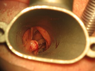
Intubation granuloma is a benign growth of granulation tissue in the larynx or trachea, which arises from tissue trauma due to endotracheal intubation. This medical condition is described as a common late complication of tracheal intubation, specifically caused by irritation to the mucosal tissue of the airway during insertion or removal of the patient’s intubation tube.

References

  1. CME Module 10: Recent Developments in Supraglottic Airway Devices Archived 2012-11-27 at the Wayback Machine , University of Toronto, Department of Anesthesia Website retrieved 21 May 2013
  2. 1 2 3 4 5 6 7 Asai, T; Shingu, K (December 2005). "The laryngeal tube". British Journal of Anaesthesia. 95 (6): 729–36. doi: 10.1093/bja/aei269 . PMID   16286348.
  3. Sunde, Geir Arne; Brattebø, Guttorm; Ødegården, Terje; Kjernlie, Dag Frode; Rødne, Emma; Heltne, Jon-Kenneth (1 October 2012). "Laryngeal tube in 347 out-of-hospital cardiac arrest patients - an acceptable airway tool, but not the final answer?". Resuscitation. 83 (1): e15. doi:10.1016/j.resuscitation.2012.08.039. PMID   21889472.
  4. Asai, T.; Murao, K.; Shingu, K. (1 November 2000). "Efficacy of the laryngeal tube during intermittent positive-pressure ventilation". Anaesthesia. 55 (11): 1099–1102. doi: 10.1046/j.1365-2044.2000.01710.x . PMID   11069337.
  5. Asai, T.; Shingu, K.; Cook, T. (1 August 2003). "Use of the laryngeal tube in 100 patients". Acta Anaesthesiologica Scandinavica. 47 (7): 828–832. doi:10.1034/j.1399-6576.2003.00183.x. PMID   12859303. S2CID   863261.
  6. Doerges, Volker; Ocker, Hartmut; Wenzel, Volker; Schmucker, Peter (1 May 2000). "The Laryngeal Tube: A New Simple Airway Device". Anesthesia & Analgesia. 90 (5): 1220–1222. doi: 10.1097/00000539-200005000-00042 . PMID   10781484. S2CID   5537072.
  7. Asai, T.; Kawashima, A.; Hidaka, I.; Kawachi, S. (1 November 2002). "The laryngeal tube compared with the laryngeal mask: insertion, gas leak pressure and gastric insufflation". British Journal of Anaesthesia. 89 (5): 729–732. doi: 10.1093/bja/89.5.729 . PMID   12393771.
  8. Cook, T. M. (1 September 2003). "Randomized comparison of laryngeal tube with classic laryngeal mask airway for anaesthesia with controlled ventilation". British Journal of Anaesthesia. 91 (3): 373–378. doi: 10.1093/bja/aeg192 . PMID   12925477.
  9. Ocker, H; Wenzel, V; Schmucker, P; Steinfath, M; Dörges, V (October 2002). "A comparison of the laryngeal tube with the laryngeal mask airway during routine surgical procedures". Anesthesia and Analgesia. 95 (4): 1094–7, table of contents. doi: 10.1097/00000539-200210000-00057 . PMID   12351302.
  10. Wrobel, M; Grundmann, U; Wilhelm, W; Wagner, S; Larsen, R (August 2004). "[Laryngeal tube versus laryngeal mask airway in anaesthetised non-paralysed patients. A comparison of handling and postoperative morbidity]. (German)". Der Anaesthesist. 53 (8): 702–8. doi:10.1007/s00101-004-0697-x. PMID   15167948.
  11. Brimacombe, Joseph; Keller, Christian; Brimacombe, Lawrence (1 September 2002). "A Comparison of the Laryngeal Mask Airway ProSeal™ and the Laryngeal Tube Airway in Paralyzed Anesthetized Adult Patients Undergoing Pressure-Controlled Ventilation". Anesthesia & Analgesia. 95 (3): 770–776. doi: 10.1213/00000539-200209000-00045 . PMID   12198070. S2CID   36634107.
  12. Cook, T. M. (1 November 2003). "Randomized crossover comparison of the ProSealTM laryngeal mask airway with the Laryngeal Tube(R) during anaesthesia with controlled ventilation". British Journal of Anaesthesia. 91 (5): 678–683. doi: 10.1093/bja/aeg239 . PMID   14570790.
  13. Matioc, Adrian A.; Olson, John (1 December 2004). "Use of the Laryngeal Tube™ in two unexpected difficult airway situations: lingual tonsillar hyperplasia and morbid obesity". Canadian Journal of Anesthesia. 51 (10): 1018–1021. doi:10.1007/BF03018491. PMID   15574554. S2CID   22751635.
  14. Asai, T.; Matsumoto, S.; Shingu, K.; Noguchi, T; Koga, K. (1 August 2005). "Use of the laryngeal tube after failed insertion of a laryngeal mask airway". Anaesthesia. 60 (8): 825–826. doi:10.1111/j.1365-2044.2005.04312.x. PMID   16029243. S2CID   46022613.
  15. Scheller, Bertram; Schalk, Richard; Byhahn, Christian; Peter, Norman; L’Allemand, Nanette; Kessler, Paul; Meininger, Dirk (30 June 2009). "Laryngeal tube suction II for difficult airway management in neonates and small infants". Resuscitation. 80 (7): 805–810. doi:10.1016/j.resuscitation.2009.03.029. PMID   19410354.
  16. Schalk, Richard; Byhahn, Christian; Fausel, Felix; Egner, Andreas; Oberndörfer, Dieter; Walcher, Felix; Latasch, Leo (28 February 2010). "Out-of-hospital airway management by paramedics and emergency physicians using laryngeal tubes". Resuscitation. 81 (3): 323–326. doi:10.1016/j.resuscitation.2009.11.007. PMID   20006418.
  17. "King Systems Corresponds to the FDA Regarding KLT(S)D Labeling". King Systems. Archived from the original on 4 November 2014. Retrieved 4 November 2014.
  18. Genzwuerker, H.V.; Dhonau, S; Ellinger, K (31 January 2002). "Use of the laryngeal tube for out-of-hospital resuscitation". Resuscitation. 52 (2): 221–224. doi:10.1016/S0300-9572(01)00472-5. PMID   11841891.
  19. Russi, Christopher S.; Wilcox, Cari L.; House, Hans R. (28 February 2007). "The laryngeal tube device: a simple and timely adjunct to airway management". The American Journal of Emergency Medicine. 25 (3): 263–267. doi:10.1016/j.ajem.2006.03.018. PMID   17349898.
  20. Wiese, C.H.R.; Semmel, T.; Müller, J.U.; Bahr, J.; Ocker, H.; Graf, B.M. (31 January 2009). "The use of the laryngeal tube disposable (LT-D) by paramedics during out-of-hospital resuscitation—An observational study concerning ERC guidelines 2005". Resuscitation. 80 (2): 194–198. doi:10.1016/j.resuscitation.2008.08.023. PMID   19010582.