Sciatic nerve block

Last updated
Sciatic nerve block

Gray832.png

Nerves of the right lower extremity Posterior view

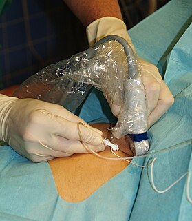
A sciatic nerve block is a nerve block that uses local anesthetic to achieve analgesia in the leg. The block works by affecting the sciatic nerve and is used for surgeries at or below the knee. [1]

Local anesthetic nerve block

Local anesthetic nerve block is a short-term nerve block involving the injection of local anesthetic as close to the nerve as possible for pain relief. The local anesthetic bathes the nerve and numbs the area of the body that is innervated by that nerve. The goal of the nerve block is to prevent pain by blocking the transmission of pain signals from the surgical site. The block provides pain relief during and after the surgery. The advantages of nerve blocks over general anesthesia include faster recovery, monitored anesthesia care vs. intubation with an airway tube, and much less postoperative pain.

Leg weight bearing and locomotive anatomical structure, usually having a columnar shape, function as "extensible struts"

A leg is a weight-bearing and locomotive anatomical structure, usually having a columnar shape. During locomotion, legs function as "extensible struts". The combination of movements at all joints can be modeled as a single, linear element capable of changing length and rotating about an omnidirectional "hip" joint.

Sciatic nerve large nerve in humans and other animals

The sciatic nerve is a large nerve in humans and other animals. It begins in the lower back and runs through the buttock and down the lower limb. It is the longest and widest single nerve in the human body, going from the top of the leg to the foot on the posterior aspect. The sciatic nerve provides the connection to the nervous system for nearly the whole of the skin of the leg, the muscles of the back of the thigh, and those of the leg and foot. It is derived from spinal nerves L4 to S3. It contains fibers from both the anterior and posterior divisions of the lumbosacral plexus.

The sciatic nerve is located in the gluteus maximus muscle, where the block is performed. [1] The sciatic nerve can be blocked at different locations. At the popliteal fossa, the sciatic nerve divides into its two branches: The tibial and the common peroneal nerve. If surgery is performed on the ankle, achilles tendon or foot a popliteal block can be performed, affecting the two branches of the sciatic nerve. It is done above the knee on the [1] posterior leg where the sciatic nerve starts splitting into the common peroneal and tibial nerves. [1]

Tibial nerve

The tibial nerve is a branch of the sciatic nerve. The tibial nerve passes through the popliteal fossa to pass below the arch of soleus.

The common fibular nerve is a nerve in the lower leg that provides sensation over the posterolateral part of the leg and the knee joint. It divides at the knee into two terminal branches: the superficial fibular nerve and deep fibular nerve, which innervate the muscles of the lateral and anterior compartments of the leg respectively. When the common fibular nerve is damaged or compressed, foot drop can be the end result.

Related Research Articles

Human leg lower extremity or limb of the human body (foot, lower leg, thigh and hip)

The human leg, in the general meaning, is the entire lower limb of the human body, including the foot, thigh and even the hip or gluteal region. However, the definition in human anatomy refers only to the section of the lower limb extending from the knee to the ankle, also known as the crus. Legs are used for standing, and all forms of locomotion including recreational such as dancing, and constitute a significant portion of a person's mass. Female legs generally have greater hip anteversion and tibiofemoral angles, but shorter femur and tibial lengths than those in males.

In human anatomy, a hamstring is one of the three posterior thigh muscles in between the hip and the knee. The hamstrings are quite susceptible to injury.

Peroneal nerve paralysis

Peroneal nerve paralysis is a paralysis on common fibular nerve that affects patient’s ability to lift the foot at the ankle. The condition was named after Friedrich Albert von Zenker. Peroneal nerve paralysis usually leads to neuromuscular disorder, peroneal nerve injury, or foot drop which can be symptoms of more serious disorders such as nerve compression. The origin of peroneal nerve palsy has been reported to be associated with musculoskeletal injury or isolated nerve traction and compression. Also it has been reported to be mass lesions and metabolic syndromes. Peroneal nerve is most commonly interrupted at the knee and possibly at the joint of hip and ankle. Most studies reported that about 30% of peroneal nerve palsy is followed from knee dislocations.

Popliteal artery

The popliteal artery is a deeply placed continuation of the femoral artery after it passes through the adductor hiatus, or opening in the distal portion of the adductor magnus muscle. It courses through the popliteal fossa and ends at the lower border of the popliteus muscle, where it branches into the anterior and posterior tibial arteries.

The biceps femoris is a muscle of the thigh located to the posterior, or back. As its name implies, it has two parts, one of which forms part of the hamstrings muscle group.

The semimembranosus is the most medial of the three hamstring muscles. It is so named because it has a flat tendon of origin. It lies posteromedially in the thigh, deep to the semitendinosus.

Semitendinosus muscle Origin : From the lower medial part of upper quadrilateral area of the ischial tuberosity

The semitendinosus is a long superficial muscle in the back of the thigh. It is so named because it has a very long tendon of insertion. It lies posteromedially in the thigh, superficial to the semimembranosus.

Plantaris muscle muscle

The plantaris is one of the superficial muscles of the superficial posterior compartment of the leg, one of the fascial compartments of the leg.

Deep peroneal nerve

The deep peroneal nerve begins at the bifurcation of the common peroneal nerve between the fibula and upper part of the peroneus longus, passes infero-medially, deep to extensor digitorum longus, to the anterior surface of the interosseous membrane, and comes into relation with the anterior tibial artery above the middle of the leg; it then descends with the artery to the front of the ankle-joint, where it divides into a lateral and a medial terminal branch.

Popliteal fossa The back of your knee

The popliteal fossa is a shallow depression located at the back of the knee joint. The bones of the popliteal fossa are the femur and the tibia. Like other flexion surfaces of large joints, it is an area where blood vessels and nerves pass relatively superficially, and with an increased amount of lymph nodes.

Foot drop gait abnormality

Foot drop is a gait abnormality in which the dropping of the forefoot happens due to weakness, irritation or damage to the common fibular nerve including the sciatic nerve, or paralysis of the muscles in the anterior portion of the lower leg. It is usually a symptom of a greater problem, not a disease in itself. Foot drop is characterized by inability or impaired ability to raise the toes or raise the foot from the ankle (dorsiflexion). Foot drop may be temporary or permanent, depending on the extent of muscle weakness or paralysis and it can occur in one or both feet. In walking, the raised leg is slightly bent at the knee to prevent the foot from dragging along the ground.

The sural nerve is a sensory nerve in the calf region (sura) of the leg. It is made up of branches of the tibial nerve and common fibular nerve, the medial cutaneous branch from the tibial nerve, and the lateral cutaneous branch from the common fibular nerve. Once formed, the nerves runs down the mid calf to the ankle and along the skin from the mid-posterior popliteal fossa to just behind to the lateral malleolus and then under the malleolus and forward along the lateral aspect of the foot.

Interosseous membrane of leg

The interosseous membrane of the leg extends between the interosseous crests of the tibia and fibula, helps stabilize the Tib-Fib relationship and separates the muscles on the front from those on the back of the leg.

Medial sural cutaneous nerve

The medial sural cutaneous nerve originates from the tibial nerve of the sciatic, descends between the two heads of the Gastrocnemius, and, about the middle of the back of the leg, pierces the deep fascia, and unites with the anastomotic ramus of the common peroneal to form the sural nerve.

Posterior compartment of thigh

The posterior compartment of the thigh is one of the fascial compartments that contains the knee flexors and hip extensors known as the hamstring muscles, as well as vascular and nervous elements, particularly the sciatic nerve.

Fibular artery

In anatomy, the fibular artery supplies blood to the lateral compartment of the leg. It arises from the tibial-fibular trunk.

References

  1. 1 2 3 4 "Lower Extremity Nerve Blocks" (PDF). NYSORA. Retrieved 4 August 2017.