Malate synthase

Last updated
malate synthase
3oyz.jpg
Malate synthase homotrimer, Haloferax volcanii
Identifiers
EC no. 2.3.3.9
CAS no. 9013-48-3
Databases
IntEnz IntEnz view
BRENDA BRENDA entry
ExPASy NiceZyme view
KEGG KEGG entry
MetaCyc metabolic pathway
PRIAM profile
PDB structures RCSB PDB PDBe PDBsum
Gene Ontology AmiGO / QuickGO
Search
PMC articles
PubMed articles
NCBI proteins

In enzymology, a malate synthase (EC 2.3.3.9) is an enzyme that catalyzes the chemical reaction

Contents

acetyl-CoA + H2O + glyoxylate (S)-malate + CoA

The 3 substrates of this enzyme are acetyl-CoA, H2O, and glyoxylate, whereas its two products are (S)-malate and CoA. This enzyme participates in pyruvate metabolism and glyoxylate and dicarboxylate metabolism.

Nomenclature

This enzyme belongs to the family of transferases, specifically acyltransferases that convert acyl groups into alkyl groups on transfer. The systematic name of this enzyme class is acetyl-CoA:glyoxylate C-acetyltransferase (thioester-hydrolysing, carboxymethyl-forming). Other names in common use include L-malate glyoxylate-lyase (CoA-acetylating), glyoxylate transacetylase, glyoxylate transacetase, glyoxylic transacetase, malate condensing enzyme, malate synthetase, malic synthetase, and malic-condensing enzyme.

Structure

Crystallographic structure of malate synthase enzyme (left) and expand view of the active site (right) complexed with its product, malate, and a coordinating magnesium cation. Malate synthase structure including active site.png
Crystallographic structure of malate synthase enzyme (left) and expand view of the active site (right) complexed with its product, malate, and a coordinating magnesium cation.

Malate synthases fall into two major families, isoforms A and G. Isoform G is monomeric with a size of about 80-kD and found exclusively in bacteria. [2] Isoform A is about 65 kD per subunit and can form homomultimers in eukaryotes. [3] This enzyme contains a central TIM barrel sandwiched between an N-terminal alpha-helical clasp and an alpha/beta domain stemming from two insertions into the TIM barrel sequence. The enzyme terminates with a C-terminal five-helix plug. The active site, where the acetyl-CoA and glyoxylate bind to the enzyme, lies between the TIM barrel and C-terminal plug. [4] Upon binding, the acetyl-CoA molecule forms a J-shape inserted into the binding pocket, by an intramolecular hydrogen bond between N7 of the adenine ring and a hydroxyl group on the pantetheine tail. [4] In addition, a critical magnesium ion within the active site coordinates with glyoxylate, glutamic acid 427, aspartic acid 455, and two water molecules. [4] The amino acids interacting with acetyl CoA upon binding are highly conserved. [2] Sequence identity is high within each class of isoforms, but between both classes sequence identity drops to about 15%. [5] The alpha/beta domain, which has no apparent function, is not seen in isoform A. [6]

Active site of malate synthase bound to pyruvate and acetyl-CoA (ACO), which is shown in its bent J configuration. The octahedral coordinating Mg cation is shown in green, water molecules as red dots, and polar contacts as dashed yellow lines. Malate synthase active site.png
Active site of malate synthase bound to pyruvate and acetyl-CoA (ACO), which is shown in its bent J configuration. The octahedral coordinating Mg cation is shown in green, water molecules as red dots, and polar contacts as dashed yellow lines.

Mechanism

The mechanism of malate synthase is an aldol reaction followed by thioester hydrolysis. Initially, aspartate 631 acts as a catalytic base, abstracting a proton from the alpha carbon of acetyl-CoA and creating an enolate that is stabilized by arginine 338. [6] This is considered to be the rate-determining step of the mechanism. [7] Then, the newly formed enolate acts as a nucleophile that attacks the aldehyde of glyoxylate, imparting a negative charge on the oxygen which is stabilized by arginine 338 and the coordinating magnesium cation. This malyl-CoA intermediate then undergoes hydrolysis at the acyl-CoA portion, generating a carboxylate anion. [2] The enzyme finally releases malate and coenzyme A.

Function

The role of malate synthase in the glyoxylate cycle. Glyoxylate cycle.png
The role of malate synthase in the glyoxylate cycle.

The citric acid cycle (also known as the tricarboxylic acid cycle or the Krebs cycle) is used by aerobic organisms to produce energy via the oxidation of acetyl-CoA, which is derived from pyruvate (a product of glycolysis). The citric acid cycle accepts acetyl-CoA and metabolizes it to form carbon dioxide. A related cycle, called the glyoxylate cycle, is found in many bacteria and plants. In plants, the glyoxylate cycle takes place in glyoxysomes. [8] In this cycle, isocitrate lyase and malate synthase skip over the decarboxylation steps of the citric acid cycle. In other words, malate synthase works together with isocitrate lyase in the glyoxylate cycle to bypass two oxidative steps of Krebs cycle and permit carbon incorporation from acetate or fatty acids in many microorganisms. [9] Together, these two enzymes serve to produce succinate (which exits the cycle to be used for synthesis of sugars) and malate (which remains in the glyoxylate cycle). During this process, acetyl-CoA and water are used as substrates. As a result, the cell does not lose 2 molecules of carbon dioxide as it does in the Krebs cycle. The glyoxylate cycle, facilitated by malate synthase and isocitrate lyase, allows plants and bacteria to subsist on acetyl-CoA or other two carbon compounds. For example, Euglena gracilis , a single-celled eukaryotic alga, consumes ethanol to form acetyl-CoA and subsequently, carbohydrates. [10] Within germinating plants, the glyoxylate cycle allows the conversion of reserve lipids into carbohydrates within glyoxysomes. [11]

Evolutionary history

Malate synthase is found as an octamer of identical subunits (each roughly 60kDa) in some plants, including maize. It is found as a homotetramer in the fungus Candida and as a homodimer in eubacteria. Malate synthase is fused to the C-terminus of isocitrate lyase in C. elegans , resulting in a single bifunctional protein. While there is currently not sufficient sequence information to determine the exact evolutionary history of malate synthase, plant, fungal, and C. elegans sequences are distinct and show no homologues from archaebacteria. [12]

Activity in humans

Traditionally, malate synthases are described in bacteria as part of the glyoxylate cycle, and malate synthase activity had not been reported for a human protein prior to a study by Strittmatter, et al. In this study, CLYBL was found to be a human mitochondrial enzyme with malate synthase activity. It is found in multiple eukaryotic taxa and is conserved in bacteria. CLYBL differs from other malate synthases in that it lacks a large portion of the C-terminal domain and shows lower specific activity and efficiency. [13] CLYBL is linked to the vitamin B12 metabolism pathway because it is strongly co-expressed with MUT, MMAA, and MMAB, three members of the mitochondrial B12 pathway. [13] Furthermore, a loss of function polymorphism, that leads to a loss of the CLYBL protein, is simultaneously associated with low levels of B12 in human plasma. [13] While the exact mechanism of CLYBL’s involvement in B12 metabolism is not well understood, it is thought to convert citramalyl-CoA into pyruvate and acetyl-CoA. Without this conversion, itaconyl-CoA, a precursor to citramalyl-CoA, builds up in the cell leads to the inactivation of vitamin B12. This inactivation inhibits the methionine cycle, which leads to reduced serine, glycine, one-carbon, and folate metabolism. [14] [15]

Clinical significance

Because the glyoxylate cycle occurs in bacteria and fungi, studying the mechanisms of malate synthase (as well as isocitrate lyase) is important for understanding human, animal, and plant pathogenesis. Studying malate synthase can shed light on the metabolic pathways that allow pathogens to survive inside a host as well as elucidate possible treatments. [16] Many studies have been conducted on malate synthase activity in pathogens, including Mycobacterium tuberculosis , Pseudomonas aeruginosa , Brucella melitensis , and Escherichia coli .

Mycobacterium tuberculosis

Malate synthase and the glyoxylate pathway is especially important to M. tuberculosis , allowing long-term persistence of its infection. [2] When cells of M. tuberculosis become phagocytosed, the bacterium upregulates genes encoding the glyoxylate shunt enzymes. [17] Mycobacterium tuberculosis is one of the most well studied pathogens in connection to the enzyme malate synthase. The structure and kinetics of Mycobacterium tuberculosis malate synthase have been well categorized. [18] [2] Malate synthase is essential to Mycobacterium tuberculosis survival because it allows the bacteria to assimilate acetyl-CoA into long-chain carbohydrates and survive in harsh environments. Beyond this, malate synthase prevents toxicity from buildup of glyoxylate produced by isocitrate lyase. [19] Downregulation of malate synthase results in reduced stress tolerance, persistence, and growth of Mycobacterium tuberculosis inside macrophages. [20] The enzyme can be inhibited by small molecules (although inhibition is microenvironment dependent), which suggests that these may be used as new chemotherapies. [21]

Pseudomonas aeruginosa

Pseudomonas aeruginosa causes severe infections in humans and is labeled as a critical threat by the World Health Organization because of its resistance to multiple therapies. The glyoxylate shunt is essential for Pseudomonas aeruginosa growth in a host organism. In 2017, McVey, et al. examined the 3D structure of P. aeruginosa malate synthase G. They found that it is a monomer composed of four domains and is highly conserved in other pathogens. They further utilized computational analysis to identify two binding pockets that may serve as drug targets. [22]

Brucella melitensis

Brucella melitensis is a pathogenic bacterium that causes fever and inflammation of the epididymis in sheep and cattle and can be transmitted to humans through the consumption of unpasteurized milk. Malate synthase has been identified as a potential virulence factor in this bacterium. In 2016, Adi, et al. constructed a 3D crystallized structure of the protein to identify catalytic domains and investigate potential inhibitors. They identified five inhibitors with non-oral toxicity that served as drugs against the bacteria, suggesting possible treatment routes for brucellosis. [23]

Escherichia coli

In Escherichia coli , the genes encoding the enzymes required for the glyoxylate cycle are expressed from the polycistronic ace operon. This operon contains genes coding for malate synthase (aceB), isocitrate lyase (aceA), and isocitrate dehydrogenase kinase/phosphatase (aceK). [24]

Structural Studies

As of early 2018, several structures have been solved for malate synthases, including those with PDB accession codes 2GQ3, 1D8C, 3OYX, 3PUG, 5TAO, 5H8M, 2JQX, 1P7T, and 1Y8B. [25]

Related Research Articles

<span class="mw-page-title-main">Citric acid cycle</span> Chemical reactions to release energy in cells

The citric acid cycle —also known as the Krebs cycle, Szent-Györgyi-Krebs cycle or the TCA cycle (tricarboxylic acid cycle)—is a series of chemical reactions to release stored energy through the oxidation of acetyl-CoA derived from carbohydrates, fats, and proteins. The Krebs cycle is used by organisms that respire (as opposed to organisms that ferment) to generate energy, either by anaerobic respiration or aerobic respiration. In addition, the cycle provides precursors of certain amino acids, as well as the reducing agent NADH, that are used in numerous other reactions. Its central importance to many biochemical pathways suggests that it was one of the earliest components of metabolism. Even though it is branded as a 'cycle', it is not necessary for metabolites to follow only one specific route; at least three alternative segments of the citric acid cycle have been recognized.

Gluconeogenesis (GNG) is a metabolic pathway that results in the generation of glucose from certain non-carbohydrate carbon substrates. It is a ubiquitous process, present in plants, animals, fungi, bacteria, and other microorganisms. In vertebrates, gluconeogenesis occurs mainly in the liver and, to a lesser extent, in the cortex of the kidneys. It is one of two primary mechanisms – the other being degradation of glycogen (glycogenolysis) – used by humans and many other animals to maintain blood sugar levels, avoiding low levels (hypoglycemia). In ruminants, because dietary carbohydrates tend to be metabolized by rumen organisms, gluconeogenesis occurs regardless of fasting, low-carbohydrate diets, exercise, etc. In many other animals, the process occurs during periods of fasting, starvation, low-carbohydrate diets, or intense exercise.

<span class="mw-page-title-main">Biological carbon fixation</span> Conversion of carbon to organic compounds

Biological carbon fixation or сarbon assimilation is the process by which inorganic carbon is converted to organic compounds by living organisms. The compounds are then used to store energy and as structure for other biomolecules. Carbon is primarily fixed through photosynthesis, but some organisms use a process called chemosynthesis in the absence of sunlight.

<span class="mw-page-title-main">Oxaloacetic acid</span> Organic compound

Oxaloacetic acid (also known as oxalacetic acid or OAA) is a crystalline organic compound with the chemical formula HO2CC(O)CH2CO2H. Oxaloacetic acid, in the form of its conjugate base oxaloacetate, is a metabolic intermediate in many processes that occur in animals. It takes part in gluconeogenesis, the urea cycle, the glyoxylate cycle, amino acid synthesis, fatty acid synthesis and the citric acid cycle.

In molecular biology, biosynthesis is a multi-step, enzyme-catalyzed process where substrates are converted into more complex products in living organisms. In biosynthesis, simple compounds are modified, converted into other compounds, or joined to form macromolecules. This process often consists of metabolic pathways. Some of these biosynthetic pathways are located within a single cellular organelle, while others involve enzymes that are located within multiple cellular organelles. Examples of these biosynthetic pathways include the production of lipid membrane components and nucleotides. Biosynthesis is usually synonymous with anabolism.

<span class="mw-page-title-main">Isocitrate dehydrogenase</span> Class of enzymes

Isocitrate dehydrogenase (IDH) (EC 1.1.1.42) and (EC 1.1.1.41) is an enzyme that catalyzes the oxidative decarboxylation of isocitrate, producing alpha-ketoglutarate (α-ketoglutarate) and CO2. This is a two-step process, which involves oxidation of isocitrate (a secondary alcohol) to oxalosuccinate (a ketone), followed by the decarboxylation of the carboxyl group beta to the ketone, forming alpha-ketoglutarate. In humans, IDH exists in three isoforms: IDH3 catalyzes the third step of the citric acid cycle while converting NAD+ to NADH in the mitochondria. The isoforms IDH1 and IDH2 catalyze the same reaction outside the context of the citric acid cycle and use NADP+ as a cofactor instead of NAD+. They localize to the cytosol as well as the mitochondrion and peroxisome.

<span class="mw-page-title-main">Mitochondrial matrix</span> Space within the inner membrane of the mitochondrion

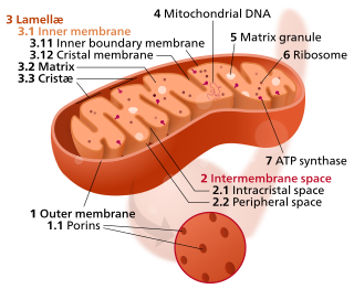
In the mitochondrion, the matrix is the space within the inner membrane. The word "matrix" stems from the fact that this space is viscous, compared to the relatively aqueous cytoplasm. The mitochondrial matrix contains the mitochondrial DNA, ribosomes, soluble enzymes, small organic molecules, nucleotide cofactors, and inorganic ions.[1] The enzymes in the matrix facilitate reactions responsible for the production of ATP, such as the citric acid cycle, oxidative phosphorylation, oxidation of pyruvate, and the beta oxidation of fatty acids.

Glyoxysomes are specialized peroxisomes found in plants (particularly in the fat storage tissues of germinating seeds) and also in filamentous fungi. Seeds that contain fats and oils include corn, soybean, sunflower, peanut and pumpkin. As in all peroxisomes, in glyoxysomes the fatty acids are oxidized to acetyl-CoA by peroxisomal β-oxidation enzymes. When the fatty acids are oxidized hydrogen peroxide (H2O2) is produced as oxygen (O2) is consumed. Thus the seeds need oxygen to germinate. Besides peroxisomal functions, glyoxysomes possess additionally the key enzymes of the glyoxylate cycle (isocitrate lyase and malate synthase) which accomplish the glyoxylate cycle bypass.

<span class="mw-page-title-main">Glyoxylate cycle</span>

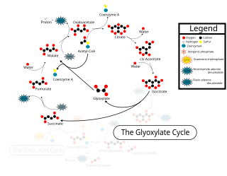
The glyoxylate cycle, a variation of the tricarboxylic acid cycle, is an anabolic pathway occurring in plants, bacteria, protists, and fungi. The glyoxylate cycle centers on the conversion of acetyl-CoA to succinate for the synthesis of carbohydrates. In microorganisms, the glyoxylate cycle allows cells to use two carbons, such as acetate, to satisfy cellular carbon requirements when simple sugars such as glucose or fructose are not available. The cycle is generally assumed to be absent in animals, with the exception of nematodes at the early stages of embryogenesis. In recent years, however, the detection of malate synthase (MS) and isocitrate lyase (ICL), key enzymes involved in the glyoxylate cycle, in some animal tissue has raised questions regarding the evolutionary relationship of enzymes in bacteria and animals and suggests that animals encode alternative enzymes of the cycle that differ in function from known MS and ICL in non-metazoan species.

<span class="mw-page-title-main">Mixed acid fermentation</span> Biochemical conversion of six-carbon sugars into acids in bacteria

In biochemistry, mixed acid fermentation is the metabolic process by which a six-carbon sugar is converted into a complex and variable mixture of acids. It is an anaerobic (non-oxygen-requiring) fermentation reaction that is common in bacteria. It is characteristic for members of the Enterobacteriaceae, a large family of Gram-negative bacteria that includes E. coli.

<span class="mw-page-title-main">Phosphoenolpyruvate carboxylase</span> Class of enzymes

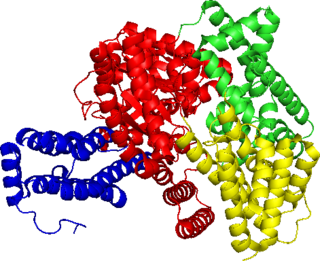
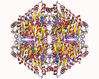
Phosphoenolpyruvate carboxylase (also known as PEP carboxylase, PEPCase, or PEPC; EC 4.1.1.31, PDB ID: 3ZGE) is an enzyme in the family of carboxy-lyases found in plants and some bacteria that catalyzes the addition of bicarbonate (HCO3) to phosphoenolpyruvate (PEP) to form the four-carbon compound oxaloacetate and inorganic phosphate:

Propionyl-CoA is a coenzyme A derivative of propionic acid. It is composed of a 24 total carbon chain and its production and metabolic fate depend on which organism it is present in. Several different pathways can lead to its production, such as through the catabolism of specific amino acids or the oxidation of odd-chain fatty acids. It later can be broken down by propionyl-CoA carboxylase or through the methylcitrate cycle. In different organisms, however, propionyl-CoA can be sequestered into controlled regions, to alleviate its potential toxicity through accumulation. Genetic deficiencies regarding the production and breakdown of propionyl-CoA also have great clinical and human significance.

In biochemistry, fatty acid synthesis is the creation of fatty acids from acetyl-CoA and NADPH through the action of enzymes called fatty acid synthases. This process takes place in the cytoplasm of the cell. Most of the acetyl-CoA which is converted into fatty acids is derived from carbohydrates via the glycolytic pathway. The glycolytic pathway also provides the glycerol with which three fatty acids can combine to form triglycerides, the final product of the lipogenic process. When only two fatty acids combine with glycerol and the third alcohol group is phosphorylated with a group such as phosphatidylcholine, a phospholipid is formed. Phospholipids form the bulk of the lipid bilayers that make up cell membranes and surrounds the organelles within the cells.

<span class="mw-page-title-main">Isocitrate lyase</span>

Isocitrate lyase, or ICL, is an enzyme in the glyoxylate cycle that catalyzes the cleavage of isocitrate to succinate and glyoxylate. Together with malate synthase, it bypasses the two decarboxylation steps of the tricarboxylic acid cycle and is used by bacteria, fungi, and plants.

<span class="mw-page-title-main">Methylisocitrate lyase</span>

The enzyme methylisocitrate lyase catalyzes the chemical reaction

<span class="mw-page-title-main">Oxalyl-CoA decarboxylase</span>

The enzyme oxalyl-CoA decarboxylase (OXC) (EC 4.1.1.8), primarily produced by the gastrointestinal bacterium Oxalobacter formigenes, catalyzes the chemical reaction

<span class="mw-page-title-main">N-acetylglucosamine-6-phosphate deacetylase</span>

In enzymology, N-acetylglucosamine-6-phosphate deacetylase (EC 3.5.1.25), also known as GlcNAc-6-phosphate deacetylase or NagA, is an enzyme that catalyzes the deacetylation of N-acetylglucosamine-6-phosphate (GlcNAc-6-P) to glucosamine-6-phosphate (GlcN-6-P):

Glyoxylate and dicarboxylate metabolism describes a variety of reactions involving glyoxylate or dicarboxylates. Glyoxylate is the conjugate base of glyoxylic acid, and within a buffered environment of known pH such as the cell cytoplasm these terms can be used almost interchangeably, as the gain or loss of a hydrogen ion is all that distinguishes them, and this can occur in the aqueous environment at any time. Likewise dicarboxylates are the conjugate bases of dicarboxylic acids, a general class of organic compounds containing two carboxylic acid groups, such as oxalic acid or succinic acid.

Isocitrate lyase family is a family of evolutionarily related proteins.

<span class="mw-page-title-main">Methylcitrate cycle</span>

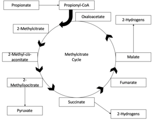
The methylcitrate cycle, or the MCC, is the mechanism by which propionyl-CoA is formed, generated by β-oxidation of odd-chain fatty acids, and broken down to its final products, succinate and pyruvate. The methylcitrate cycle is closely related to both the citric acid cycle and the glyoxylate cycle, in that they share substrates, enzymes and products. The methylcitrate cycle functions overall to detoxify bacteria of toxic propionyl-CoA, and plays an essential role in propionate metabolism in bacteria. Incomplete propionyl-CoA metabolism may lead to the buildup of toxic metabolites in bacteria, and thus the function of the methylcitrate cycle is an important biological process.

References

  1. PDB: 5T8G ; Huang HL, Krieger IV, Parai MK, Gawandi VB, Sacchettini JC (December 2016). "Mycobacterium tuberculosis Malate Synthase Structures with Fragments Reveal a Portal for Substrate/Product Exchange". The Journal of Biological Chemistry. 291 (53): 27421–32. doi: 10.1074/jbc.m116.750877 . PMC   5207166 . PMID   27738104.
  2. 1 2 3 4 5 Smith CV, Huang CC, Miczak A, Russell DG, Sacchettini JC, Höner zu Bentrup K (January 2003). "Biochemical and structural studies of malate synthase from Mycobacterium tuberculosis". The Journal of Biological Chemistry. 278 (3): 1735–43. doi: 10.1074/jbc.M209248200 . PMID   12393860.
  3. Durchschlag H, Biedermann G, Eggerer H (February 1981). "Large-scale purification and some properties of malate synthase from baker's yeast". European Journal of Biochemistry. 114 (2): 255–62. doi:10.1111/j.1432-1033.1981.tb05144.x. PMID   7011808.
  4. 1 2 3 Anstrom DM, Kallio K, Remington SJ (September 2003). "Structure of the Escherichia coli malate synthase G:pyruvate:acetyl-coenzyme A abortive ternary complex at 1.95 A resolution". Protein Science. 12 (9): 1822–32. doi:10.1110/ps.03174303. PMC   2323980 . PMID   12930982.
  5. Serrano JA, Bonete MJ (August 2001). "Sequencing, phylogenetic and transcriptional analysis of the glyoxylate bypass operon (ace) in the halophilic archaeon Haloferax volcanii". Biochimica et Biophysica Acta (BBA) - Gene Structure and Expression. 1520 (2): 154–62. doi:10.1016/s0167-4781(01)00263-9. PMID   11513957.
  6. 1 2 Howard BR, Endrizzi JA, Remington SJ (March 2000). "Crystal structure of Escherichia coli malate synthase G complexed with magnesium and glyoxylate at 2.0 A resolution: mechanistic implications". Biochemistry. 39 (11): 3156–68. doi:10.1021/bi992519h. PMID   10715138.
  7. Clark JD, O'Keefe SJ, Knowles JR (August 1988). "Malate synthase: proof of a stepwise Claisen condensation using the double-isotope fractionation test". Biochemistry. 27 (16): 5961–71. doi:10.1021/bi00416a020. PMID   2847778.
  8. Stryer L, Berg JM, Tymoczko JL (2003). Biochemistry (fifth ed.). New York: Freeman. ISBN   978-0-7167-4684-3. OCLC   48055706.
  9. Kornberg HL, Sadler JR (December 1961). "The metabolism of C2-compounds in micro-organisms. VIII. A dicarboxylic acid cycle as a route for the oxidation of glycollate by Escherichia coli". The Biochemical Journal. 81: 503–13. doi:10.1042/bj0810503. PMC   1243371 . PMID   14458448.
  10. Nakazawa M (2017). "C2 metabolism in Euglena". Advances in Experimental Medicine and Biology. 979: 39–45. doi:10.1007/978-3-319-54910-1_3. ISBN   978-3-319-54908-8. PMID   28429316.
  11. Cioni M, Pinzauti G, Vanni P (1981). "Comparative biochemistry of the glyoxylate cycle". Comparative Biochemistry and Physiology B. 70: 1–26. doi:10.1016/0305-0491(81)90118-8.
  12. Schnarrenberger C, Martin W (February 2002). "Evolution of the enzymes of the citric acid cycle and the glyoxylate cycle of higher plants. A case study of endosymbiotic gene transfer". European Journal of Biochemistry. 269 (3): 868–83. doi: 10.1046/j.0014-2956.2001.02722.x . PMID   11846788.
  13. 1 2 3 Strittmatter L, Li Y, Nakatsuka NJ, Calvo SE, Grabarek Z, Mootha VK (May 2014). "CLYBL is a polymorphic human enzyme with malate synthase and β-methylmalate synthase activity". Human Molecular Genetics. 23 (9): 2313–23. doi:10.1093/hmg/ddt624. PMC   3976331 . PMID   24334609.
  14. Shen H, Campanello GC, Flicker D, Grabarek Z, Hu J, Luo C, Banerjee R, Mootha VK (November 2017). "The Human Knockout Gene CLYBL Connects Itaconate to Vitamin B12". Cell. 171 (4): 771–782.e11. doi:10.1016/j.cell.2017.09.051. PMC   5827971 . PMID   29056341.
  15. Reid MA, Paik J, Locasale JW (November 2017). "A Missing Link to Vitamin B12 Metabolism". Cell. 171 (4): 736–737. doi: 10.1016/j.cell.2017.10.030 . PMID   29100069.
  16. Dunn MF, Ramírez-Trujillo JA, Hernández-Lucas I (October 2009). "Major roles of isocitrate lyase and malate synthase in bacterial and fungal pathogenesis". Microbiology. 155 (Pt 10): 3166–75. doi: 10.1099/mic.0.030858-0 . PMID   19684068.
  17. Höner Zu Bentrup K, Miczak A, Swenson DL, Russell DG (December 1999). "Characterization of activity and expression of isocitrate lyase in Mycobacterium avium and Mycobacterium tuberculosis". Journal of Bacteriology. 181 (23): 7161–7. doi:10.1128/JB.181.23.7161-7167.1999. PMC   103675 . PMID   10572116.
  18. Quartararo CE, Blanchard JS (August 2011). "Kinetic and chemical mechanism of malate synthase from Mycobacterium tuberculosis". Biochemistry. 50 (32): 6879–87. doi:10.1021/bi2007299. PMC   3153559 . PMID   21728344.
  19. Puckett S, Trujillo C, Wang Z, Eoh H, Ioerger TR, Krieger I, Sacchettini J, Schnappinger D, Rhee KY, Ehrt S (March 2017). "Glyoxylate detoxification is an essential function of malate synthase required for carbon assimilation in Mycobacterium tuberculosis". Proceedings of the National Academy of Sciences of the United States of America. 114 (11): E2225–E2232. doi: 10.1073/pnas.1617655114 . PMC   5358392 . PMID   28265055.
  20. Singh KS, Sharma R, Keshari D, Singh N, Singh SK (September 2017). "Down-regulation of malate synthase in Mycobacterium tuberculosis H37Ra leads to reduced stress tolerance, persistence and survival in macrophages". Tuberculosis. 106: 73–81. doi:10.1016/j.tube.2017.07.006. PMID   28802408.
  21. May EE, Leitão A, Tropsha A, Oprea TI (December 2013). "A systems chemical biology study of malate synthase and isocitrate lyase inhibition in Mycobacterium tuberculosis during active and NRP growth". Computational Biology and Chemistry. 47: 167–80. doi:10.1016/j.compbiolchem.2013.07.002. PMC   4010430 . PMID   24121675.
  22. McVey AC, Medarametla P, Chee X, Bartlett S, Poso A, Spring DR, Rahman T, Welch M (October 2017). "Structural and Functional Characterization of Malate Synthase G from Opportunistic Pathogen Pseudomonas aeruginosa". Biochemistry. 56 (41): 5539–5549. doi: 10.1021/acs.biochem.7b00852 . PMID   28985053.
  23. Adi PJ, Yellapu NK, Matcha B (December 2016). "Modeling, molecular docking, probing catalytic binding mode of acetyl-CoA malate synthase G in Brucella melitensis 16M". Biochemistry and Biophysics Reports. 8: 192–199. doi:10.1016/j.bbrep.2016.08.020. PMC   5613768 . PMID   28955956.
  24. Cortay JC, Bleicher F, Duclos B, Cenatiempo Y, Gautier C, Prato JL, Cozzone AJ (September 1989). "Utilization of acetate in Escherichia coli: structural organization and differential expression of the ace operon". Biochimie. 71 (9–10): 1043–9. doi:10.1016/0300-9084(89)90109-0. PMID   2512996.
  25. Bank, RCSB Protein Data. "RCSB PDB: Homepage". www.rcsb.org. Retrieved 2018-03-05.