Related Research Articles
Dermatophyte is a common label for a group of fungus of Arthrodermataceae that commonly causes skin disease in animals and humans. Traditionally, these anamorphic mold genera are: Microsporum, Epidermophyton and Trichophyton. There are about 40 species in these three genera. Species capable of reproducing sexually belong in the teleomorphic genus Arthroderma, of the Ascomycota. As of 2019 a total of nine genera are identified and new phylogenetic taxonomy has been proposed.

Tinea corporis is a fungal infection of the body, similar to other forms of tinea. Specifically, it is a type of dermatophytosis that appears on the arms and legs, especially on glabrous skin; however, it may occur on any superficial part of the body.

Tinea capitis is a cutaneous fungal infection (dermatophytosis) of the scalp. The disease is primarily caused by dermatophytes in the genera Trichophyton and Microsporum that invade the hair shaft. The clinical presentation is typically single or multiple patches of hair loss, sometimes with a 'black dot' pattern, that may be accompanied by inflammation, scaling, pustules, and itching. Uncommon in adults, tinea capitis is predominantly seen in pre-pubertal children, more often boys than girls.

Tinea barbae is a fungal infection of the hair. Tinea barbae is due to a dermatophytic infection around the bearded area of men. Generally, the infection occurs as a follicular inflammation, or as a cutaneous granulomatous lesion, i.e. a chronic inflammatory reaction. It is one of the causes of folliculitis. It is most common among agricultural workers, as the transmission is more common from animal-to-human than human-to-human. The most common causes are Trichophyton mentagrophytes and T. verrucosum.

Dermatophytosis, also known as ringworm, is a fungal infection of the skin. Typically it results in a red, itchy, scaly, circular rash. Hair loss may occur in the area affected. Symptoms begin four to fourteen days after exposure. Multiple areas can be affected at a given time.

The KOH Test for Candida albicans, also known as a potassium hydroxide preparation or KOH prep, is a quick, inexpensive fungal test to differentiate dermatophytes and Candida albicans symptoms from other skin disorders like psoriasis and eczema.

Tinea nigra, also known as superficial phaeohyphomycosis and Tinea nigra palmaris et plantaris, is a superficial fungal infection, a type of phaeohyphomycosis rather than a tinea, that causes usually a single 1–5 cm dark brown-black, non-scaly, flat, painless patch on the palms of the hands and the soles of the feet of healthy people. There may be multiple spots. The macules occasionally extend to the fingers, toes, and nails, and may be reported on the chest, neck, or genital area. Tinea nigra infections can present with multiple macules that can be mottled or velvety in appearance, and may be oval or irregular in shape. The macules can be anywhere from a few mm to several cm in size.

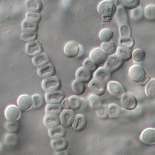
White piedra is a mycosis of the hair caused by several species of fungi in the genus Trichosporon. It is characterized by soft nodules composed of yeast cells and arthroconidia that encompass hair shafts.

Piedraia hortae is a superficial fungus that exists in the soils of tropical and subtropical environments and affects both sexes of all ages. The fungus grows very slowly, forming dark hyphae, which contain chlamydoconidia cells and black colonies when grown on agar. Piedraia hortae is a dermatophyte and causes a superficial fungal infection known as black piedra, which causes the formation of black nodules on the hair shaft and leads to progressive weakening of the hair. The infection usually infects hairs on the scalp and beard, but other varieties tend to grow on pubic hairs. The infection is usually treated with cutting or shaving of the hair and followed by the application of anti-fungal and topical agents. The fungus is used for cosmetic purposes to darken hair in some societies as a symbol of attractiveness.

Tinea incognita or Tinea incognito is a fungal infection of the skin masked and often exacerbated by application of a topical immunosuppressive agent. The usual agent is a topical corticosteroid. As the skin fungal infection has lost some of the characteristic features due to suppression of inflammation, it may have a poorly defined border and florid growth. Occasionally, secondary infection with bacteria occurs with concurrent pustules and impetigo.

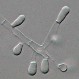
Microsporum audouinii is an anthropophilic fungus in the genus Microsporum. It is a type of dermatophyte that colonizes keratinized tissues causing infection. The fungus is characterized by its spindle-shaped macroconidia, clavate microconidia as well as its pitted or spiny external walls.

Trichophyton tonsurans is a fungus in the family Arthrodermataceae that causes ringworm infection of the scalp. It was first recognized by David Gruby in 1844. Isolates are characterized as the "–" or negative mating type of the Arthroderma vanbreuseghemii complex. This species is thought to be conspecific with T. equinum, although the latter represents the "+" mating strain of the same biological species Despite their biological conspecificity, clones of the two mating types appear to have undergone evolutionary divergence with isolates of the T. tonsurans-type consistently associated with Tinea capitis whereas the T. equinum-type, as its name implies, is associated with horses as a regular host. Phylogenetic relationships were established in isolates from Northern Brazil, through fingerprinting polymorphic RAPD and M13 markers. There seems to be lower genomic variability in the T. tonsurans species due to allopatric divergence. Any phenotypic density is likely due to environmental factors, not genetic characteristics of the fungus.

Pityriasis amiantacea is an eczematous condition of the scalp in which thick tenaciously adherent scale infiltrates and surrounds the base of a group of scalp hairs. It does not result in scarring or alopecia.
Non scarring hair loss, also known as noncicatricial alopecia is the loss of hair without any scarring being present. There is typically little inflammation and irritation, but hair loss is significant. This is in contrast to scarring hair loss during which hair follicles are replaced with scar tissue as a result of inflammation. Hair loss may be spread throughout the scalp (diffuse) or at certain spots (focal). The loss may be sudden or gradual with accompanying stress.

Majocchi's granuloma is a skin condition characterized by deep, pustular plaques, and is a form of tinea corporis. It is a localized form of fungal folliculitis. Lesions often have a pink and scaly central component with pustules or folliculocentric papules at the periphery. The name comes from Professor Domenico Majocchi, who discovered the disorder in 1883. Majocchi was a professor of dermatology at the University of Parma and later the University of Bologna. The most common dermatophyte is called Trichophyton rubrum.

Favus or tinea favosa is the severe form of tinea capitis, a skin infectious disease caused by the dermatophyte fungus Trichophyton schoenleinii. Typically the species affects the scalp, but occasionally occurs as onychomycosis, tinea barbae, or tinea corporis.

Trichophyton verrucosum, commonly known as the cattle ringworm fungus, is a dermatophyte largely responsible for fungal skin disease in cattle, but is also a common cause of ringworm in donkeys, dogs, goat, sheep, and horses. It has a worldwide distribution, however human infection is more common in rural areas where contact with animals is more frequent, and can cause severe inflammation of the afflicted region. Trichophyton verrucosum was first described by Emile Bodin in 1902.

Epidermophyton floccosum is a filamentous fungus that causes skin and nail infections in humans. This anthropophilic dermatophyte can lead to diseases such as tinea pedis, tinea cruris, tinea corporis and onychomycosis. Diagnostic approaches of the fungal infection include physical examination, culture testing, and molecular detection. Topical antifungal treatment, such as the use of terbinafine, itraconazole, voriconazole, and ketoconazole, is often effective.
References
- ↑ James, William D.; Berger, Timothy G.; et al. (2006). Andrews' Diseases of the Skin: clinical Dermatology. Saunders Elsevier. ISBN 0-7216-2921-0.
- ↑ " Piedra " at Dorland's Medical Dictionary
- ↑ Veasey JV, Avila RB, Miguel BAF, Muramatu LH. White piedra, black piedra, tinea versicolor, and tinea nigra: contribution to the diagnosis of superficial mycosis. An Bras Dermatol. 2017 May-Jun;92(3):413-416. doi : 10.1590/abd1806-4841.20176018 PMID 29186263