
Leprosy, also known as Hansen's disease (HD), is a long-term infection by the bacteria Mycobacterium leprae or Mycobacterium lepromatosis. Infection can lead to damage of the nerves, respiratory tract, skin, and eyes. This nerve damage may result in a lack of ability to feel pain, which can lead to the loss of parts of a person's extremities from repeated injuries or infection through unnoticed wounds. An infected person may also experience muscle weakness and poor eyesight. Leprosy symptoms may begin within one year, but, for some people, symptoms may take 20 years or more to occur.

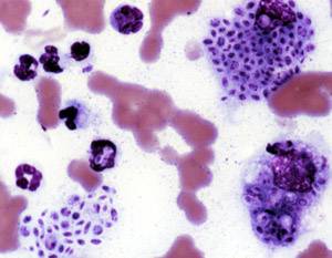
Blastomycosis, also known as Gilchrist's disease, is a fungal infection, typically of the lungs, which can spread to brain, stomach, intestine and skin, where it appears as crusting purplish warty plaques with a roundish bumpy edge and central depression. Only about half of people with the disease have symptoms, which can include fever, cough, night sweats, muscle pains, weight loss, chest pain, and feeling tired. Symptoms usually develop between three weeks and three months after breathing in the spores. In 25% to 40% of cases, the infection also spreads to other parts of the body, such as the skin, bones or central nervous system. Although blastomycosis is especially dangerous for those with weak immune systems, most people diagnosed with blastomycosis have healthy immune systems.

In infectious disease ecology and epidemiology, a natural reservoir, also known as a disease reservoir or a reservoir of infection, is the population of organisms or the specific environment in which an infectious pathogen naturally lives and reproduces, or upon which the pathogen primarily depends for its survival. A reservoir is usually a living host of a certain species, such as an animal or a plant, inside of which a pathogen survives, often without causing disease for the reservoir itself. By some definitions a reservoir may also be an environment external to an organism, such as a volume of contaminated air or water.

Fungal infection, also known as mycosis, is a disease caused by fungi. Different types are traditionally divided according to the part of the body affected; superficial, subcutaneous, and systemic. Superficial fungal infections include common tinea of the skin, such as tinea of the body, groin, hands, feet and beard, and yeast infections such as pityriasis versicolor. Subcutaneous types include eumycetoma and chromoblastomycosis, which generally affect tissues in and beneath the skin. Systemic fungal infections are more serious and include cryptococcosis, histoplasmosis, pneumocystis pneumonia, aspergillosis and mucormycosis. Signs and symptoms range widely. There is usually a rash with superficial infection. Fungal infection within the skin or under the skin may present with a lump and skin changes. Pneumonia-like symptoms or meningitis may occur with a deeper or systemic infection.

Chromoblastomycosis is a long-term fungal infection of the skin and subcutaneous tissue.
Sporotrichosis, also known as rose handler's disease, is a fungal infection that may be localised to skin, lungs, bone and joint, or become systemic. It presents with firm painless nodules that later ulcerate. Following initial exposure to Sporothrix schenckii, the disease typically progresses over a period of a week to several months. Serious complications may develop in people who have a weakened immune system.

Camelpox is a disease of camels caused by the camelpox virus (CMPV) of the family Poxviridae, subfamily Chordopoxvirinae, and the genus Orthopoxvirus. It causes skin lesions and a generalized infection. Approximately 25% of young camels that become infected will die from the disease, while infection in older camels is generally more mild. Although rare, the infection may spread to the hands of those that work closely with camels.

Paracoccidioidomycosis (PCM), also known as South American blastomycosis, is a fungal infection that can occur as a mouth and skin type, lymphangitic type, multi-organ involvement type (particularly lungs), or mixed type. If there are mouth ulcers or skin lesions, the disease is likely to be widespread. There may be no symptoms, or it may present with fever, sepsis, weight loss, large glands, or a large liver and spleen.

Blastomyces dermatitidis is a dimorphic fungus that causes blastomycosis, an invasive and often serious fungal infection found occasionally in humans and other animals. It lives in soil and wet, decaying wood, often in an area close to a waterway such as a lake, river or stream. Indoor growth may also occur, for example, in accumulated debris in damp sheds or shacks. The fungus is endemic to parts of eastern North America, particularly boreal northern Ontario, southeastern Manitoba, Quebec south of the St. Lawrence River, parts of the U.S. Appalachian mountains and interconnected eastern mountain chains, the west bank of Lake Michigan, the state of Wisconsin, and the entire Mississippi Valley including the valleys of some major tributaries such as the Ohio River. In addition, it occurs rarely in Africa both north and south of the Sahara Desert, as well as in the Arabian Peninsula and the Indian subcontinent. Though it has never been directly observed growing in nature, it is thought to grow there as a cottony white mold, similar to the growth seen in artificial culture at 25 °C (77 °F). In an infected human or animal, however, it converts in growth form and becomes a large-celled budding yeast. Blastomycosis is generally readily treatable with systemic antifungal drugs once it is correctly diagnosed; however, delayed diagnosis is very common except in highly endemic areas.
Lacazia is a genus of fungi containing the single species Lacazia loboi, which is responsible for Lobo's disease. It is a member of the order Onygenales.

Herpes simplex is a viral infection caused by the herpes simplex virus. Infections are categorized based on the part of the body infected.
Sealpox is a cutaneous (skin) condition caused by a Parapoxvirus, usually affecting seal handlers who have been bitten by infected harbor or grey seals. First identified in 1969, it wasn't unequivocally proven to be transmissible to humans until 2005, though such transmission had been reported at least as early as 1987. It causes lesions that closely resemble those caused by orf. As many as 2% of seals in marine mammal rehabilitation facilities in North America may have it.
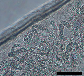
Dirofilaria repens is a filarial nematode that affects dogs and other carnivores such as cats, wolves, coyotes, foxes, and sea lions, as well as muskrats. It is transmitted by mosquitoes. Although humans may become infected as aberrant hosts, the worms fail to reach adulthood while infecting a human body.
The walleye epidermal hyperplasia viruses are two species of retroviruses classified under Epsilonretrovirus, a genus in the family of Retroviridae. There are three genome sequenced and identified exogenous retroviruses of this genus which include two known types associated with walleye epidermal hyperplasia disease. Both viral types are confirmed to be the causative agents of the neoplastic condition in the freshwater fish species, the North American walleye (Sander vitreus). The specific association of retroviral infection with proliferative lesions in fish is based on the presence of retrovirus-like particles and reverse transcriptase activity from neoplastic tissue. Although both virus types have been observed in lesions of diseased fish, each cell of the infected tissue is host to a specific virus. Transmission studies have also shown that WEHV-2 has been the more proliferative agent of the condition as compared to WEHV-1.
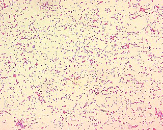
Brucella ceti is a gram negative bacterial pathogen of the Brucellaceae family that causes brucellosis in cetaceans. Brucella ceti has been found in both classes of cetaceans, mysticetes and odontocetes. Brucellosis in some dolphins and porpoises can result in serious clinical signs including fetal abortions, male infertility, neurobrucellosis, cardiopathies, bone and skin lesions, stranding events, and death.
The critical community size (CCS) is the minimum size of a closed population within which a human-to-human, non-zoonotic pathogen can persist indefinitely.
Phaeohyphomycosis is a diverse group of fungal infections, caused by dematiaceous fungi whose morphologic characteristics in tissue include hyphae, yeast-like cells, or a combination of these. It can be associated an array of melanistic filamentous fungi including Alternaria species,Exophiala jeanselmei, and Rhinocladiella mackenziei.
Trichophyton verrucosum, commonly known as the cattle ringworm fungus, is a dermatophyte largely responsible for fungal skin disease in cattle, but is also a common cause of ringworm in donkeys, dogs, goat, sheep, and horses. It has a worldwide distribution, however human infection is more common in rural areas where contact with animals is more frequent, and can cause severe inflammation of the afflicted region. Trichophyton verrucosum was first described by Emile Bodin in 1902.

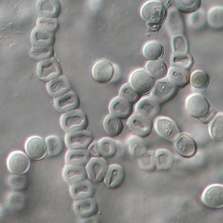
Trichosporon asteroides is an asexual basidiomycetous fungus first described from human skin but now mainly isolated from blood and urine. T. asteroides is a hyphal fungus with a characteristically yeast-like appearance due to the presence of slimy arthroconidia. Infections by this species usually respond to treatment with azoles and amphotericin B.
Arthrographis kalrae is an ascomycetous fungus responsible for human nail infections described in 1938 by Cochet as A. langeronii. A. kalrae is considered a weak pathogen of animals including human restricted to the outermost keratinized layers of tissue. Infections caused by this species are normally responsive to commonly used antifungal drugs with only very rare exceptions.

