Pseudallescheria boydii

Last updated

Pseudallescheria boydii
Pseudallescheria boydii.jpg
Crushed cleistothecium of Pseudallescheria boydii mounted in Melzer's reagent, showing dextrinoid reaction of ascospores
Scientific classification
Kingdom:
Division:
Class:
Order:
Family:
Genus:
Species:
P. boydii
Binomial name
Pseudallescheria boydii
(Shear) McGinnis, A.A.Padhye & Ajello (1982)
Synonyms [1]
  • Allescheria boydiiShear (1922)
  • Petriellidium boydii(Shear) Malloch (1970)
  • Verticillium graphii Harz & Bezold (1889)
  • Sporocybe chartoikoon Beij. (1913)
  • Acladium castellanii Pinoy (1916)
  • Pseudallescheria sheariiNegroni & I.Fisch. (1944)
  • Scedosporium boydii

Pseudallescheria boydii is a species of fungus classified in the Ascomycota. [2] It is associated with some forms of eumycetoma/maduromycosis [3] and is the causative agent of pseudallescheriasis. Typically found in stagnant and polluted water, it has been implicated in the infection of immunocompromised and near-drowned pneumonia patients. Treatment of infections with P. boydii is complicated by resistance to many of the standard antifungal agents normally used to treat infections by filamentous fungi. [4]

Contents

Fungal pneumonia caused by this mold was the cause of death in three athletes submerged in the Yarkon River after a bridge collapsed during the 1997 Maccabiah Games. [5]

Taxonomy

The fungus was originally described by American mycologist Cornelius Lott Shear in 1922 as a species of Allescheria . Shear obtained cultures from a patient of the Medical Department of the University of Texas. The microbe was apparently associated with a penetrating thorn the patient had incurred in his ankle while running barefoot 12 years before. The diseased area was found to contain hyphae-containing granules that, when cultured, led to the growth of the organism. Shear considered the fungus most closely related to Eurotiopsis gayoni (now called Allescheria gayoni ). The specific epithet boydii refers to Dr. Mark F. Boyd, who sent Lott the specimen. [6] David Malloch moved the species to the newly created genus Petriellidium in 1970. [7] The genus name of Petriellidium was in honour of Lionello Petri (1875-1946), who was an Italian botanist (Mycology) and Phytopathologist from Florence. [8] The species was then transferred to the genus Pseudallescheria in 1982 when examination of the type specimens of Petriellidium and Pseudallescheria revealed that they were the same genus. [9]

Ecology

An ability to tolerate minimal aeration and high osmotic pressure [10] enables P. boydii to grow on soil, polluted and stagnant water and manure. [11] Although this fungus is commonly found in temperate climates, it is thermotolerant and can survive in tropical climates and in environments with low oxygen pressure. [10] Growth of P. boydii can be seen in environments where nitrogen-containing compounds are common, usually due to human pollution. Its ability to use natural gas and other volatile organic compounds suggests a capacity for bioremediation. [10]

Growth and morphology

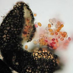
Pseudallescheria boydii is a saprotrophic fungus with broad hyphae growing up to 2–5  μm in width. [12] Colonies change in colour from white to pale brown and develop a cottony texture with maturity. [11] [13] After a 2–3 week incubation period, cleistothecia may form [13] containing asci filled with eight fusiform, one-celled ascospores [14] measuring 12–18 × 9–13 μm in diameter. [15] This fungus grows on most standard media, maturing in 7 days. [15] Its primary nutrients are the sugars xylose, [11] arabinose, [11] glucose, [11] sucrose, [16] ribitol, [16] xylitol [16] and L-arabinitol. [16] It cannot assimilate maltose or lactose; however, it is able to assimilate urea, asparagine, potassium nitrate and ammonium nitrate. [10] The optimal temperature for growth is 25 °C (77 °F) and the fungus is generally considered to be mesophilic, [13] although it can grow at higher temperatures (up to 37 °C (99 °F)) as well. [10] Asexual reproduction manifests in one of two forms: the Scedosporium type (the most common type) and the Graphium type. Scedosporium apiospermum forms greyish-white colonies with a grey-black reverse. The conidia are single-celled, pale brown and oval in form. Their size ranges from 4–9 x 6–10 μm and their development is annellidic. [15]

Pathogenicity

Pseudallescheria boydii is an emerging opportunistic pathogen. [11] Immune response is characterized by TLR2 recognition of P. boydii derived α-glucans, while TLR4 mediates the recognition of P. boydii derived rhamnomannans. [17] Human infection takes one of two forms: mycetoma (99% of infections), a chronic, subcutaneous disease, [11] and pseudallescheriasis, which includes all other forms of the disease commonly presented in the central nervous system, lungs, joints and bone. [18] The former can also be distinguished by the presence of sclerotia, or granules, which are typically absent in pseudallescheriasis-type infections. [15] Infection is initiated via inhalation or traumatic implantation in the skin. [18] Infection can lead to arthritis, [11] otitis, [11] endocarditis, [11] sinusitis, and other manifestations. [11] Masses of hyphae can form "fungus balls" in the lungs. [11] While "fungus balls" can also form in other organs, they are commonly derived from host necrotic tissue resulting from nodular infarction and thrombosis of lung vessels following infection. [10]

This species is second in prevalence after Aspergillus fumigatus as a fungal pathogen in cystic fibrosis patients. It causes allergic bronchopulmonary disease and chronic lung lesions that resemble aspergillosis. [15] Infections can also occur in immunocompetent individuals, usually in the lungs and upper respiratory tract. [10] Infections in the CNS, which are rare, present as neutrophilic meningitis or multiple brain abscesses [19] and have a mortality rate of up to 75%. [15] Infections have also been observed in animals, notably corneal infection, abdominal mycetoma and disseminated infections in dogs and horses. [13] Transient colonization is more likely than disease. However, invasive pseudoallescheriasis can be found in patients with prolonged neutropenia, high-dose corticosteroid therapy and allotransplantation of bone marrow. [18] Pseudallescheria boydii has also been implicated in pneumonia subsequent to near-drowning events with infection developing anywhere between a few weeks to several months after exposure yielding high mortality. Dissemination of the organism to the central nervous system has been observed in some cases. [20] This species is also known as a non-invasive colonist of the external ear and airways of patients with poor lung or sinus clearance, and the first documented case of human pseudallescheriasis involved the ear canal. [21] It has also been implicated in infection of joints following traumatic injury, and these infections can progress to osteomyelitis. Infections of the skin and cornea have also been reported. Typical host-related risk factors for infection include lymphopenia, steroid treatment, serum albumin levels of < 3 mg/dL and neutropenia. [22]

Diagnosis

Detection and diagnosis of S. apiospermum is possible through isolation of the fungus in culture or through cytology and histopathology in the tissues of diseased individuals. [10] In mycetoma-type infections, a confluence of symptoms is necessary for diagnosis, including tumefaction, draining sinuses and extrusion of grains. Furthermore, P. boydii grains and hyphae should be cultured and observed microscopically after staining with H&E, periodic acid–Schiff stain, Tissue Gram or Grocott's methenamine silver stain. [10] A radiological diagnosis may be helpful in elucidating the extent of the disease in terms of bone and soft tissue involvement. Scedosporium-caused eumycetomas have been found to have thick-walled cavities and grains appearing as hyperreflective echoes on scans, while actinomycetomas show fine echoes at the bottom of cavities. [10]

Direct detection is possible in samples histochemically stained in 20% KOH followed by fluorescence microscopy with antibody. The characteristic shape, texture and colour of tissues can help identify S. apiospermum grains, which are often surrounded by an eosinophilic zone. [10] Histopathologically, hyalohyphomycotic fungi like Scedosporium spp., Aspergillus spp., Fusarium spp. and Petriella spp. are similar in that they show septation of hyphae at regular intervals, have dichotomous branching and invade blood vessels. However, Scedosporium presents more irregular branching, sometimes with terminal or intercalary chlamydospores. [10] In serum, Scedosporium infections can be detected by counterimmunoelectrophoresis. [23] Molecular diagnostics appear to be promising in complementing current conventional diagnostic methods. [10]

Culture detection is accomplished by rinsing "grains" in 70% ethanol and sterile saline solution to avoid bacterial contamination prior to inoculation on growth medium. Selection of Scedosporium growth can be achieved on Leonian's agar supplemented with 10 g/mL benomyl, or on media containing cycloheximide or amphotericin B. [10] Optimal incubation is at a temperature of 25–35 °C (77–95 °F). [10]

Treatment

Pseudallescheria boydii is resistant to amphotericin B [18] and nearly all other antifungal drugs. Consequently, there is currently no consistently effective antifungal therapy for this agent. [18] Miconazole has shown the best in vivo activity; however, itraconazole, fluconazole, ketoconazole and voriconazole have also been used in treatment, albeit with less success. [15] [20] In an in vitro environment, terbinafine has been found to work in synergy with azoles against P. boydii. Echinocandins, such as caspofungin and sordarins, have shown promise in in vitro assays. CMT-3, a chemically modified tetracycline, has also shown to be active in vitro against P. boydii. [13]

Epidemiology

In the United States, P. boydii is the most common causal agent of eumycetoma, and tends to be more common in men than in women, particularly in the 20- to 45-year-old age group. [10] In the United States, the incidence of infection by S. apiospermum between 1993 and 1998 was 0.82; this figure increased to 1.33 by 2005. [10] Pseudallescheria boydii infection was implicated in the deaths of three athletes injured during the opening ceremony of the 1997 Maccabiah Games when the Maccabiah bridge collapsed in the Yarkon River. [5]

Related Research Articles

<span class="mw-page-title-main">Eumycetoma</span> Human and animal fungal infection

Eumycetoma, also known as Madura foot, is a persistent fungal infection of the skin and the tissues just under the skin, affecting most commonly the feet, although it can occur in hands and other body parts. It starts as a painless wet nodule, which may be present for years before ulceration, swelling, grainy discharge and weeping from sinuses and fistulae, followed by bone deformity.

Acremonium strictum is an environmentally widespread saprotroph species found in soil, plant debris, and rotting mushrooms. Isolates have been collected in North and Central America, Asia, Europe and Egypt. A. strictum is an agent of hyalohyphomycosis and has been identified as an increasingly frequent human pathogen in immunosuppressed individuals, causing localized, disseminated and invasive infections. Although extremely rare, A. strictum can infect immunocompetent individuals, as well as neonates. Due to the growing number of infections caused by A. strictum in the past few years, the need for new medical techniques in the identification of the fungus as well as for the treatment of human infections has risen considerably.

<i>Acrophialophora fusispora</i> Species of ascomycete fungus found in soil, air and various plants

Acrophialophora fusispora is a poorly studied ascomycete fungus found in soil, air and various plants. A. fusispora is morphologically similar to the genera Paecilomyces and Masonia, but differ in the presence of pigmented conidiophores, verticillate phialides, and frequent sympodial proliferation. Moreover, A. fusispora is distinguished by its pigmented spindle-shaped conidia, covered with spiral bands. The fungus is naturally found in soils of tropical to temperate regions. The fungus has been identified as a plant and animal pathogen, and has recently been recognized as an emerging opportunistic human pathogen. A. fusispora infection in human is rare and has few documented clinical cases, but due to the rarity of the fungus and potential misidentification, the infections may be underdiagnosed. Clinical cases of A. fusispora include cases of keratitis, pulmonary colonization and infection, and cerebral infections. The fungus also has two documented cases of infection in dogs.

Exophiala jeanselmei is a saprotrophic fungus in the family Herpotrichiellaceae. Four varieties have been discovered: Exophiala jeanselmei var. heteromorpha, E. jeanselmei var. lecanii-corni, E. jeanselmei var. jeanselmei, and E. jeanselmei var. castellanii. Other species in the genus Exophiala such as E. dermatitidis and E. spinifera have been reported to have similar annellidic conidiogenesis and may therefore be difficult to differentiate.

Pathogenic fungi are fungi that cause disease in humans or other organisms. Although fungi are eukaryotic, many pathogenic fungi are microorganisms. Approximately 300 fungi are known to be pathogenic to humans; their study is called "medical mycology". Fungal infections are estimated to kill more people than either tuberculosis or malaria—about two million people per year.

<span class="mw-page-title-main">Geotrichosis</span> Medical condition

Geotrichosis is a mycosis caused by Geotrichum candidum.

<i>Lomentospora prolificans</i> Species of fungus

Lomentospora prolificans is an emerging opportunistic fungal pathogen that causes a wide variety of infections in immunologically normal and immunosuppressed people and animals. It is resistant to most antifungal drugs and infections are often fatal. Drugs targeting the Class II dihydroorotate dehydrogenase (DHODH) proteins of L. prolificans, Scedosporium, Aspergillus and other deadly moulds are the basis for at least one new therapy, Olorofim, which is currently in phase 2b clinical trials and has received breakthrough status by FDA. For information on all DHODH proteins, please see Dihydroorotate dehydrogenase.

Saksenaea vasiformis is an infectious fungus associated with cutaneous or subcutaneous lesions following trauma. It causes opportunistic infections as the entry of the fungus is through open spaces of cutaneous barrier ranging in severity from mild to severe or fatal. It lives in soils worldwide, but is considered as a rare human pathogen since only 38 cases were reported as of 2012. Saksenaea vasiformis usually fails to sporulate on the routine culture media, creating a challenge for early diagnosis, which is essential for a good prognosis. Infections are usually treated using a combination of amphotericin B and surgery. Saksenaea vasiformis is one of the few fungi known to cause necrotizing fasciitis or "flesh-eating disease".

Mycetoma is a chronic infection in the skin caused by either bacteria (actinomycetoma) or fungi (eumycetoma), typically resulting in a triad of painless firm skin lumps, the formation of weeping sinuses, and a discharge that contains grains. 80% occur in feet.

<i>Exophiala dermatitidis</i> Species of fungus

Exophiala dermatitidis is a thermophilic black yeast, and a member of the Herpotrichiellaceae. While the species is only found at low abundance in nature, metabolically active strains are commonly isolated in saunas, steam baths, and dish washers. Exophiala dermatitidis only rarely causes infection in humans, however cases have been reported around the world. In East Asia, the species has caused lethal brain infections in young and otherwise healthy individuals. The fungus has been known to cause cutaneous and subcutaneous phaeohyphomycosis, and as a lung colonist in people with cystic fibrosis in Europe. In 2002, an outbreak of systemic E. dermatitidis infection occurred in women who had received contaminated steroid injections at North Carolina hospitals.

<i>Ochroconis gallopava</i> Species of fungus

Ochroconis gallopava, also called Dactylaria gallopava or Dactylaria constricta var. gallopava, is a member of genus Dactylaria. Ochroconis gallopava is a thermotolerant, darkly pigmented fungus that causes various infections in fowls, turkeys, poults, and immunocompromised humans first reported in 1986. Since then, the fungus has been increasingly reported as an agent of human disease especially in recipients of solid organ transplants. Ochroconis gallopava infection has a long onset and can involve a variety of body sites. Treatment of infection often involves a combination of antifungal drug therapy and surgical excision.

<i>Apophysomyces variabilis</i> Species of fungus

Apophysomyces variabilis is an emerging fungal pathogen that can cause serious and sometimes fatal infection in humans. This fungus is a soil-dwelling saprobe with tropical to subtropical distribution. It is a zygomycete that causes mucormycosis, an infection in humans brought about by fungi in the order Mucorales. Infectious cases have been reported globally in locations including the Americas, Southeast Asia, India, and Australia. Apophysomyces variabilis infections are not transmissible from person to person.

Coniochaeta hoffmannii, also known as Lecythophora hoffmannii, is an ascomycete fungus that grows commonly in soil. It has also been categorized as a soft-rot fungus capable of bringing the surface layer of timber into a state of decay, even when safeguarded with preservatives. Additionally, it has pathogenic properties, although it causes serious infection only in rare cases. A plant pathogen lacking a known sexual state, C. hoffmannii has been classified as a "dematiaceous fungus" despite its contradictory lack of pigmentation; both in vivo and in vitro, there is no correlation between its appearance and its classification.

Scedosporiosis is the general name for any mycosis - i.e., fungal infection - caused by a fungus from the genus Scedosporium. Current population-based studies suggest Scedosporium prolificans and Scedosporium apiospermum to be among the most common infecting agents from the genus, although infections caused by other members thereof are not unheard of. The latter is an asexual form (anamorph) of another fungus, Pseudallescheria boydii. The former is a “black yeast”, currently not characterized as well, although both of them have been described as saprophytes.

Histoplasma duboisii is a saprotrophic fungus responsible for the invasive infection known as African histoplasmosis. This species is a close relative of Histoplasma capsulatum, the agent of classical histoplasmosis, and the two occur in similar habitats. Histoplasma duboisii is restricted to continental Africa and Madagascar, although scattered reports have arisen from other places usually in individuals with an African travel history. Like, H. capsulatum, H. duboisii is dimorphic – growing as a filamentous fungus at ambient temperature and a yeast at body temperature. It differs morphologically from H. capsulatum by the typical production of a large-celled yeast form. Both agents cause similar forms of disease, although H. duboisii predominantly causes cutaneous and subcutaneous disease in humans and non-human primates. The agent responds to many antifungal drug therapies used to treat serious fungal diseases.

<i>Scedosporium</i> Genus of fungi

Scedosporium is a genus of fungi in the family Microascaceae.

<i>Neoscytalidium dimidiatum</i> Species of fungus

Neoscytalidium dimidiatum was first described in 1933 as Hendersonula toruloidea from diseased orchard trees in Egypt. Decades later, it was determined to be a causative agent of human dermatomycosis-like infections and foot infections predominantly in tropical areas; however the fungus is considered to be widespread. A newer name, Scytalidium dimidiatum, was applied to a synanamorph of Nattrassia mangiferae, otherwise known as Neofusicoccum mangiferae. Substantial confusion has arisen in the literature on this fungus resulting from the use of multiple different names including Torula dimidiata, Fusicoccum dimidiatum, Scytalidium dimidiatum, and Hendersonula toruloidea. Additionally, Scytalidium lignicola and Scytalidium lignicolum are often considered earlier names of N. dimidiatum.

Sarocladium kiliense is a saprobic fungus that is occasionally encountered as a opportunistic pathogen of humans, particularly immunocompromised and individuals. The fungus is frequently found in soil and has been linked with skin and systemic infections. This species is also known to cause disease in the green alga, Cladophora glomerata as well as various fruit and vegetable crops grown in warmer climates.

Arthrographis kalrae is an ascomycetous fungus responsible for human nail infections described in 1938 by Cochet as A. langeronii. A. kalrae is considered a weak pathogen of animals including human restricted to the outermost keratinized layers of tissue. Infections caused by this species are normally responsive to commonly used antifungal drugs with only very rare exceptions.

Cladophialophora arxii is a black yeast shaped dematiaceous fungus that is able to cause serious phaeohyphomycotic infections. C. arxii was first discovered in 1995 in Germany from a 22-year-old female patient suffering multiple granulomatous tracheal tumours. It is a clinical strain that is typically found in humans and is also capable of acting as an opportunistic fungus of other vertebrates Human cases caused by C. arxii have been reported from all parts of the world such as Germany and Australia.

References

  1. "Pseudallescheria boydii (Shear) McGinnis, A.A. Padhye & Ajello, Mycotaxon 14 (1): 97 (1982)". MycoBank. International Mycological Association. Retrieved 2014-11-23.
  2. Pseudallescheria at the U.S. National Library of Medicine Medical Subject Headings (MeSH)
  3. Janda-Ulfig, Katarzyna; Ulfig, Krzysztof; Cano, Josep; Guarro, Josep (June 2008). "A study of the growth of Pseudallescheria boydii isolates from sewage sludge and clinical sources on tributyrin, rapeseed oil, biodiesel oil and diesel oil". Annals of Agricultural and Environmental Medicine. 15 (1): 45–49. PMID   18581978 . Retrieved 2024-02-11.
  4. Wiederhold, N.P.; Lewis, R.E. (June 2009). "Antifungal activity against Scedosporium species and novel assays to assess antifungal pharmacodynamics against filamentous fungi". Medical Mycology. 47 (4): 422–32. doi: 10.1080/13693780802510224 . PMID   19058049.
  5. 1 2 Tal, Alon (2002). Pollution in a Promised Land: An Environmental History of Israel. University of California Press. ISBN   978-0-520-23428-4.
  6. Shear, C.L. (1922). "Life history of an undescribed ascomycete isolated from a granular mycetoma of man". Mycologia. 14 (5): 239–43. doi:10.2307/3753469. JSTOR   3753469.
  7. Malloch, D. (1970). "New concepts in the Microascaceae illustrated by two species". Mycologia. 62 (4): 727–740. doi:10.2307/3757662. JSTOR   3757662.
  8. Burkhardt, Lotte (2022). Eine Enzyklopädie zu eponymischen Pflanzennamen [Encyclopedia of eponymic plant names](pdf) (in German). Berlin: Botanic Garden and Botanical Museum, Freie Universität Berlin. doi:10.3372/epolist2022. ISBN   978-3-946292-41-8. S2CID   246307410 . Retrieved January 27, 2022.
  9. McGinnis, M.R.; Padhye, A.A.; Ajello, L. (1982). "Pseudallescheria Negroni et Fischer, 1943 and its later synonym Petrielldium Malloch, 1970". Mycotaxon. 14 (1): 94–102.
  10. 1 2 3 4 5 6 7 8 9 10 11 12 13 14 15 16 17 Cortez, K. J.; Roilides, E.; Quiroz-Telles, F.; Meletiadis, J.; Antachopoulos, C.; Knudsen, T.; Buchanan, W.; Milanovich, J.; Sutton, D. A.; Fothergill, A.; Rinaldi, M. G.; Shea, Y. R.; Zaoutis, T.; Kottilil, S.; Walsh, T. J. (17 January 2008). "Infections Caused by Scedosporium spp". Clinical Microbiology Reviews. 21 (1): 157–197. doi:10.1128/CMR.00039-07. PMC   2223844 . PMID   18202441.
  11. 1 2 3 4 5 6 7 8 9 10 11 12 de Hoog, G. S.; Guarro, J.; Gené, J.; Figueras, M. J. (2000). Atlas of Clinical Fungi (Second ed.). American Society for Microbiology. ISBN   978-90-70351-43-4.
  12. Salfelder, K.; Liscano, T.R. de; Sauerteig, E. (1990). Atlas of fungal pathology. Dordrecht: Kluwer Academic Publishers. ISBN   978-0792389354.
  13. 1 2 3 4 5 Patterson, Thomas F.; McGinnis, Michael R. "Pseudallescheria spp". www. doctorfungus.org. Archived from the original on 28 October 2014. Retrieved 28 October 2014.
  14. Gilgado, F; Cano, J; Gené, J; Guarro, J (2005). "Molecular phylogeny of the Pseudallescheria boydii species complex: proposal of two new species". Journal of Clinical Microbiology. 43 (10): 4930–42. doi:10.1128/jcm.43.10.4930-4942.2005. PMC   1248451 . PMID   16207945.
  15. 1 2 3 4 5 6 7 Prober, Charles G.; Long, Sarah; Fischer, Marc (2012). Principles and practice of pediatric infectious disease (4th ed.). Edinburgh: Elsevier/Saunders. ISBN   9781437727029.
  16. 1 2 3 4 de Hoog, GS; Marvin-Sikkema, FD; Lahpoor, GA; Gottschall, JC; Prins, RA; Guého, E (1994). "Ecology and physiology of the emerging opportunistic fungi Pseudallescheria boydii and Scedosporium prolificans". Mycoses. 37 (3–4): 71–8. doi:10.1111/j.1439-0507.1994.tb00780.x. PMID   7845423. S2CID   22605873.
  17. Figueiredo, Rodrigo Tinoco; Bittencourt, Vera Carolina B.; Lopes, Lívia Cristina L.; Sassaki, Guilherme; Barreto-Bergter, Eliana (2012). "Toll-like receptors (TLR2 and TLR4) recognize polysaccharides of Pseudallescheria boydii cell wall". Carbohydrate Research. 356: 260–264. doi:10.1016/j.carres.2012.02.028. PMID   22507831.
  18. 1 2 3 4 5 Bennett, John E.; Dolin, Raphael; Blaser, Martin J. (2000). Mandell, Douglas, and Bennett's principles and practice of infectious diseases (5th ed.). Philadelphia: Churchill Livingstone. ISBN   978-0-443-07593-3.
  19. Kershaw, P.; Freeman, R.; Templeton, D.; DeGirolami, P. C.; DeGirolami, U.; Tarsy, D.; Hoffmann, S.; Eliopoulos, G.; Karchmer, A. W. (1 April 1990). "Pseudallescheria boydii Infection of the Central Nervous System". Archives of Neurology. 47 (4): 468–472. doi:10.1001/archneur.1990.00530040126029. PMID   2181980.
  20. 1 2 Ender, Peter T.; Dolan, Matthew J. (1997). "Pneumonia Associated with Near-Drowning". Clinical Infectious Diseases. 25 (4): 896–907. doi: 10.1086/515532 . PMID   9356805.
  21. Rippon, John Willard (1988). Medical mycology : the pathogenic fungi and the pathogenic actinomycetes (3rd ed.). Philadelphia, PA: Saunders. ISBN   978-0721624440.
  22. Ajello, L.; Hay, R. J. (1997). Medical Mycology Vol 4 Topley & Wilson's Microbiology and Infectious Infections (9th ed.). London: Arnold.
  23. Thornton, C. R. (25 March 2009). "Tracking the Emerging Human Pathogen Pseudallescheria boydii by Using Highly Specific Monoclonal Antibodies". Clinical and Vaccine Immunology. 16 (5): 756–764. doi:10.1128/CVI.00061-09. PMC   2681584 . PMID   19321690.