Zygomycosis

Last updated

Zygomycosis
Periorbital fungal infection known as mucormycosis, or phycomycosis PHIL 2831 lores.jpg
Group of periorbital fungal infections including mucormycosis (shown) and phycomycosis
Specialty Infectious diseases   OOjs UI icon edit-ltr-progressive.svg

Zygomycosis is the broadest term to refer to infections caused by bread mold fungi of the zygomycota phylum. However, because zygomycota has been identified as polyphyletic, and is not included in modern fungal classification systems, the diseases that zygomycosis can refer to are better called by their specific names: mucormycosis [1] (after Mucorales), phycomycosis [2] (after Phycomycetes) and basidiobolomycosis (after Basidiobolus ). [3] These rare yet serious and potentially life-threatening fungal infections usually affect the face or oropharyngeal (nose and mouth) cavity. [4] Zygomycosis type infections are most often caused by common fungi found in soil and decaying vegetation. While most individuals are exposed to the fungi on a regular basis, those with immune disorders (immunocompromised) are more prone to fungal infection. [2] [5] [6] These types of infections are also common after natural disasters, such as tornadoes or earthquakes, where people have open wounds that have become filled with soil or vegetative matter. [7]

Contents

The condition may affect the gastrointestinal tract or the skin, often beginning in the nose and paranasal sinuses. It is one of the most rapidly spreading fungal infections in humans. Treatment consists of prompt and intensive antifungal drug therapy and surgery to remove the infected tissue.

Symptoms and signs

In the primary cutaneous form, the lesions are usually painful and necrotic, with black eschar, accompanied by a fever. Patients will usually present with a history of poorly controlled diabetes or malignancy. [8] Myocutaneous infections may lead to amputation. Pulmonary tract infections seen with lung transplant patients, who are at high risk for fatal invasive mycoses. [9] Rhinocerebral infection is characterized by paranasal swelling with necrotic tissues. Patient may have hemorrhagic exudates (tissue fluid from lesions tinged with blood) from the nose and eyes as the fungi penetrate through blood vessels and other anatomical structures. [10]

Causes

Micrograph showing a zygomycetes infection Zygomycosis.jpg
Micrograph showing a zygomycetes infection

Pathogenic zygomycosis is caused by species in two orders: Mucorales or Entomophthorales, with the former causing far more disease than the latter. [11] These diseases are known as "mucormycosis" and "entomophthoramycosis", respectively. [12]

Diagnosis

Diagnosis is done with potassium hydroxide (KOH) preparation in tissue. On light microscopy, there will be broad, ribbon-like septate hyphae with 90 degree angles on branches. [10] KOH wet mount of the black eschar will show fungal aseptate hyphae with right angle branching. Periodic Acid Schiff (PAS) staining will reveal similar broad hyphae in the dermis and cutis. Fungal culture can also confirm the organism. [13] Diagnosis remains difficult due to the lack of laboratory tests as mortality remains high. In 2005, a multiplex PCR test was able to identify five species of Rhizopus and may prove useful as a screening method for visceral mucormycosis in the future. [14]

The clinical approach to diagnosis includes radiologic, where more than ten nodules and pleural effusion are associated to pulmonary forms of the disease. In CT, a reverse halo sign is noted. Direct microscopy and histopathology, and cultures remain the gold standards for diagnoses. [15] Zygomycophyta share close clinical and radiological features to Aspergillosis. Invasive procedures such as bronchial endoscopy and lung biopsy may be necessary to confirm pulmonary diagnosis are no validated indirect tests are available. Quantitative polymerase chain reaction to detect serum DNA of the pathogen shows promise. [16]

Treatment

The condition may affect the gastrointestinal tract or the skin. In non-trauma cases, it usually begins in the nose and paranasal sinuses and is one of the most rapidly spreading fungal infections in humans. [2] Common symptoms include thrombosis and tissue necrosis. [17]

Due to the organisms' rapid growth and invasion, zygomycosis presents with a high fatality rate. Treatment must begin immediately with debridement of the necrotic tissue plus amphotericin B. [10] Complete excision of the infectious tissue may be required as suspected dead tissue must be excised aggressively. [13] [18] [19] In a documented case, conservative surgical drainage and intravenous amphotericin B in an insulin-dependent diabetic was proven effective in sino-orbital infection. [20] The prognosis varies vastly depending upon an individual patient's circumstances. [17]

Epidemiology

Zygomycosis has been found in survivors of the 2004 Indian Ocean earthquake and tsunami and in survivors of the 2011 Joplin, Missouri tornado. [21]

Other animals

The term oomycosis is used to describe oomycete infections. [22] These are more common in animals, notably dogs and horses. These are heterokonts, not true fungi. Types include pythiosis (caused by Pythium insidiosum ) and lagenidiosis.

Zygomycosis has been described in a cat, where fungal infection of the tracheobronchus led to respiratory disease requiring euthanasia. [23]

Related Research Articles

Nocardiosis is an infectious disease affecting either the lungs or the whole body. It is due to infection by a bacterium of the genus Nocardia, most commonly Nocardia asteroides or Nocardia brasiliensis.

<span class="mw-page-title-main">Aspergilloma</span> Medical condition

An aspergilloma is a clump of mold which exists in a body cavity such as a paranasal sinus or an organ such as the lung. By definition, it is caused by fungi of the genus Aspergillus.

Phycomycosis is an uncommon condition affecting the gastrointestinal tract and skin, most commonly found in dogs and horses. The condition is caused by various molds and fungi, with individual forms including pythiosis, zygomycosis, and lagenidiosis. Pythiosis, the most common type, is caused by Pythium, a type of water mould. Zygomycosis can be caused by two types of zygomycetes: Entomophthorales and Mucorales. The latter type of zygomycosis is also referred to as mucormycosis. Lagenidiosis is caused by a Lagenidium species, which like Pythium is a water mould. Since both pythiosis and lagenidiosis are caused by organisms from the Oomycetes and not the kingdom fungi, they are sometimes collectively referred to as oomycosis.

<span class="mw-page-title-main">Pythiosis</span> Tropical disease caused by the oomycete Pythium insidiosum

Pythiosis is a rare and deadly tropical disease caused by the oomycete Pythium insidiosum. Long regarded as being caused by a fungus, the causative agent was not discovered until 1987. It occurs most commonly in horses, dogs, and humans, with isolated cases in other large mammals. The disease is contracted after exposure to stagnant fresh water such as swamps, ponds, lakes, and rice paddies. P. insidiosum is different from other members of the genus in that human and horse hair, skin, and decaying animal and plant tissue are chemoattractants for its zoospores. Additionally, it is the only member in the genus known to infect mammals, while other members are pathogenic to plants and are responsible for some well-known plant diseases.

<span class="mw-page-title-main">Aspergillosis</span> Medical condition

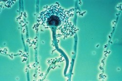
Aspergillosis is a fungal infection of usually the lungs, caused by the genus Aspergillus, a common mould that is breathed in frequently from the air, but does not usually affect most people. It generally occurs in people with lung diseases such as asthma, cystic fibrosis or tuberculosis, or COVID-19 or those who are immunocompromized such as those who have had a stem cell or organ transplant or those who take medications such as steroids and some cancer treatments which suppress the immune system. Rarely, it can affect skin.

<i>Basidiobolus ranarum</i> Species of fungus

Basidiobolus ranarum is a filamentous fungus with worldwide distribution. The fungus was first isolated by Eidam in 1886. It can saprophytically live in the intestines of mainly cold-blooded vertebrates and on decaying fruits and soil. The fungus prefers glucose as a carbon source and grows rapidly at room temperature. Basidiobolus ranarum is also known as a cause of subcutaneous zygomycosis, usually causing granulomatous infections on a host's limbs. Infections are generally geographically limited to tropical and subtropical regions such as East and West Africa. Subcutaneous zygomycosis caused by B. ranarum is a rare disease and predominantly affects children and males. Common subcutaneous zygomycosis shows characteristic features and is relatively easy to be diagnosed; while, certain rare cases might show non-specific clinical features that might pose a difficulty on its identification. Although disease caused by this fungus is known to resolve spontaneously on its own, there are a number of treatments available.

<i>Apophysomyces</i> Genus of fungi

Apophysomyces is a genus of filamentous fungi that are commonly found in soil and decaying vegetation. Species normally grow in tropical to subtropical regions.

<span class="mw-page-title-main">Conidiobolomycosis</span> Fungal infection

Conidiobolomycosis is a rare long-term fungal infection that is typically found just under the skin of the nose, sinuses, cheeks and upper lips. It may present with a nose bleed or a blocked or runny nose. Typically there is a firm painless swelling which can slowly extend to the nasal bridge and eyes, sometimes causing facial disfigurement.

<span class="mw-page-title-main">Mucormycosis</span> Fungal infection

Mucormycosis, also known as black fungus, is a serious fungal infection that comes under fulminant fungal sinusitis, usually in people who are immunocompromised. It is curable only when diagnosed early. Symptoms depend on where in the body the infection occurs. It most commonly infects the nose, sinuses, eyes and brain resulting in a runny nose, one-sided facial swelling and pain, headache, fever, blurred vision, bulging or displacement of the eye (proptosis), and tissue death. Other forms of disease may infect the lungs, stomach and intestines, and skin. The fatality rate is about 54%.

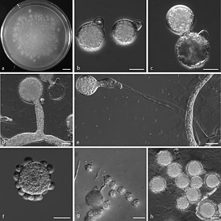
Saksenaea vasiformis is an infectious fungus associated with cutaneous or subcutaneous lesions following trauma. It causes opportunistic infections as the entry of the fungus is through open spaces of cutaneous barrier ranging in severity from mild to severe or fatal. It lives in soils worldwide, but is considered as a rare human pathogen since only 38 cases were reported as of 2012. Saksenaea vasiformis usually fails to sporulate on the routine culture media, creating a challenge for early diagnosis, which is essential for a good prognosis. Infections are usually treated using a combination of amphotericin B and surgery. Saksenaea vasiformis is one of the few fungi known to cause necrotizing fasciitis or "flesh-eating disease".

Entomophthoramycosis is a mycosis caused by Entomophthorales.

Rhizomucor pusillus is a species of Rhizomucor. It can cause disease in humans. R. pusillus is a grey mycelium fungi most commonly found in compost piles. Yellow-brown spores grow on a stalk to reproduce more fungal cells.

<i>Apophysomyces variabilis</i> Species of fungus

Apophysomyces variabilis is an emerging fungal pathogen that can cause serious and sometimes fatal infection in humans. This fungus is a soil-dwelling saprobe with tropical to subtropical distribution. It is a zygomycete that causes mucormycosis, an infection in humans brought about by fungi in the order Mucorales. Infectious cases have been reported globally in locations including the Americas, Southeast Asia, India, and Australia. Apophysomyces variabilis infections are not transmissible from person to person.

<i>Lichtheimia corymbifera</i> Species of fungus

Lichtheimia corymbifera is a thermophilic fungus in the phylum Zygomycota. It normally lives as a saprotrophic mold, but can also be an opportunistic pathogen known to cause pulmonary, CNS, rhinocerebral, or cutaneous infections in animals and humans with impaired immunity.

Cunninghamella bertholletiae is a species of zygomycetous fungi in the order Mucorales. It is found globally, with increased prevalence in Mediterranean and subtropical climates. It typically grows as a saprotroph and is found in a wide variety of substrates, including soil, fruits, vegetables, nuts, crops, and human and animal waste. Although infections are still rare, C. betholletiae is emerging as an opportunistic human pathogen, predominantly in immunocompromised people, leukemia patients, and people with uncontrolled diabetes. Cunninghamella bertholletiae infections are often highly invasive, and can be more difficult to treat with antifungal drugs than infections with other species of the Mucorales, making prompt and accurate recognition and diagnosis of mycoses caused by this fungus an important medical concern.

<span class="mw-page-title-main">Fungal sinusitis</span> Inflammation of the paranasal sinuses due to fungal infection

Fungal sinusitis or fungal rhinosinusitis is the inflammation of the lining mucosa of the paranasal sinuses due to a fungal infection. It occurs in people with reduced immunity. The maxillary sinus is the most commonly involved. Fungi responsible for fungal sinusitis are Aspergillus fumigatus (90%), Aspergillus flavus, and Aspergillus niger. Fungal sinusitis occurs most commonly in middle-aged populations. Diabetes mellitus is the most common risk factor involved.

<i>Rhizopus oryzae</i> Species of fungus

Rhizopus oryzae is a filamentous heterothallic microfungus that occurs as a saprotroph in soil, dung, and rotting vegetation. This species is very similar to Rhizopus stolonifer, but it can be distinguished by its smaller sporangia and air-dispersed sporangiospores. It differs from R. oligosporus and R. microsporus by its larger columellae and sporangiospores. The many strains of R. oryzae produce a wide range of enzymes such as carbohydrate digesting enzymes and polymers along with a number of organic acids, ethanol and esters giving it useful properties within the food industries, bio-diesel production, and pharmaceutical industries. It is also an opportunistic pathogen of humans causing mucormycosis.

<i>Rhizopus stolonifer</i> Species of fungus

Rhizopus stolonifer is commonly known as black bread mold. It is a member of Zygomycota and considered the most important species in the genus Rhizopus. It is one of the most common fungi in the world and has a global distribution although it is most commonly found in tropical and subtropical regions. It is a common agent of decomposition of stored foods. Like other members of the genus Rhizopus, R. stolonifer grows rapidly, mostly in indoor environments.

Mycotypha microspora, also known as Microtypha microspora, is a filamentous fungus in the division Zygomycota. It was discovered in a Citrus aurantium peel in 1932 by E. Aline Fenner, who proposed a new genus Mycotypha to accommodate it. Mycotypha africana, which is another species in the genus Mycotypha, is closely related to M. microspora. The fungus has subsequently been isolated from both outdoor and indoor settings around the world, and is typically found in soil and dung. The species rarely causes infections in humans, but has recently been involved in the clinical manifestation of the life-threatening disease mucormycosis.

Lichtheimia ramosa is a saprotrophic zygomycete, typically found in soil or dead plant material. It is a thermotolerant fungus that has also been known to act as an opportunistic pathogen–infecting both humans and animals.

References

  1. Toro, Carlos; del Palacio, Amalia; Álvarez, Carmen; Rodríguez-Peralto, José Luis; Carabias, Esperanza; Cuétara, Soledad; Carpintero, Yolanda; Gómez, César (1998). "Zigomicosis cutánea por Rhizopus arrhizus en herida quirúrgica" [Cutaneous zygomycosis caused by Rhizopus arrhizus in a surgical wound]. Revista Iberoamericana de Micología (in Spanish). 15 (2): 94–6. PMID   17655419.
  2. 1 2 3 Auluck, Ajit (2007). "Maxillary necrosis by mucormycosis. a case report and literature review" (PDF). Medicina Oral Patologia Oral y Cirugia Bucal. 12 (5): E360–4. PMID   17767099.
  3. Centers for Disease Control and Prevention (1999). "Gastrointestinal Basidiobolomycosis — Arizona, 1994–1999". Morbidity and Mortality Weekly Report. 48 (32): 710–3. PMID   21033182.
  4. Nancy F Crum-Cianflone; MD MPH. "Mucormycosis". eMedicine. Retrieved 19 May 2008.
  5. MedlinePlus Encyclopedia : Mucormycosis
  6. Ettinger, Stephen J.; Feldman, Edward C. (1995). Textbook of Veterinary Internal Medicine (4th ed.). W.B. Saunders Company. ISBN   0-7216-6795-3.[ page needed ]
  7. Draper, Bill; Suhr, Jim (11 June 2011). "Survivors of Joplin tornado develop rare infection". Seattle Post-Intelligencer . Associated Press.[ permanent dead link ]
  8. Rodríguez-Lobato E, Ramírez-Hobak L, Aquino-Matus JE, Ramírez-Hinojosa JP, Lozano-Fernández VH, Xicohtencatl-Cortes J, Hernández-Castro R, Arenas R (April 2017). "Primary Cutaneous Mucormycosis Caused by Rhizopus oryzae: A Case Report and Review of Literature". Mycopathologia. 182 (3–4): 387–392. doi:10.1007/s11046-016-0084-6. PMID   27807669.
  9. Mattner F, Weissbrodt H, Strueber M (2004). "Two case reports: fatal Absidia corymbifera pulmonary tract infection in the first postoperative phase of a lung transplant patient receiving voriconazole prophylaxis, and transient bronchial Absidia corymbifera colonization in a lung transplant patient". Scand J Infect Dis. 36 (4): 312–4. doi:10.1080/00365540410019408. PMID   15198193.
  10. 1 2 3 Moscatello, Kim (2013). USMLE Step 1: Immunology and Microbiology Lecture Notes. Chicago: Kaplan Publishing. pp. 430–1. ISBN   978-1625232557.
  11. Ribes, J. A.; Vanover-Sams, C. L.; Baker, D. J. (2000). "Zygomycetes in Human Disease". Clinical Microbiology Reviews. 13 (2): 236–301. doi:10.1128/CMR.13.2.236. PMC   100153 . PMID   10756000.
  12. Prabhu, R. M.; Patel, R. (2004). "Mucormycosis and entomophthoramycosis: A review of the clinical manifestations, diagnosis and treatment". Clinical Microbiology and Infection. 10: 31–47. doi: 10.1111/j.1470-9465.2004.00843.x . PMID   14748801.
  13. 1 2 Li H, Hwang SK, Zhou C, Du J, Zhang J (August 2013). "Gangrenous cutaneous mucormycosis caused by Rhizopus oryzae: a case report and review of primary cutaneous mucormycosis in China over Past 20 years". Mycopathologia. 176 (1–2): 123–8. doi:10.1007/s11046-013-9654-z. PMID   23615822.
  14. Nagao K, Ota T, Tanikawa A, Takae Y, Mori T, Udagawa S, Nishikawa T (July 2005). "Genetic identification and detection of human pathogenic Rhizopus species, a major mucormycosis agent, by multiplex PCR based on internal transcribed spacer region of rRNA gene". J Dermatol Sci. 39 (1): 23–31. doi:10.1016/j.jdermsci.2005.01.010. PMID   15978416.
  15. Skiada A, Lass-Floerl C, Klimko N, Ibrahim A, Roilides E, Petrikkos G (April 2018). "Challenges in the diagnosis and treatment of mucormycosis". Med Mycol. 56 (suppl_1): 93–101. doi:10.1093/mmy/myx101. PMC   6251532 . PMID   29538730.
  16. Danion F, Aguilar C, Catherinot E, Alanio A, DeWolf S, Lortholary O, Lanternier F (October 2015). "Mucormycosis: New Developments into a Persistently Devastating Infection". Semin Respir Crit Care Med. 36 (5): 692–705. doi:10.1055/s-0035-1562896. PMID   26398536.
  17. 1 2 Spellberg, B.; Edwards, J.; Ibrahim, A. (2005). "Novel Perspectives on Mucormycosis: Pathophysiology, Presentation, and Management". Clinical Microbiology Reviews. 18 (3): 556–69. doi:10.1128/CMR.18.3.556-569.2005. PMC   1195964 . PMID   16020690.
  18. Spellberg, Brad; Walsh, Thomas J.; Kontoyiannis, Dimitrios P.; Edwards, Jr.; Ibrahim, Ashraf S. (2009). "Recent Advances in the Management of Mucormycosis: From Bench to Bedside". Clinical Infectious Diseases. 48 (12): 1743–51. doi:10.1086/599105. PMC   2809216 . PMID   19435437.
  19. Grooters, A (2003). "Pythiosis, lagenidiosis, and zygomycosis in small animals". Veterinary Clinics of North America: Small Animal Practice. 33 (4): 695–720. doi:10.1016/S0195-5616(03)00034-2. PMID   12910739.
  20. Rosenberger RS, West BC, King JW (1983). "Survival from sino-orbital mucormycosis due to Rhizopus rhizopodiformis". Am J Med Sci. 286 (3): 25–30. doi:10.1097/00000441-198311000-00004. PMID   6356916.
  21. "Joplin toll rises to 151; some suffer from fungus". Associated Press. 10 June 2011 via Medical Xpress.
  22. "Merck Veterinary Manual" . Retrieved 4 April 2009.
  23. Snyder, Katherine D.; Spaulding, Kathy; Edwards, John (2010). "Imaging diagnosis—tracheobronchial zygomycosis in a cat". Veterinary Radiology & Ultrasound. 51 (6): 617–20. doi:10.1111/j.1740-8261.2010.01720.x. PMID   21158233.