Phycomycosis

Last updated
Phycomycosis
Causes Pythium insidiosum

Phycomycosis is an uncommon condition of the gastrointestinal tract and skin most commonly found in dogs and horses. The condition is caused by a variety of molds and fungi, and individual forms include pythiosis, zygomycosis, and lagenidiosis. Pythiosis is the most common type and is caused by Pythium , a type of water mould. Zygomycosis can also be caused by two types of zygomycetes, Entomophthorales (such as Basidiobolus and Conidiobolus ) and Mucorales (such as Mucor , Mortierella , Absidia , Rhizopus , Rhizomucor , and Saksenaea ). [1] The latter type of zygomycosis is also referred to as mucormycosis. Lagenidiosis is caused by a Lagenidium species, which like Pythium is a water mould. Since both pythiosis and lagenidiosis are caused by organisms from the class Oomycetes, they are sometimes collectively referred to as oomycosis.

Contents

Pythiosis

Ulcerative and destructive skin lesion on a dog caused by Pythium insidiosum Pythiosis 2.jpg
Ulcerative and destructive skin lesion on a dog caused by Pythium insidiosum

Pythiosis is caused by Pythium insidiosum and occurs most commonly in dogs and horses, but is also found in cats, cattle, and humans. In the United States it is most commonly found in the Gulf states, especially Louisiana, but has been found in midwest and eastern states. It is also found in southeast Asia, eastern Australia, New Zealand, and South America. Pythiosis occurs in areas with mild winters due to the organism surviving in standing water that does not reach freezing temperatures. [2] Pythium occupies swamps in late summer and infects dogs who drink water containing it. The disease is typically found in young, large breed dogs. [1]

It is suspected that pythiosis is caused by invasion of the organism into wounds, either in the skin or in the gastrointestinal tract. [2] The disease grows slowly in the stomach and small intestine, eventually forming large lumps of granulation tissue. It can also invade surrounding lymph nodes. Symptoms include vomiting, diarrhea, depression, weight loss, and a mass in the abdomen. Pythiosis of the skin in dogs is very rare, and appears as ulcerated lumps. Primary infection can also occur in the bones and lungs.
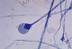
Pythium hyphae Pythiosis hyphae.jpg
Pythium hyphae

In horses, subcutaneous pythiosis is the most common form and infection occurs through a wound while standing in water containing the pathogen. [3] The disease is also known as leeches, swamp cancer, and bursatti. Lesions are most commonly found on the lower limbs, abdomen, chest, and genitals. They are granulomatous and itchy, and may be ulcerated or fistulated. The lesions often contain yellow, firm masses of dead tissue known as kunkers. [4] It is possible with chronic infection for the disease to spread to underlying bone. [5]

In humans it can cause arteritis, keratitis, and periorbital cellulitis. [6]

In cats pythioisis is almost always confined to the skin as hairless and edematous lesions. It is usually found on the limbs, perineum, and at the base of the tail. [7] Lesions may also develop in the nasopharynx. [4]

Pythium insidiosum is different from other members of the genus in that human and horse hair, skin, and decaying animal tissue are chemoattractants for its zoospores, in addition to decaying plant tissue. [3]

Zygomycosis

A mature sporangium of a Mucor sp. fungus Mature sporangium of a Mucor sp. fungus.jpg
A mature sporangium of a Mucor sp. fungus

Zygomycosis usually is a disease of the skin, but can also occur in the sinuses or gastrointestinal tract. In humans it is most prevalent in immunocompromised patients (HIV/AIDS, the elderly, SCID, etc) and patients in acidosis (diabetes, burns), particularly after barrier injury to the skin or mucus membranes. Zygomycosis caused by Mucorales causes a rapidly progressing disease of sudden onset in sick or immunocompromised animals. Entomophthorales cause chronic, local infections in otherwise healthy animals. The important species that cause entomophthoromycosis are Conidiobolus coronatus, C. incongruous, and Basidiobolus ranarum . Conidiobolus infections of the upper respiratory system have been reported in humans, sheep, horses, and dogs, and Basidiobolus has been reported less commonly in humans and dogs. [8] Horses are one of the most common domestic animals to be affected by entomophthoromycosis. C. coronatus causes lesions in the nasal and oral mucosa of horses that may cause nasal discharge or difficulty breathing. B. ranarum causes large circular nodules on the upper body and neck of horses. [9] Entomophthorales is found in soil and decaying plant matter, and specifically Basidiobolus can be contracted from insects and the feces of reptiles or amphibians. [6]

Zygomycosis of the sinuses can extend from the sinuses into the orbit and the cranial vault, leading to rhinocerebral mucormycosis.[ citation needed ]

Lagenidiosis

The best known species of Lagenidium is Lagenidium giganteum , a parasite of mosquito larvae used in biological control of mosquitoes. Two different species cause disease exclusively in dogs: L. caninum and L. karlingii. Lagenidium is found in the southeastern United States in lakes and ponds. Lagenidiosis causes progressive skin and subcutaneous lesions in the legs, groin, trunk, and near the tail. The lesions are firm nodules or ulcerated regions with draining tracts. Regional lymph nodes are usually swollen. Spread of the disease to distant lymph nodes, large blood vessels, and the lungs may occur. [6] An aneurysm of a great vessel can rupture and cause sudden death. [4] L. caninum is the more aggressive species and is more likely to spread to other organs than L. karlingii. [10]

Diagnosis and treatment

Diagnosis is through biopsy or culture, although an ELISA test has been developed for Pythium insidiosum in animals. [11] Treatment is very difficult and includes surgery when possible. Postoperative recurrence is common. Antifungal drugs show only limited effect on the disease, but itraconazole and terbinafine hydrochloride are often used for two to three months following surgery. [6] Humans with Basidiobolus infections have been treated with amphotericin B and potassium iodide. [8] For pythiosis and lagenidiosis, a new drug targeting water moulds called caspofungin is available, but it is very expensive. [6] Immunotherapy has been used successfully in humans and horses with pythiosis. [11] The prognosis for any type of phycomycosis is poor.

Related Research Articles

<span class="mw-page-title-main">Gastric dilatation volvulus</span> Medical condition in dogs

Gastric dilatation volvulus (GDV), also known as gastric dilation, twisted stomach, or gastric torsion, is a medical condition that affects dogs in which the stomach becomes overstretched and rotated by excessive gas content. The word bloat is often used as a general term to mean gas distension without stomach torsion, or to refer to GDV.

<span class="mw-page-title-main">Zygomycosis</span> Medical condition

Zygomycosis is the broadest term to refer to infections caused by bread mold fungi of the zygomycota phylum. However, because zygomycota has been identified as polyphyletic, and is not included in modern fungal classification systems, the diseases that zygomycosis can refer to are better called by their specific names: mucormycosis, phycomycosis and basidiobolomycosis. These rare yet serious and potentially life-threatening fungal infections usually affect the face or oropharyngeal cavity. Zygomycosis type infections are most often caused by common fungi found in soil and decaying vegetation. While most individuals are exposed to the fungi on a regular basis, those with immune disorders (immunocompromised) are more prone to fungal infection. These types of infections are also common after natural disasters, such as tornadoes or earthquakes, where people have open wounds that have become filled with soil or vegetative matter.

Skin disorders are among the most common health problems in dogs, and have many causes. The condition of a dog's skin and coat is also an important indicator of its general health. Skin disorders of dogs vary from acute, self-limiting problems to chronic or long-lasting problems requiring life-time treatment. Skin disorders may be primary or secondary in nature, making diagnosis complicated.

<span class="mw-page-title-main">Dog health</span> Health of dogs

The health of dogs is a well studied area in veterinary medicine.

Panosteitis, sometimes shortened to pano among breeders, is an occasionally seen long bone condition in large breed dogs. It manifests with sudden, unexplained pain and lameness that may shift from leg to leg, usually between 5 and 14 months of age, earning the nickname "growing pains. " Signs such as fever, weight loss, anorexia, and lethargy can also be seen. The cause is unknown, but genetics, stress, infection, metabolism, or an autoimmune component may be factors. It has also been suggested that rapid growth and high-protein food are involved in the pathogenesis. Whole blood analysis may show an elevated white blood cell count; this finding lends support to the theory that panosteitis is due to an infection.

<span class="mw-page-title-main">Lymphoma in animals</span> Type of cancer in animals

Lymphoma (lymphosarcoma) in animals is a type of cancer defined by a proliferation of malignant lymphocytes within solid organs such as the lymph nodes, bone marrow, liver and spleen. The disease also may occur in the eye, skin, and gastrointestinal tract.

Canid alphaherpesvirus 1 (CaHV-1), formerly Canine herpesvirus (CHV), is a virus of the family Herpesviridae which most importantly causes a fatal hemorrhagic disease in puppies less than two to three weeks old. It is known to exist in the United States, Canada, Australia, Japan, England and Germany. CHV was first recognized in the mid-1960s from a fatal disease in puppies.

<span class="mw-page-title-main">Protothecosis</span> Medical condition

Protothecosis, otherwise known as Algaemia, is a disease found in dogs, cats, cattle, and humans caused by a type of green alga known as Prototheca that lacks chlorophyll and enters the human or animal bloodstream. It and its close relative Helicosporidium are unusual in that they are actually green algae that have become parasites. The two most common species are Prototheca wickerhamii and Prototheca zopfii. Both are known to cause disease in dogs, while most human cases are caused by P. wickerhami. Prototheca is found worldwide in sewage and soil. Infection is rare despite high exposure, and can be related to a defective immune system. In dogs, females and Collies are most commonly affected.

<span class="mw-page-title-main">Pythiosis</span> Tropical disease caused by the oomycete Pythium insidiosum

Pythiosis is a rare and deadly tropical disease caused by the oomycete Pythium insidiosum. Long regarded as being caused by a fungus, the causative agent was not discovered until 1987. It occurs most commonly in horses, dogs, and humans, with isolated cases in other large mammals. The disease is contracted after exposure to stagnant fresh water such as swamps, ponds, lakes, and rice paddies. P. insidiosum is different from other members of the genus in that human and horse hair, skin, and decaying animal and plant tissue are chemoattractants for its zoospores. Additionally, it is the only member in the genus known to infect mammals, while other members are pathogenic to plants and are responsible for some well-known plant diseases.

Hypertrophic osteopathy is a bone disease secondary to cancer in the lungs.

<span class="mw-page-title-main">Corneal ulcers in animals</span> Veterinary inflammatory condition of the cornea

A corneal ulcer, or ulcerative keratitis, is an inflammatory condition of the cornea involving loss of its outer layer. It is very common in dogs and is sometimes seen in cats. In veterinary medicine, the term corneal ulcer is a generic name for any condition involving the loss of the outer layer of the cornea, and as such is used to describe conditions with both inflammatory and traumatic causes.

Lagenidiosis is a type of infectious disease.

<span class="mw-page-title-main">Mammary tumor</span>

A mammary tumor is a neoplasm originating in the mammary gland. It is a common finding in older female dogs and cats that are not spayed, but they are found in other animals as well. The mammary glands in dogs and cats are associated with their nipples and extend from the underside of the chest to the groin on both sides of the midline. There are many differences between mammary tumors in animals and breast cancer in humans, including tumor type, malignancy, and treatment options. The prevalence in dogs is about three times that of women. In dogs, mammary tumors are the second most common tumor over all and the most common tumor in female dogs with a reported incidence of 3.4%. Multiple studies have documented that spaying female dogs when young greatly decreases their risk of developing mammary neoplasia when aged. Compared with female dogs left intact, those spayed before puberty have 0.5% of the risk, those spayed after one estrous cycle have 8.0% of the risk, and dogs spayed after two estrous cycles have 26.0% of the risk of developing mammary neoplasia later in life. Overall, unspayed female dogs have a seven times greater risk of developing mammary neoplasia than do those that are spayed. While the benefit of spaying decreases with each estrous cycle, some benefit has been demonstrated in female dogs even up to 9 years of age. There is a much lower risk in male dogs and a risk in cats about half that of dogs.

A vaccine-associated sarcoma (VAS) or feline injection-site sarcoma (FISS) is a type of malignant tumor found in cats which has been linked to certain vaccines. VAS has become a concern for veterinarians and cat owners alike and has resulted in changes in recommended vaccine protocols. These sarcomas have been most commonly associated with rabies and feline leukemia virus vaccines, but other vaccines and injected medications have also been implicated.

<span class="mw-page-title-main">Basidiobolomycosis</span> Fungal disease

Basidiobolomycosis is a fungal disease caused by Basidiobolus ranarum. It may appear as one or more painless firm nodules in the skin which becomes purplish with an edge that appears to be slowly growing outwards. A serious but less common type affects the stomach and intestine, which usually presents with abdominal pain, fever and a mass.

<i>Basidiobolus ranarum</i> Species of fungus

Basidiobolus ranarum is a filamentous fungus with worldwide distribution. The fungus was first isolated by Eidam in 1886. It can saprophytically live in the intestines of mainly cold-blooded vertebrates and on decaying fruits and soil. The fungus prefers glucose as a carbon source and grows rapidly at room temperature. Basidiobolus ranarum is also known as a cause of subcutaneous zygomycosis, usually causing granulomatous infections on a host's limbs. Infections are generally geographically limited to tropical and subtropical regions such as East and West Africa. Subcutaneous zygomycosis caused by B. ranarum is a rare disease and predominantly affects children and males. Common subcutaneous zygomycosis shows characteristic features and is relatively easy to be diagnosed; while, certain rare cases might show non-specific clinical features that might pose a difficulty on its identification. Although disease caused by this fungus is known to resolve spontaneously on its own, there are a number of treatments available.

<span class="mw-page-title-main">Veterinary chiropractic</span>

Veterinary chiropractic, also known as animal chiropractic, is chiropractic for animals – a type of spinal manipulation. Veterinary chiropractors typically treat horses, racing greyhounds, and pets. Veterinary chiropractic is a controversial method due to a lack of evidence as to the efficacy of chiropractic methods. Contrary to traditional medicine, chiropractic therapies are alternative medicine. There is some degree of risk associated with even skilled manipulation in animals as the potential for injury exists with any technique used. The founder of chiropractic, Daniel David Palmer, used the method on animals, partly to challenge claims that the placebo effect was responsible for favorable results in humans. Chiropractic treatment of large animals dates back to the early 1900s. As of 2019, many states in the US provide statutory or regulatory guidelines for the practice of chiropractic and related treatments on animals, generally requiring some form of veterinary involvement.

Entomophthoramycosis is a mycosis caused by Entomophthorales.

<i>Pythium insidiosum</i> Species of single-celled organism

Pythium insidiosum is a species of Pythium and a member of the class oomycota. Pythium insidiosum is mainly found in standing water and occasionally soil. Unlike most Pythium species, which are generally pathogens of terrestrial plants, Pythium insidiosum is a pathogen of mammals. It causes pythiosis, mainly in horses, dogs, and humans. It can also cause disease in cats. It is a non-transmissible disease and occurs mainly in tropical climate, endemic to Thailand, affecting mainly humans and horses and in Brazil, affecting mainly horses. Infection can occur in healthy mammals. The pathogen is well-adapted to mammalian body temperature, with an optimum temperature for growth of 34–36 °C (93–97 °F).

<i>Lagenidium giganteum forma caninum</i> Animal pathogenic oomycete

Lagenidium giganteum forma caninum is an oomycete, a fungal-like organism known as water-mold, and belonging to the genus Lagenidium that causes lagenidiosis in some mammal species, characterized by progressive, severe and invasive cutaneous, subcutaneous, and disseminated infection. Clinical and pathological aspects of the disease resemble those seen in pythiosis. The first cases of lagenidiosis in mammals were officially reported in dogs in 2003. Since then, it has become increasingly recognized in dogs and cats as a cause of skin lesions.

References

  1. 1 2 Ettinger, Stephen J.; Feldman, Edward C. (1995). Textbook of Veterinary Internal Medicine (4th ed.). W.B. Saunders Company. ISBN   0-7216-6795-3.
  2. 1 2 Helman R, Oliver J (1999). "Pythiosis of the digestive tract in dogs from Oklahoma". J Am Anim Hosp Assoc. 35 (2): 111–4. doi:10.5326/15473317-35-2-111. PMID   10102178.
  3. 1 2 Liljebjelke K, Abramson C, Brockus C, Greene C (2002). "Duodenal obstruction caused by infection with Pythium insidiosum in a 12-week-old puppy". J Am Vet Med Assoc. 220 (8): 1188–91, 1162. doi: 10.2460/javma.2002.220.1188 . PMID   11990966.
  4. 1 2 3 "Oomycosis". The Merck Veterinary Manual. 2006. Retrieved 2007-02-03.
  5. Worster A, Lillich J, Cox J, Rush B (2000). "Pythiosis with bone lesions in a pregnant mare". J Am Vet Med Assoc. 216 (11): 1795–8, 1760. doi: 10.2460/javma.2000.216.1795 . PMID   10844973.
  6. 1 2 3 4 5 Grooters A (2003). "Pythiosis, lagenidiosis, and zygomycosis in small animals". Vet Clin North Am Small Anim Pract. 33 (4): 695–720, v. doi:10.1016/S0195-5616(03)00034-2. PMID   12910739.
  7. Wolf, Alice (2005). "Opportunistic fungal infections". In August, John R. (ed.). Consultations in Feline Internal Medicine Vol. 5. Elsevier Saunders. ISBN   0-7216-0423-4.
  8. 1 2 Greene C, Brockus C, Currin M, Jones C (2002). "Infection with Basidiobolus ranarum in two dogs". J Am Vet Med Assoc. 221 (4): 528–32, 500. doi: 10.2460/javma.2002.221.528 . PMID   12184703.
  9. "Zygomycosis". The Merck Veterinary Manual. 2006. Retrieved 2007-02-03.
  10. Todd-Jenkins, Karen (September 2007). "A new disease: clinically interesting for all the right reasons". Veterinary Forum. Veterinary Learning Systems. 24 (9): 18–20.
  11. 1 2 Hensel P, Greene C, Medleau L, Latimer K, Mendoza L (2003). "Immunotherapy for treatment of multicentric cutaneous pythiosis in a dog". J Am Vet Med Assoc. 223 (2): 215–8, 197. doi:10.2460/javma.2003.223.215. PMID   12875449.