Potassium channel tetramerisation domain

Last updated
K+ channel tetramerisation domain
PDB 1t1d EBI.jpg
[1]
Identifiers
SymbolK_tetra
Pfam PF02214
InterPro IPR003131
SCOP2 1t1d / SCOPe / SUPFAM
OPM superfamily 8
OPM protein 2a79
Available protein structures:
Pfam   structures / ECOD  
PDB RCSB PDB; PDBe; PDBj
PDBsum structure summary
PDB 3kvt S:12-102 1nn7 A:43-132 1s1g B:42-131

1dsx G:35-119 1qdv B:35-126 1qdw H:35-119 2a79 B:35-126 1exb E:39-129 1a68 :68-152 1eof A:68-159 1eod A:68-159 1eoe A:68-159

Contents

1t1d A:68-159

K+ channel tetramerisation domain is the N-terminal, cytoplasmic tetramerisation domain (T1) of voltage-gated K+ channels. It defines molecular determinants for subfamily-specific assembly of alpha-subunits into functional tetrameric channels. It is distantly related to the BTB/POZ domain Pfam PF00651.

Potassium channels

Potassium channels are the most diverse group of the ion channel family. [2] [3] They are important in shaping the action potential, and in neuronal excitability and plasticity. [4] The potassium channel family is composed of several functionally distinct isoforms, which can be broadly separated into 2 groups: [5] the practically non-inactivating 'delayed' group and the rapidly inactivating 'transient' group.

These are all highly similar proteins, with only small amino acid changes causing the diversity of the voltage-dependent gating mechanism, channel conductance and toxin binding properties. Each type of K+ channel is activated by different signals and conditions depending on their type of regulation: some open in response to depolarisation of the plasma membrane; others in response to hyperpolarisation or an increase in intracellular calcium concentration; some can be regulated by binding of a transmitter, together with intracellular kinases; while others are regulated by GTP-binding proteins or other second messengers. [6] In eukaryotic cells, K+ channels are involved in neural signalling and generation of the cardiac rhythm, act as effectors in signal transduction pathways involving G protein-coupled receptors (GPCRs) and may have a role in target cell lysis by cytotoxic T-lymphocytes. [7] In prokaryotic cells, they play a role in the maintenance of ionic homeostasis. [8]

Alpha subunits of the channels

All K+ channels discovered so far possess a core of alpha subunits, each comprising either one or two copies of a highly conserved pore loop domain (P-domain). The P-domain contains the sequence (T/SxxTxGxG), which has been termed the K+ selectivity sequence. In families that contain one P-domain, four subunits assemble to form a selective pathway for K+ across the membrane. However, it remains unclear how the 2 P-domain subunits assemble to form a selective pore. The functional diversity of these families can arise through homo- or hetero-associations of alpha subunits or association with auxiliary cytoplasmic beta subunits. K+ channel subunits containing one pore domain can be assigned into one of two superfamilies: those that possess six transmembrane (TM) domains and those that possess only two TM domains. The six TM domain superfamily can be further subdivided into conserved gene families: the voltage-gated (Kv) channels; the KCNQ channels (originally known as KvLQT channels); the EAG-like K+ channels; and three types of calcium (Ca)-activated K+ channels (BK, IK and SK). [8] [9] The 2TM domain family comprises inward-rectifying K+ channels. In addition, there are K+ channel alpha-subunits that possess two P-domains. These are usually highly regulated K+ selective leak channels.

The Kv family can be divided into several subfamilies on the basis of sequence similarity and function. Four of these subfamilies, Kv1 (Shaker), Kv2 (Shab), Kv3 (Shaw) and Kv4 (Shal), consist of pore-forming alpha subunits that associate with different types of beta subunit. Each alpha subunit comprises six hydrophobic TM domains with a P-domain between the fifth and sixth, which partially resides in the membrane. The fourth TM domain has positively charged residues at every third residue and acts as a voltage sensor, which triggers the conformational change that opens the channel pore in response to a displacement in membrane potential. [10] More recently, 4 new electrically-silent alpha subunits have been cloned: Kv5 (KCNF), Kv6 (KCNG), Kv8 and Kv9 (KCNS). These subunits do not themselves possess any functional activity, but appear to form heteromeric channels with Kv2 subunits, and thus modulate Shab channel activity. [11] When highly expressed, they inhibit channel activity, but at lower levels show more specific modulatory actions.

Tetramerization domain

The N-terminal, cytoplasmic tetramerization domain (T1) of voltage-gated potassium channels encodes molecular determinants for subfamily-specific assembly of alpha-subunits into functional tetrameric channels. [12] This domain is found in a subset of a larger group of proteins that contain the BTB/POZ domain.

Human proteins containing this domain

BTBD10; KCNA1; KCNA10; KCNA2; KCNA3; KCNA4; KCNA5; KCNA6; KCNA7; KCNB1; KCNB2; KCNC1; KCNC2; KCNC3; KCNC4; KCND1; KCND2; KCND3; KCNF1; KCNG1; KCNG2; KCNG3; KCNG4; KCNRG; KCNS1; KCNS2; KCNS3; KCNV1; KCNV2; KCTD1; KCTD10; KCTD11; KCTD12; KCTD13; KCTD14; KCTD15; KCTD16; KCTD17; KCTD18; KCTD19; KCTD2; KCTD20; KCTD21; KCTD3; KCTD4; KCTD5; KCTD6; KCTD7; KCTD8; KCTD9; SHKBP1; TNFAIP1;

Related Research Articles

<span class="mw-page-title-main">Ion channel</span> Pore-forming membrane protein

Ion channels are pore-forming membrane proteins that allow ions to pass through the channel pore. Their functions include establishing a resting membrane potential, shaping action potentials and other electrical signals by gating the flow of ions across the cell membrane, controlling the flow of ions across secretory and epithelial cells, and regulating cell volume. Ion channels are present in the membranes of all cells. Ion channels are one of the two classes of ionophoric proteins, the other being ion transporters.

<span class="mw-page-title-main">BK channel</span> Family of transport proteins

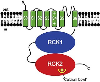
BK channels (big potassium), are large conductance calcium-activated potassium channels, also known as Maxi-K, slo1, or Kca1.1. BK channels are voltage-gated potassium channels that conduct large amounts of potassium ions (K+) across the cell membrane, hence their name, big potassium. These channels can be activated (opened) by either electrical means, or by increasing Ca2+ concentrations in the cell. BK channels help regulate physiological processes, such as circadian behavioral rhythms and neuronal excitability. BK channels are also involved in many processes in the body, as it is a ubiquitous channel. They have a tetrameric structure that is composed of a transmembrane domain, voltage sensing domain, potassium channel domain, and a cytoplasmic C-terminal domain, with many X-ray structures for reference. Their function is to repolarize the membrane potential by allowing for potassium to flow outward, in response to a depolarization or increase in calcium levels.

<span class="mw-page-title-main">Potassium channel</span> Ion channel that selectively passes K+

Potassium channels are the most widely distributed type of ion channel found in virtually all organisms. They form potassium-selective pores that span cell membranes. Potassium channels are found in most cell types and control a wide variety of cell functions.

<span class="mw-page-title-main">Voltage-gated ion channel</span> Type of ion channel transmembrane protein

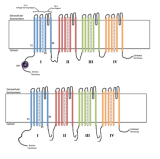
Voltage-gated ion channels are a class of transmembrane proteins that form ion channels that are activated by changes in the electrical membrane potential near the channel. The membrane potential alters the conformation of the channel proteins, regulating their opening and closing. Cell membranes are generally impermeable to ions, thus they must diffuse through the membrane through transmembrane protein channels. They have a crucial role in excitable cells such as neuronal and muscle tissues, allowing a rapid and co-ordinated depolarization in response to triggering voltage change. Found along the axon and at the synapse, voltage-gated ion channels directionally propagate electrical signals. Voltage-gated ion-channels are usually ion-specific, and channels specific to sodium (Na+), potassium (K+), calcium (Ca2+), and chloride (Cl) ions have been identified. The opening and closing of the channels are triggered by changing ion concentration, and hence charge gradient, between the sides of the cell membrane.

<span class="mw-page-title-main">Dendrotoxin</span> Chemical compound

Dendrotoxins are a class of presynaptic neurotoxins produced by mamba snakes (Dendroaspis) that block particular subtypes of voltage-gated potassium channels in neurons, thereby enhancing the release of acetylcholine at neuromuscular junctions. Because of their high potency and selectivity for potassium channels, dendrotoxins have proven to be extremely useful as pharmacological tools for studying the structure and function of these ion channel proteins.

<span class="mw-page-title-main">Kv1.1</span>

Potassium voltage-gated channel subfamily A member 1 also known as Kv1.1 is a shaker related voltage-gated potassium channel that in humans is encoded by the KCNA1 gene. Isaacs syndrome is a result of an autoimmune reaction against the Kv1.1 ion channel.

<span class="mw-page-title-main">Voltage-gated potassium channel</span> Class of transport proteins

Voltage-gated potassium channels (VGKCs) are transmembrane channels specific for potassium and sensitive to voltage changes in the cell's membrane potential. During action potentials, they play a crucial role in returning the depolarized cell to a resting state.

<span class="mw-page-title-main">Two-pore-domain potassium channel</span> Class of transport proteins

The two-pore-domain or tandem pore domain potassium channels are a family of 15 members that form what is known as leak channels which possess Goldman-Hodgkin-Katz (open) rectification. These channels are regulated by several mechanisms including signaling lipids, oxygen tension, pH, mechanical stretch, and G-proteins. Two-pore-domain potassium channels correspond structurally to a inward-rectifier potassium channel α-subunits. Each inward-rectifier potassium channel α-subunit is composed of two transmembrane α-helices, a pore helix and a potassium ion selectivity filter sequence and assembles into a tetramer forming the complete channel. The two-pore domain potassium channels instead are dimers where each subunit is essentially two α-subunits joined together.

<span class="mw-page-title-main">KCNE1</span> Protein-coding gene in the species Homo sapiens

Potassium voltage-gated channel subfamily E member 1 is a protein that in humans is encoded by the KCNE1 gene.

<span class="mw-page-title-main">Calcium-activated potassium channel subunit alpha-1</span> Voltage-gated potassium channel protein

Calcium-activated potassium channel subunit alpha-1 also known as large conductance calcium-activated potassium channel, subfamily M, alpha member 1 (KCa1.1), or BK channel alpha subunit, is a voltage gated potassium channel encoded by the KCNMA1 gene and characterized by their large conductance of potassium ions (K+) through cell membranes.

<span class="mw-page-title-main">KCND2</span> Protein-coding gene in the species Homo sapiens

Potassium voltage-gated channel subfamily D member 2 is a protein that in humans is encoded by the KCND2 gene. It contributes to the cardiac transient outward potassium current (Ito1), the main contributing current to the repolarizing phase 1 of the cardiac action potential.

<span class="mw-page-title-main">KCNA4</span> Protein-coding gene in the species Homo sapiens

Potassium voltage-gated channel subfamily A member 4 also known as Kv1.4 is a protein that in humans is encoded by the KCNA4 gene. It contributes to the cardiac transient outward potassium current (Ito1), the main contributing current to the repolarizing phase 1 of the cardiac action potential.

<span class="mw-page-title-main">KCNB1</span> Protein-coding gene in the species Homo sapiens

Potassium voltage-gated channel, Shab-related subfamily, member 1, also known as KCNB1 or Kv2.1, is a protein that, in humans, is encoded by the KCNB1 gene.

<span class="mw-page-title-main">KCNAB1</span> Protein-coding gene in the species Homo sapiens

Voltage-gated potassium channel subunit beta-1 is a protein that in humans is encoded by the KCNAB1 gene.

<span class="mw-page-title-main">KCNAB2</span> Protein-coding gene in the species Homo sapiens

Voltage-gated potassium channel subunit beta-2 is a protein that in humans is encoded by the KCNAB2 gene.

<span class="mw-page-title-main">KCNMB2</span> Protein-coding gene in the species Homo sapiens

Calcium-activated potassium channel subunit beta-2 is a protein that in humans is encoded by the KCNMB2 gene.

<span class="mw-page-title-main">KCNMB3</span> Protein-coding gene in the species Homo sapiens

Calcium-activated potassium channel subunit beta-3 is a protein that in humans is encoded by the KCNMB3 gene.

<span class="mw-page-title-main">KCNS3</span> Protein-coding gene in the species Homo sapiens

Potassium voltage-gated channel subfamily S member 3 (Kv9.3) is a protein that in humans is encoded by the KCNS3 gene. KCNS3 gene belongs to the S subfamily of the potassium channel family. It is highly expressed in pulmonary artery myocytes, placenta, and parvalbumin-containing GABA neurons in brain cortex. In humans, single-nucleotide polymorphisms of the KCNS3 gene are associated with airway hyperresponsiveness, whereas decreased KCNS3 mRNA expression is found in the prefrontal cortex of patients with schizophrenia.

<span class="mw-page-title-main">KCNMB4</span> Protein-coding gene in humans

Calcium-activated potassium channel subunit beta-4 is a protein that in humans is encoded by the KCNMB4 gene.

<span class="mw-page-title-main">HVCN1</span> Protein-coding gene in the species Homo sapiens

Voltage-gated hydrogen channel 1 is a protein that in humans is encoded by the HVCN1 gene.

References

  1. Bixby KA, Nanao MH, Shen NV, et al. (January 1999). "Zn2+-binding and molecular determinants of tetramerization in voltage-gated K+ channels". Nature Structural & Molecular Biology. 6 (1): 38–43. doi:10.1038/4911. PMID   9886290. S2CID   5414052.
  2. Perney TM, Kaczmarek LK (1991). "The molecular biology of K+ channels". Curr. Opin. Cell Biol. 3 (4): 663–670. doi:10.1016/0955-0674(91)90039-2. PMID   1772658.
  3. Williams JB, Luneau C, Smith JS, Wiedmann R (1991). "Shaw-like rat brain potassium channel cDNA's with divergent 3' ends". FEBS Lett. 288 (1): 163–167. doi: 10.1016/0014-5793(91)81026-5 . PMID   1879548. S2CID   20731463.
  4. Jan LY, Jan YN, Tempel BL (1988). "Cloning of a probable potassium channel gene from mouse brain". Nature. 332 (6167): 837–839. Bibcode:1988Natur.332..837T. doi:10.1038/332837a0. PMID   2451788. S2CID   4262757.
  5. Stuhmer W, Ruppersberg JP, Schroter KH, Sakmann B, Stocker M, Giese KP, Perschke A, Baumann A, Pongs O (1989). "Molecular basis of functional diversity of voltage-gated potassium channels in mammalian brain". EMBO J. 8 (11): 3235–3244. doi:10.1002/j.1460-2075.1989.tb08483.x. PMC   401447 . PMID   2555158.
  6. Jan LY, Jan YN, Schwarz TL, Tempel BL, Papazian DM (1988). "Multiple potassium-channel components are produced by alternative splicing at the Shaker locus in Drosophila". Nature. 331 (6152): 137–142. Bibcode:1988Natur.331..137S. doi:10.1038/331137a0. PMID   2448635. S2CID   4245558.
  7. Mattei MG, Lesage F, Lazdunski M, Romey G, Barhanin J, Attali B, Honore E, Ricard P, Schmid-Alliana A (1992). "Cloning, functional expression, and regulation of two K+ channels in human T lymphocytes". J. Biol. Chem. 267 (12): 8650–8657. PMID   1373731.
  8. 1 2 Miller C (2000). "An overview of the potassium channel family". Genome Biol. 1 (4): reviews0004.1. doi: 10.1186/gb-2000-1-4-reviews0004 . PMC   138870 . PMID   11178249.
  9. Ashcroft FM (2000). "Voltage-gated K+ channels". Ion Channels and Disease: 97–123. doi:10.1016/b978-012065310-2/50007-2. ISBN   9780120653102.
  10. Sansom MS (2000). "Potassium channels: watching a voltage-sensor tilt and twist". Curr. Biol. 10 (5): R206–9. doi: 10.1016/S0960-9822(00)00354-7 . PMID   10712896. S2CID   323242.
  11. Duprat F, Lazdunski M, Heurteaux C, Salinas M, Hugnot JP (1997). "New modulatory alpha subunits for mammalian Shab K+ channels". J. Biol. Chem. 272 (39): 24371–24379. doi: 10.1074/jbc.272.39.24371 . PMID   9305895.
  12. Kreusch A, Choe S, Bixby KA, Nanao MH, Shen NV, Bellamy H, Pfaffinger PJ (1999). "Zn2+-binding and molecular determinants of tetramerization in voltage-gated K+ channels". Nature Structural & Molecular Biology. 6 (1): 38–43. doi:10.1038/4911. PMID   9886290. S2CID   5414052.

Further reading

This article incorporates text from the public domain Pfam and InterPro: IPR003131