Nasolacrimal duct obstruction

Last updated
Nasolacrimal duct obstruction
Other namesDacryocystitis
Tear system.svg
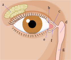
Tear system consists of lacrimal gland (a), punctums (b,e), canalicules (c,f), lacrimal sac (d). Tear is then drained through nasolacrimal duct (g) into nasal cavity
Specialty Ophthalmology
Differential diagnosis Tears arising from lacrimal sac fistula. [1]

Nasolacrimal duct obstruction is the obstruction of the nasolacrimal duct and may be either congenital or acquired. Obstruction of the nasolacrimal duct leads to the excess overflow of tears called epiphora. [2]

Contents

Sign and symptoms

Excessive tearing is the most common complaint of patients with nasolacrimal duct obstruction, followed by acute or chronic infections. [3] Pain at the side of the nose suggests dacryocystitis.

Nasolacrimal duct obstruction is more common with increasing age and more common in females than males. [3]

Cause

Involutional stenosis

Involutional stenosis is probably the most common cause of nasolacrimal duct obstruction in older people. It affects women twice as frequently as men. Although the inciting event in this process is unknown, clinicopathologic study suggests that compression of the lumen of the nasolacrimal duct is caused by inflammatory infiltrates and edema. This may be the result of an unidentified infection or possibly an autoimmune disease.[ citation needed ]

Dacryolith

Dacryoliths or cast formation, within the lacrimal sac can also produce obstruction of the nasolacrimal duct.

Sinus disease

Sinus disease often occurs in conjunction with, and in other instances may contribute to the development of nasolacrimal duct obstruction. Patients should be asked about previous sinus surgery, as the nasolacrimal duct is sometimes damaged when the maxillary sinus ostium is being enlarged anteriorly.

Trauma

Naso-orbital fractures may involve the nasolacrimal duct. Early treatment by fracture reduction with stenting of the entire lacrimal drainage system should be considered. However, such injuries are often not recognized or are initially neglected as more serious injuries are managed. In such cases, late treatment of persistent epiphora usually requires dacryocystorhinostomy.

Inflammatory disease

Granulomatous disease, including sarcoidosis, granulomatosis with polyangiitis, and midline granuloma, may also lead to nasolacrimal duct obstruction.

Lacrimal plugs

As with similar cases of canalicular obstruction, dislodged punctal and canalicular plugs can migrate to and occlude the nasolacrimal duct.

Neoplasm

Neoplasm should be considered in any patient presenting with nasolacrimal duct obstruction. In patients with atypical presentations, including younger age and male gender, further workup is appropriate. Bloody punctual discharge or lacrimal sac distension above the medial canthal tendon is also highly suggestive of neoplasm.

Congenital

Congenital nasolacrimal duct obstruction, or dacryostenosis, occurs when the lacrimal duct has failed to open at the time of birth, most often due to an imperforate membrane at the valve of Hasner. [4] Around 6% of infants have congenital nasolacrimal duct obstruction, or dacryostenosis, usually experiencing a persistent watery eye even when not crying. If a secondary infection occurs (Dacryocystitis), purulent (yellow / green) discharge may be present.

Most cases resolve spontaneously, with antibiotics reserved only if conjunctivitis occurs. Lacrimal sac massage has been proposed as helping to open the duct, though this is not always successful. [5] The aim of massage is to generate enough hydrostatic pressure (downward, toward the nose) to "pop" open any obstruction. Additional massage may then be performed up toward the lacrimal punctum, in order to express any infectious material out of the nasolacrimal sac. When discharge or crusting is present, the lids should be gently cleaned using cooled pre-boiled water or saline.

Referral to an ophthalmologist is indicated if symptoms are still present at 12 months, or sooner if significant symptoms or recurrent infections occur. Nasolacrimal duct probing may be performed in the office setting (usually from 4 to 8 months of age) or under general anesthesia in an operating room for older patients. The success rate of probing is higher for younger children. A silastic tube or stent may be employed along with probing to maintain tear duct patency. [6] A systematic review comparing immediate probing with deferred probing found that in children with unilateral nasolacrimal duct obstruction, immediate probing resulted in a higher success rate of treatment compared to deferred probing. [7]

Diagnosis

Evaluation is in the form of a dye disappearance test followed by irrigation test. By using this sequence (with modifications) as a guide, the physician can frequently streamline diagnostic testing.

Dye disappearance test

The dye disappearance test is useful for assessing the presence or absence of adequate lacrimal outflow, especially in unilateral cases. It is more heavily relied upon in children, in whom lacrimal irrigation is impossible without deep sedation. Using a drop of sterile 2% fluorescein solution or a moistened fluorescein strip, the examiner instills fluorescein into the conjunctival fornices of each eye and then observes the tear film, preferably with the cobalt blue filter of the slit lamp. Persistence of significant dye and, particularly asymmetric clearance of the dye from the tear meniscus over a 5-minute period indicate an obstruction. If the dye disappearance test result is normal, severe lacrimal drainage dysfunction is highly unlikely. The Jones tests are variations of the dye disappearance test.

Irrigation test

Flushing the nasolacrimal duct in a cat. Traenengangspuelung.jpg
Flushing the nasolacrimal duct in a cat.

In irrigation test, a lacrimal irrigation cannula is passed into the punctum and advanced through the canaliculus to the lacrimal fossa. Clear water or saline is then irrigated through the cannula. If fluid passes into the nose without reflux out of the opposite canaliculus, the system is patent. If no fluid passes but it all comes back through either punctum, nasolacrimal duct obstruction is present.

Management

Intubation and stenting

Some clinicians believe that partial stenosis of the nasolacrimal duct with symptomatic epiphora sometimes responds to surgical intubation of the entire lacrimal drainage system. This procedure should be performed only if the tubes can be passed easily. In complete nasolacrimal duct obstruction, intubation alone is not effective, and a dacryocystorhinostomy should be considered.

Dacryocystorhinostomy

A dacryocystorhinostomy is the treatment of choice for most patients with acquired nasolacrimal duct obstruction. Surgical indications include recurrent dacryocystitis, chronic mucoid reflux, painful distension of the lacrimal sac, and bothersome epiphora. For patients with dacryocystitis, active infection should be cleared, if possible, before a dacryocystorhinostomy is performed. [2]

See also

Related Research Articles

<span class="mw-page-title-main">Tears</span> Clear liquid secreted from glands in eyes of mammals

Tears are a clear liquid secreted by the lacrimal glands found in the eyes of all land mammals. Tears are made up of water, electrolytes, proteins, lipids, and mucins that form layers on the surface of eyes. The different types of tears—basal, reflex, and emotional—vary significantly in composition.

<span class="mw-page-title-main">Eye surgery</span> Surgery performed on the eye or its adnexa

Eye surgery, also known as ophthalmic or ocular surgery, is surgery performed on the eye or its adnexa, by an ophthalmologist. Eye surgery is part of ophthalmology. The eye is a fragile organ, and requires due care before, during, and after a surgical procedure to minimize or prevent further damage. An eye surgeon is responsible for selecting the appropriate surgical procedure for the patient, and for taking the necessary safety precautions. Mentions of eye surgery can be found in several ancient texts dating back as early as 1800 BC, with cataract treatment starting in the fifth century BC. It continues to be a widely practiced class of surgery, with various techniques having been developed for treating eye problems.

<span class="mw-page-title-main">Meckel's diverticulum</span> Medical condition

A Meckel's diverticulum, a true congenital diverticulum, is a slight bulge in the small intestine present at birth and a vestigial remnant of the omphalomesenteric duct. It is the most common malformation of the gastrointestinal tract and is present in approximately 2% of the population, with males more frequently experiencing symptoms.

<span class="mw-page-title-main">Dry eye syndrome</span> Medical condition

Dry eye syndrome, also known as keratoconjunctivitis sicca, is the condition of having dry eyes. Symptoms include dryness in the eye, irritation, redness, discharge, blurred vision, and easily fatigued eyes. Symptoms range from mild and occasional to severe and continuous. Dry eye syndrome can lead to blurred vision, instability of the tear film, increased risk of damage to the ocular surface such as scaring of the cornea, and changes in the eye including the neurosensory system.

<span class="mw-page-title-main">Nasolacrimal duct</span> Carries tears from the lacrimal sac of the eye into the nasal cavity.

The nasolacrimal duct carries tears from the lacrimal sac of the eye into the nasal cavity. The duct begins in the eye socket between the maxillary and lacrimal bones, from where it passes downwards and backwards. The opening of the nasolacrimal duct into the inferior nasal meatus of the nasal cavity is partially covered by a mucosal fold.

<span class="mw-page-title-main">Schirmer's test</span>

Schirmer's test determines whether the eye produces enough tears to keep it moist. This test is used when a person experiences very dry eyes or excessive watering of the eyes. It can cause damage to the cornea. A negative test result is normal. Both eyes normally secrete the same amount of tears.

Vaginal atresia is a condition in which the vagina is abnormally closed or absent. The main causes can either be complete vaginal hypoplasia, or a vaginal obstruction, often caused by an imperforate hymen or, less commonly, a transverse vaginal septum. It results in uterovaginal outflow tract obstruction. This condition does not usually occur by itself within an individual, but coupled with other developmental disorders within the female. The disorders that are usually coupled with a female who has vaginal atresia are Mayer-Rokitansky-Küster-Hauser syndrome, Bardet-Biedl syndrome, or Fraser syndrome. One out of every 5,000 women have this abnormality.

An imperforate lacrimal punctum is a congenital disorder of dogs involving the lack of an opening to the nasolacrimal duct in the conjunctiva. Dogs normally have two lacrimal puncta, the superior and inferior. This condition can affect either or both. Symptoms include excessive tearing and tear staining of the hair around the eye. Affected breeds include the American Cocker Spaniel, Bedlington Terrier, Golden Retriever, Poodle, and Samoyed. Imperforate lacrimal puncta can be corrected by surgical opening of the punctum.

<span class="mw-page-title-main">Lacrimal sac</span> Upper, dilated end of the nasolacrimal duct

The lacrimal sac or lachrymal sac is the upper dilated end of the nasolacrimal duct, and is lodged in a deep groove formed by the lacrimal bone and frontal process of the maxilla. It connects the lacrimal canaliculi, which drain tears from the eye's surface, and the nasolacrimal duct, which conveys this fluid into the nasal cavity. Lacrimal sac occlusion leads to dacryocystitis.

<span class="mw-page-title-main">Sialography</span> Radiological technique

Sialography is the radiographic examination of the salivary glands. It usually involves the injection of a small amount of contrast medium into the salivary duct of a single gland, followed by routine X-ray projections.

Oculoplastics, or oculoplastic surgery, includes a wide variety of surgical procedures that deal with the orbit, eyelids, tear ducts, and the face. It also deals with the reconstruction of the eye and associated structures.

<span class="mw-page-title-main">Dacryocystorhinostomy</span>

Dacryocystorhinostomy (DCR) is a surgical procedure to restore the flow of tears into the nose from the lacrimal sac when the nasolacrimal duct does not function.

<span class="mw-page-title-main">Dacryocystitis</span> Medical condition

Dacryocystitis is an infection of the lacrimal sac, secondary to obstruction of the nasolacrimal duct at the junction of lacrimal sac. The term derives from the Greek dákryon (tear), cysta (sac), and -itis (inflammation). It causes pain, redness, and swelling over the inner aspect of the lower eyelid and epiphora. When nasolacrimal duct obstruction is secondary to a congenital barrier it is referred to as dacryocystocele. It is most commonly caused by Staphylococcus aureus and Streptococcus pneumoniae. The most common complication is corneal ulceration, frequently in association with S. pneumoniae. The mainstays of treatment are oral antibiotics, warm compresses, and relief of nasolacrimal duct obstruction by dacryocystorhinostomy.

A punctal plug, also known as tear duct plug or lacrimal plug, is a small medical device that is inserted into the tear duct (puncta) of an eye to block the duct. This prevents the drainage of liquid from the eye. They are used to treat dry eye.

<span class="mw-page-title-main">Epiphora (medicine)</span> Medical condition

Epiphora is an overflow of tears onto the face, other than caused by normal crying. It is a clinical sign or condition that constitutes insufficient tear film drainage from the eyes, in that tears will drain down the face rather than through the nasolacrimal system.

<span class="mw-page-title-main">Retropharyngeal abscess</span> Medical condition

Retropharyngeal abscess (RPA) is an abscess located in the tissues in the back of the throat behind the posterior pharyngeal wall. Because RPAs typically occur in deep tissue, they are difficult to diagnose by physical examination alone. RPA is a relatively uncommon illness, and therefore may not receive early diagnosis in children presenting with stiff neck, malaise, difficulty swallowing, or other symptoms listed below. Early diagnosis is key, while a delay in diagnosis and treatment may lead to death. Parapharyngeal space communicates with retropharyngeal space and an infection of retropharyngeal space can pass down behind the esophagus into the mediastinum. RPAs can also occur in adults of any age.

<span class="mw-page-title-main">Dacryocystocele</span> Medical condition

Dacryocystocele (Dacryocystitis) or timo cyst is a benign, bluish-gray mass in the inferomedial canthus that develops within a few days or weeks after birth. The uncommon condition forms as a result as a consequence of narrowing or obstruction of the nasolacrimal duct, usually during prenatal development. Nasolacrimal duct obstruction disrupts the lacrimal drainage system, eventually creating a swelling cyst in the lacrimal sac area by the nasal cavity. The location of the cyst can cause respiratory dysfunction, compromising the airway. The obstruction ultimately leads to epiphora, an abundance of tear production.

<span class="mw-page-title-main">Conjunctivochalasis</span> Medical condition

Conjunctivochalasis, also known as Mechanical Dry Eye (MDE), is a common eye surface condition characterized by the presence of excess folds of the conjunctiva located between the globe of the eye and the eyelid margin.

<span class="mw-page-title-main">Frontal sinus trephination</span>

Frontal sinus trephination is a surgical procedure where in a small opening is made in the floor of the frontal sinus facilitating drainage of its contents. Drainage of the frontal sinus is done through the floor of frontal sinus above the inner canthus.

<span class="mw-page-title-main">Dacryoscintigraphy</span>

Dacryoscintigraphy (DSG), also known as lacrimal scintigraphy, is a nuclear medicine technique for imaging the lacrimal apparatus. It is used to identify obstructions, for example in the lacrimal duct, nasal cavity or nasolacrimal duct.

References

  1. Nerad, Jeffrey A.; Carter, Keith D.; Alford, Mark (2008). "Disorders of the Lacrimal System: Congenital Obstruction". Oculoplastic and Reconstructive Surgery. Elsevier. pp. 131–137. doi:10.1016/b978-0-323-05386-0.50010-7. ISBN   978-0-323-05386-0. This tearing is different (from Dacryocystitis), as it originates from the fistula located below the eyelid on the cheek (may be associated with nasolacrimal duct obstruction).
  2. 1 2 Myron Yanoff; Jay S. Duker (2009). Ophthalmology (3rd ed.). Mosby Elsevier. pp. 1482–1487. ISBN   9780323043328.
  3. 1 2 Jawaheer L, MacEwen CJ, Anijeet D (2017). "Endonasal versus external dacryocystorhinostomy for nasolacrimal duct obstruction". Cochrane Database Syst Rev. 2017 (2): CD007097. doi:10.1002/14651858.CD007097.pub3. PMC   6464401 . PMID   28231605.
  4. Blocked tear ducts in infants, Pediatric Views, June 2006 http://www.childrenshospital.org/views/june06/blocked_tear_ducts.html Archived 2012-07-08 at the Wayback Machine
  5. Young JD, MacEwen CJ (1997). "Managing congenital lacrimal obstruction in general practice". BMJ. 315 (7103): 293–6. doi:10.1136/bmj.315.7103.293. PMC   2127215 . PMID   9274552.
  6. Engel JM, Hichie-Schmidt C, Khammar A, Ostfeld BM, Vyas A, Ticho BH (2007). "Monocanalicular silastic intubation for the initial correction of congenital nasolacrimal duct obstruction". J AAPOS. 11 (2): 183–186. doi:10.1016/j.jaapos.2006.09.009. PMID   17307001.
  7. Petris C, Liu D (2017). "Probing for congenital nasolacrimal duct obstruction". Cochrane Database of Systematic Reviews. 2017 (7): CD011109. doi:10.1002/14651858.CD011109.pub2. PMC   5580992 . PMID   28700811.